Foundational Concepts
1/50
There's no tags or description
Looks like no tags are added yet.
Name | Mastery | Learn | Test | Matching | Spaced |
|---|
No study sessions yet.
51 Terms
What are the 7 levels of organisation?
Atoms, mocecules and macro-molecules (make up chemical level)
Organelles
Cells
Tissues
Organs
Systems
Organisms
What is the integumentary system?
Everything that makes up the outermost layer of an organism acting as a physical barrier from the external environment, including the skin, hair, nails and more.
Location, structure and function of the plasma membrane.
Semi-permeable phospholipid bilayer that surrounds a cell, separating its contents from the external environment, controlling what enters and exits the cell and commmunicates with other cells.
Location, structure and function of the cytoplasm.
Contains all the organelles (expect for the nucleus) and the cytosol, which is the fluid that fills the cell and the organelles are suspended in.
Location, structure and function of the cytoskeleton.
A network of protein filaments located throughout the cytoplasm that provide shape and structure to the cell.
Location, structure and function of ribosomes.
Ribosomes are small organelles that are located on the surface of the rough endoplasmic reticulum as well as freely throughout the cytoplasm and undergo protein synthesis.
Location, structure and function of the nucleus.
Rough spherically shaped organelles that are seperate to the cytoplasm and contain DNA and nucleoli that produce ribosomes.
Location, structure and function of the rough endoplasmic reticulum.
Network of membranes formed from flattened sacs that extend from the nuclear envelope. Contain ribosomes on their surface and therefore play a role in protein synthesis.
Location, structure and function of the smooth endoplasmic reticulum.
Network of smooth membranes formed from tubules that extend from the rough endoplasmic reticulum, playing a role in lipid and steroid synthesis.
Location, structure and function of the Golgi apparatus.
A series of flattened sacs found in the cytoplasm that sort, modify and package proteins recieved from the rough endoplasmic reticulum into vesicles to be transported around or out of the cell.
Location, structure and function of the mitochondria.
Double-membrane bound organelles containing ribosomes that are found within the cytoplasm and are the site of ATP production.
Location, structure and function of lysosomes.
A spherical membrane-bound vesicle found within the cytoplasm that contains digestive enzymes for the breakdown of cellular components.
Describe the 2 parts of phospholipids.
Phosphate head that is polar and hydrophilic, attracted to the extracellular environment
Fatty acid tails that are non-polar and hydrophobic, repelled from the water in the extracellular environment causing them to rotate towards each other.
Types of membrane proteins
Channels - create pores in the membrane, allowing certain ions to pass through. Can be voltage-gated, ligand-gated or ungated.
Carriers - changes shape to allow substances to pass through the membrane
Receptors - binds to chemical changes that trigger a change in cellular processes
Enzymes - catalyze chemical reactions
Explain the selective permeability of the plasma membrane.
Small non-polar, uncharged molecules can pass through due to the non-polar fatty acid tails being the central component
Protein channels and carriers increase the permeability by allowing selected ions and polar molecules to pass through
However large molecules such as proteins must pass via vesicles
What is the difference between active and passive transport across the plasma membrane?
Passive transport requires no input of energy as molecules move with their concentration gradient. Whereas active transport requires an input of energy as molecules are moving against their concentration gradient.
Name and describe the 3 types of passive transport.
Simple Diffusion - particles naturally move with their concentration gradient to achieve equilibrium.
Osmosis - water molecules naturally move with their concentration gradient to achieve equilibrium of solute concentration
Facilitated Diffsion - particles moving with their concentration gradient through carrier and channel proteins
Name and describe the 2 types of active transport.
Primary Active Transport - particles moving against their concentration gradient through carrier proteins that require ATP energy.
Vesicular Transport - transport of large particles via vesicles
Name and describe the types of vesicular transport.
Exocytosis - transport out of the cell
Endocytosis - transport into the cell
Pinocytosis - endocytosis of liquid particles
Phagocytosis - endocytosis of solid particles
What are tissues?
Groups of cells within an extracellular matrix that perform a specialised function.
What are the 4 types of tissue?
Epithelial
Connective
Muscular
Nervous
Describe epithelial tissue/epithelium.
Closely packed sheets of cells with very little extracellular matrix between that form linings and coverings of other tissues.
Describe the structure of epithelial tissue.
Free surface - the uncovered surface
Basement membrane - the deepest layer that attaches to other tissue
What are the 3 functions of epithelial tissue and examples?
form a protective barrier, eg. Outer layer of the skin
Control what enters and exits the body, eg. Lining the intestines to control what substances of food and drink enter the body
secrete substances by lining glands, eg. Lining of the sweat glands that secrete sweat onto the skin
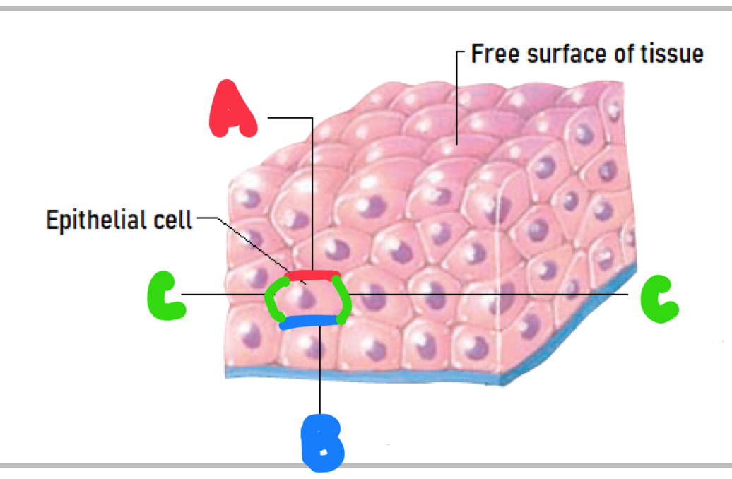
Name the different surfaces of an epithelial cell.
A - apical surface (faces the free surface)
B - basal surface (facing the basement membrane)
C - lateral surfaces (facing the side)
What are the categories of epithelial tissue based on layers?
Simple epithelium - one layer of cells and a basement membrane
Stratified epithelium - multiple layers of cells and a basement membrane
What are the layers of stratified epithelial tissue?
Apical layer (makes up the free surface)
Basal Layer (attached to the basement membrane)
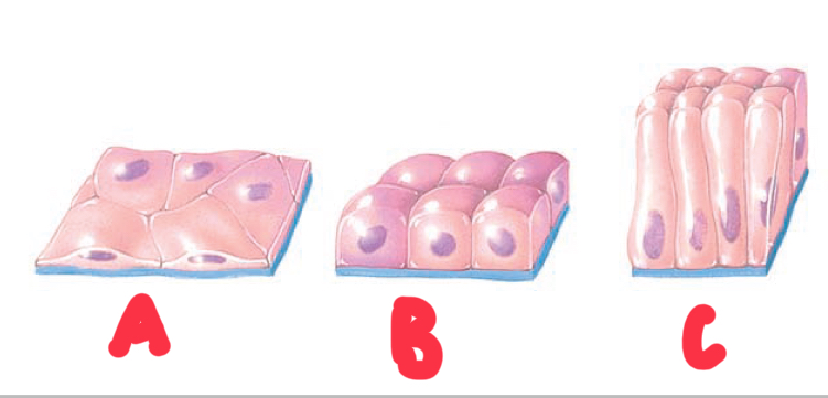
Name and describe the cell shapes of epithelial tissues.
A - squamous (flat cells)
B - cuboidal (cube-shaped cells)
C - columnar (column-shaped cells)
What is the difference between exocrine and endocrine glands?
Exocrine Glands - epithelial cells synthesise and secrete substances into ducts and carry to bodily surfaces
Endocrine Glands - epithelial cells synthesise and secrete hormones into blood vessels to be distributed throughout the body
What is the extracellular matrix made of?
Ground substance - water and dissolved solutes
Proteins fibres
Collagen fibres - strong, resist tension but also flexible
Elastic fibres - stretch and bounce back
Reticular fibres - network of proteins that support tissue structure
What are 3 differences between connective and epithelial tissue.
Extracellular matrix is more abundant in connective tissue than epithelial
Cells are well distributed in connective tissue compared to epithelial where they are closely packed together in sheets
Epithelial tissue is avascular, connective tissue is vascular
What is connective tissue?
The most abundant type of tissue in the body that connects and binds tissues and organs together.
What are the 5 classifications of connective tissue?
Loose connective tissue
Dense connective tissue
Liquids connective tissue
Cartilage
Bone
What are cell junctions?
The contact points between plasma membranes of adjacent cells, allowing cells to form functional units.
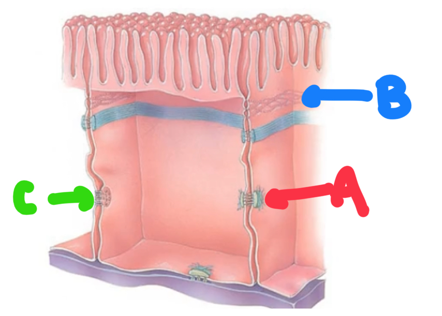
What are the 3 types of cell junctions?
A - desmosome
B - tight junction
C - gap junction
Describe gap junctions
Tunnels that connect adjacent plasma membranes, allowing substances to freely diffuse between cells.
Describe desmosomes.
Junctions that strongly adhere adjacent cells together
Describe tight junctions
Protein beads that wrap around adjacent plasma membranes, creating a fluid tight seal that prevents fluid from moving between the gaps of cells.
What are membranes?
Organs that line body structures and contain both epithelial and connective tissue.
What are the 2 types of membranes?
Mucous membranes (mucosa)
Serous membranes (serosa)
Describe mucous membranes and examples.
A layer of connective tissue underneath epithelial tissue that contains a layer of mucous on the free surface to keep the membrane moist. They line body cavities such as the oral and nasal cavity.
Describe serous membranes and examples.
Double layered membrane of connective and epithelial tissue separated by serous fluid that reduces friction. They line body cavities between body parts, eg. Ribs and lungs.
Name and describe the 4 anatomical planes of the body.
Transverse/horizontal - divides superior and inferior
Sagittal/median - divides left and right
Coronal/frontal - divides anterior and posterior
Oblique - any other plane
What do intermediate, ipsilateral and contralateral mean?
intermediate - in between 2 structures
Ipsilateral - on the same side of the body
Contralateral - on different sides of the body
Describe homeostasis.
The body’s internal environment in a state of balance in which everything is within normal ranges and there is optimal functioning.
Compare 3 differences between the effects of the endocrine and nervous system.
NS is fast-acting whereas ES is much slower
NS effects are short-lived whereas ES is long-lasting
NS affects a short range whereas ES affects a larger range
Describe a feedback loop.
A series of processes that occurs within the body to maintain homeostasis.
Name and describe the 5 elements of a feedback loop.
Variable - an element of the body
Stimulus - something that causes a change to a variable
Receptors - detect stimulus and transmit input to control center
Control Center - the brain or other structures that receive input and determine whether variable is within its set point. If not, transmits output to effectors to correct this.
Effectors - recieve output and carry out response to return variable to homeostasis
What is the difference between positive and negative feedback loops?
Negative feedback loops reverse the effects of the stimulus to return the variable to homeostasis. Whereas a positive feedback loop reinforces the effects of the stimulus to continuously increase or decrease the variable.
Describe a shut-off mechanism.
A mechanism that ceases the transmission of input and output in a positive feedback loop to return the variable to homeostasis.
What is an example of a positive feedback loop?
Stimulus - cervix stretch during childbirth
Receptors - cervic detects stretching and transmits input to brain
Control center - brain receives and processes input and transmits output to endocrine cells
Effectors - endocrine cells release oxytocin that travels to the uterus causing the uterine wall to contract and push the baby, further stretching the cervix and resulting in further transmission of input. Once the baby is born, the cervix is no longer stretched and input transmission ceases.