Wrist & Hand Examination
 Call Kai
 Learn
Learn Practice Test
 Spaced Repetition
 Match
Match Flashcards
 Knowt Play
1/47

There's no tags or description
Looks like no tags are added yet.
Name | Mastery | Learn | Test | Matching | Spaced | Call with Kai | Chat |
|---|
No analytics yet
Send a link to your students to track their progress
48 Terms
What are the major joints of the wrist and hand?
Distal Radioulnar Joint (DRUJ), Radiocarpal Joint, Intercarpal Joints, Metacarpal Joints

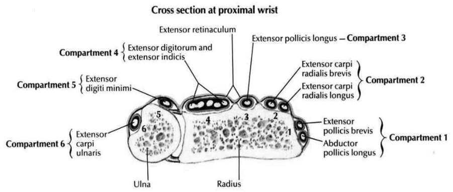
How many compartments are there in the wrist and what are they comprised of?
There are 6 compartments in the wrist:
Compartment 1: EPB; APB
Compartment 2: ECRB, ECRL
Compartment 3: EPL
Compartment 4: ED, EI
Compartment 5: EDM
Compartment 6: ECU
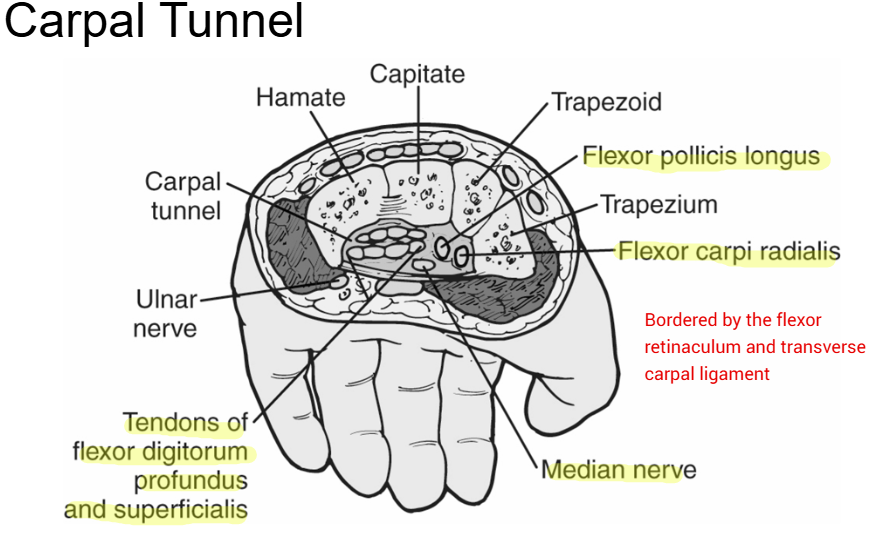
What structures are involved in the carpal tunnel?
Flexor Pollices Longus, Flexor Carpi Radialis, Tendons of Flexor Digitorum Profundis and Superficialis, Median Nerve
What should the subjective history include for hand/wrist pathologies?
-Signs and Symptoms (N/T, Crepitus, Discoloration, Weakness, Pain Description)
-MOI (FOOSH, Insidious)
-PMH
-Function Impairment, Occupation, Recreation
What should be observed when accessing wrist/hand patholigies?
-Hand in resting position (Edema, Discoloration, Deformities, MM wasting
-Wound Status
-Movements
-Facial Expressions
-Fingernails
What should be performed during the examination for wrist/hand pathologies?
-AROM/PROM
-Resisted Isos
-Grip Strength
-Special Tests
-Reflexes
-JPAs
-Palpation
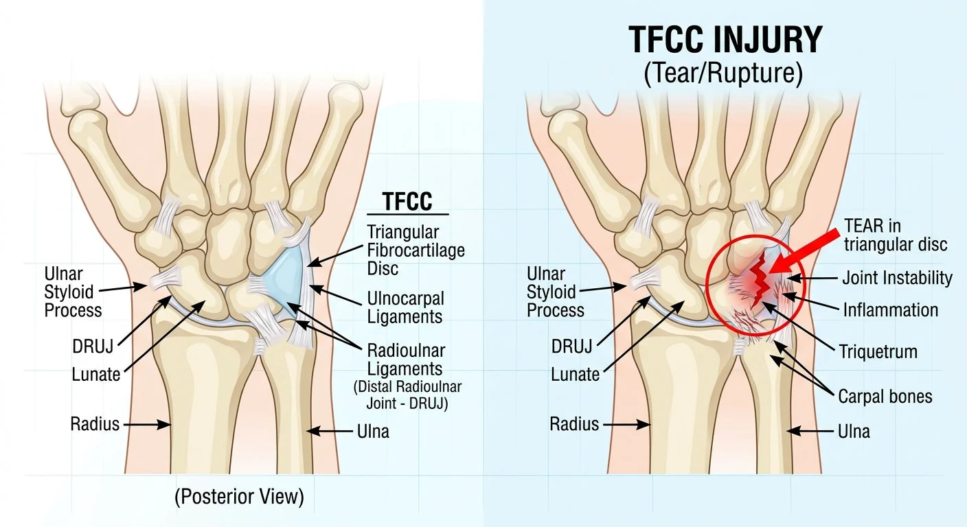
Common Pathology: What is TFCC Injury?
-TFCC or Triangular Fibrocartilage Complex injury
-MOI: FOOSH w/ supination/chronic rotational loading (racket sports)
-Ligamentous Support: Ulnolunate, Ultotriquetral, UCL, RU
Symptoms: Pain @ medial wrist at distal ulna increased with gripping and end range supination/pronation. TTP @ dorsal depression (ulna fovea). Mobility of carpals into UD elicits painful crepitation. Passive supination + UD reproduces pain.
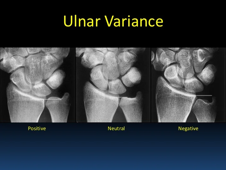
+Ulnar Variance
What is ulnar variance and what are the types?
-Length of the ulna compared to the length of the radius at the wrist.
-Neutral Ulnar Variance: End of the ulna is in line with end of the radius (Normal)
-Negative Ulnar Variance: Ulna is shorter than the radius (Keinbock’s Disease/AVN lunate)
-Positive Ulnar Radiance: Ulna is longer than the radius (TFCC Degen)
What can be used to treat TFCC injuries?
Conservative Management:
Splint/Long Arm Cast: used for 6 weeks and put in elbow flex 90 deg, wrist UD and extension. After removed, A/AAROM for flex/ext, then pron/sup/RD/UD. Progressive strengthening after 2 weeks if asymptomatic and be cautious with torsion
-Proximal Strengthening
What are the two types of wrist instabilities?
-Dissociative: Dorsal intercalated Segmental Instability (DISI), Ventral Intercalated Segmental Instability (VISI)
-Non-dissociative
What is Dorsal Intercalated Segmental Instability (DISI)
Most common dissociative instability caused by scapholunate instability also referred to as “SLAC” wrist (Scapholunate Advanced Collapse). Lunate tilts dorsally.
-MOI: FOOSH (wrist ext and UD)
-Symptoms: weakness with grip, chronic/vague pain, TTP @ scaphoid/lunate, laxity with mobility of scapholunate articulation
+Watson’s Scaphoid Shift = “clunk”
What is Watson’s Scaphoid Shift Test?
A special test used to confirm a DISI. Palpate the scaphoid tubercle and passively ulnarly deviate. Maintain scaphoid pressure and radially deviate. (+) clunk and concordant pain

What can be used to treat a DISI?
Surgical: Scapho-lunate Ligament repair
What is a Ventral Intercalated Segmental Instability (VISI)
2nd most common dissociative instability of the lunotriquetral ligament which is less common. Similar symptoms to the DISI and there is a ventral tilt of the lunate.
-MOI: FOOSH (wrist ext and UD)
-Symptoms: weakness with grip, chronic/vague pain, TTP @ triquetrum/lunate
What are Non-Dissociative Instabilities
These are the most common dynamic instabilities at the wrist and caused by insufficiency of dorsal intercarpal ligaments. They can be asymptomatic w/ volitional subluxation/reduction.
-Management: conservative treatment includes cast for 2 weeks and surgery if conservative fails
What is Carpal Tunnel Syndrome?
Inschemic compression of the median nerve which leads to increased carpal tunnel pressure due to synovial fluid, tendon tension, and stenosis.
Causes: cumulative trauma, extreme wrist flexion/extension, fingertip loading, forceful contraction of finger flexors, FOOSH, pregnancy, renal dysfunction, infection, gout, pseudogout, acromegaly, amyotrophy, RA, collagen disorders, diabetes, hemodialysis
Carpal Tunnel Syndrome vs Cervical Radoculopathy
Neck and shoulder symptoms are common with CTS and cervical OA can contribute to symptoms of CTS. Foreword head posture and decreased cervical ROM with unilateral CTS.
-Bottomline: examine both distal and proximal nerve injury and treat both.
What should be involved in Carpal Tunnel Syndrome examination?
Subjective: intermittent pain and paresthesia in median nerve distribution that radiates proximally into forearm and arm. Worse in the evening (wrist flexion) and morning stiffness
Objective: motor and sensory examination including ULTT median nerve, Phalen test, Tinel’s test, Median nerve compression test
Diagnostics: Nerve Conduction Study, EMG, Radiography
How to conservatively treat Carpal Tunnel Syndrome?
Splinting: in neutral or wrist extension that is used day/night or both. Can be rigid or flexible based on personal needs.
Activity Modification: avoiding repetitive wrist motion, sustained pinching gripping or wrist flexion, ergonomics
Exercise: median nerve gliding, tendon gliding (FDS and FDP), yoga
Pharmacological: diuretics and NSAIDS
How to surgically treat Carpal Tunnel Syndrome
-Release of TCL (Flexor Retinaculum)
-Tendon Transfers if atrophy, worsening function, failed conservative treatment
Post-Surgical: edema control, splinting, wound management/STM, tendon gliding, desensitization, monitor nerve recovery
What are other wrist neuropathies?
Ulnar Nerve: nerve compression in the cubital or Guyon tunnel due to abnormality of Pisotriquetral joint or hook of hamate. PMH can include diabetes/peripheral neuropathy and can be treated with splinting and patient education
-Radial Nerve: nerve compression caused by repetitive wrist extension leading to irritation of dorsal wrist, wrist drop (weak finger/wrist ext), and impaired sensation in dorsal 1st interosseous space.
What is DeQuervain’s Disease?
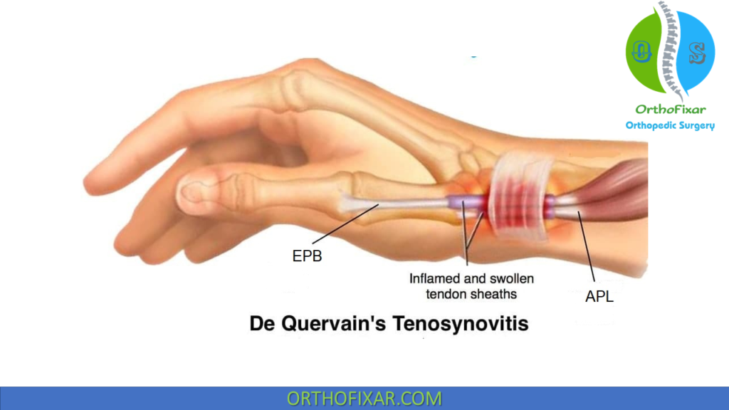
Progressive tenosynovitis of 1st compartment (APL and EPB) tendon sheaths. The extensor retinaculum is thickened and there is stenosis in the canal.
MOI: caused by overuse of thumb or RD/UD and OA
Symptoms: dull ache over the radial wrist and pain with turning doorknob/keys. “Creak” with AROM.
What objective measures can be used to diagnose DeQuervain’s Disease/
Objective: localized swelling and TTP at distal radius/styloid with pain radiating into proximal forearm and thumb. Dull ache over radial wrist worse with UD and thumb flexion/abduction. Crepitus over 1st compartment (APL and EPB).
+Finkelstein’s Test
What is conservative treatment for DeQuervain’s Disease?
-Splinting: 3-6 weeks of splinting using thumb splica splint and thumb in extension and abduction. Ensure thumb and finger opposition for continued function.
-Rest, Activity Mod, NSAIDs
-AROM and progressive strengthening
-Manual Therapy: Radial glide proximal row + ulnar glide trapezium, capitate manip, CMC joint mob, TFM 1st compartment, cervical mob, neuro mob,
-Cortisone Injection
What is Intersection Syndrome?
Commonly confused with DeQuervain’s and caused by tenosynovitis of ECLR and ECRB.
Symptoms: pain at the distal forearm 4-8 cm proximal to Lister’s tubercle and increased with wrist flexion and extension and resisted wrist extension.
Treatment: spint w/ thumb and wrist 15-20 deg extension, iontophoresis/phonophoresis, manual therapy, activity mod, patient education.

What are other tendinopathies of the hand/wrist?
EPL: rare except with RA and referred to as “Drummer Boy Palsy” caused by overuse
EIP: Overuse with pain with resisted index finger extension in wrist flexion
FCU: Most common wrist flexor tendinopathy caused by overuse and racket sports and pain with wrist flexion/UD and swelling at proximal pisiform
FCR: Common with other conditions such as OA of CMC
What is a Scaphoid Fracture?
The most commonly fractured carpal and is commonly fractured at the waist and proximal pole.
Subjective: pain in the dorsal radius and wrist pain
MOI: FOOSH w/ pronation
Objective: Axial compression, TTP over anatomic snuffbox, little swelling and loss of snuffbox concavity.
Imaging: difficult to identify on radiograph so MRI if (-) radiograph and high suspicion.
Symptoms: chronic pain, loss of motion, early OA common following fracture.
Where can the scaphoid fracture and how does it affect recovery?
The scaphoid can fracture, at the distal, waist, and proximal portions. Blood flow only goes one way and the closer the fracture is to the proximal portion the less blood flow and decreased chance of reunion.
Management:
Proximal: 16-20 weeks in slight wrist extension and radial deviation
Central Third: 6 weeks in long arm thumb spica then 6 weeks in short arm thumb spica
Distal Third: 6-8 weeks short thumb spica
What are other common wrist fractures?
Distal Radius Fracture: the most common fracture for all ages.
Colle’s Fracture: complete radius fracture with posterior displacement in the shape of a “silver fork”
Management: anatomical alignment (radius length) + soft tissue management. Closed reduction and immobilization if more serious. Commonly lose full forearm pronation/supination
Older = metaphyseal; Younger = intra-articular
What are Metacarpal Fractures?
The most commonly associated with direct trauma or indirect trauma.
CMC Fracture: axial load or stress with wrist flexion. Reverse Bennett’s: Intraarticular fracture/dislocation at base of the 5th
Metacarpal Shaft: axial load, direct trauma, torsional stress
Metacarpal Neck: Striking a solid object; “Boxer’s Fracture”
Management: Closed reduction/ immobilization or controlled mobilization (block splint)
What are the LOAF muscles of the hand and why are they important?
L: Lateral 2 lumbricals
O: Opponens Pollicis
A: Abductor Pollicis Brevis
F: Flexor Pollicis Brevis
Importance: These are the intrinsic hand muscles primarily innervated by the median nerve.
What is an ulnar drift hand deformity?
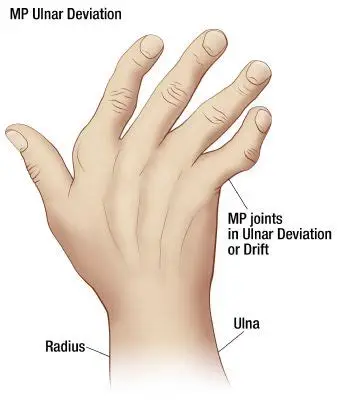
Damage to the collateral ligaments and extensor mechanism at MCP which results in the pulling of fingers into UD, pronation and palmar subluxation. Classic occurrence with RA.
What is a swan neck hand deformity?
Flexion deformity of DIP; hyperextension at PIP and the least function of all deformities. Results in destruction of the ORL.

What is a Boutonniere hand deformity?
Damage to the common extensor tendon at base of the middle phalanx. Extra effort for extension results in hyperextension at DIP. 2ns most common closed tendon sports injury.

What is a mallet finger hand deformity?
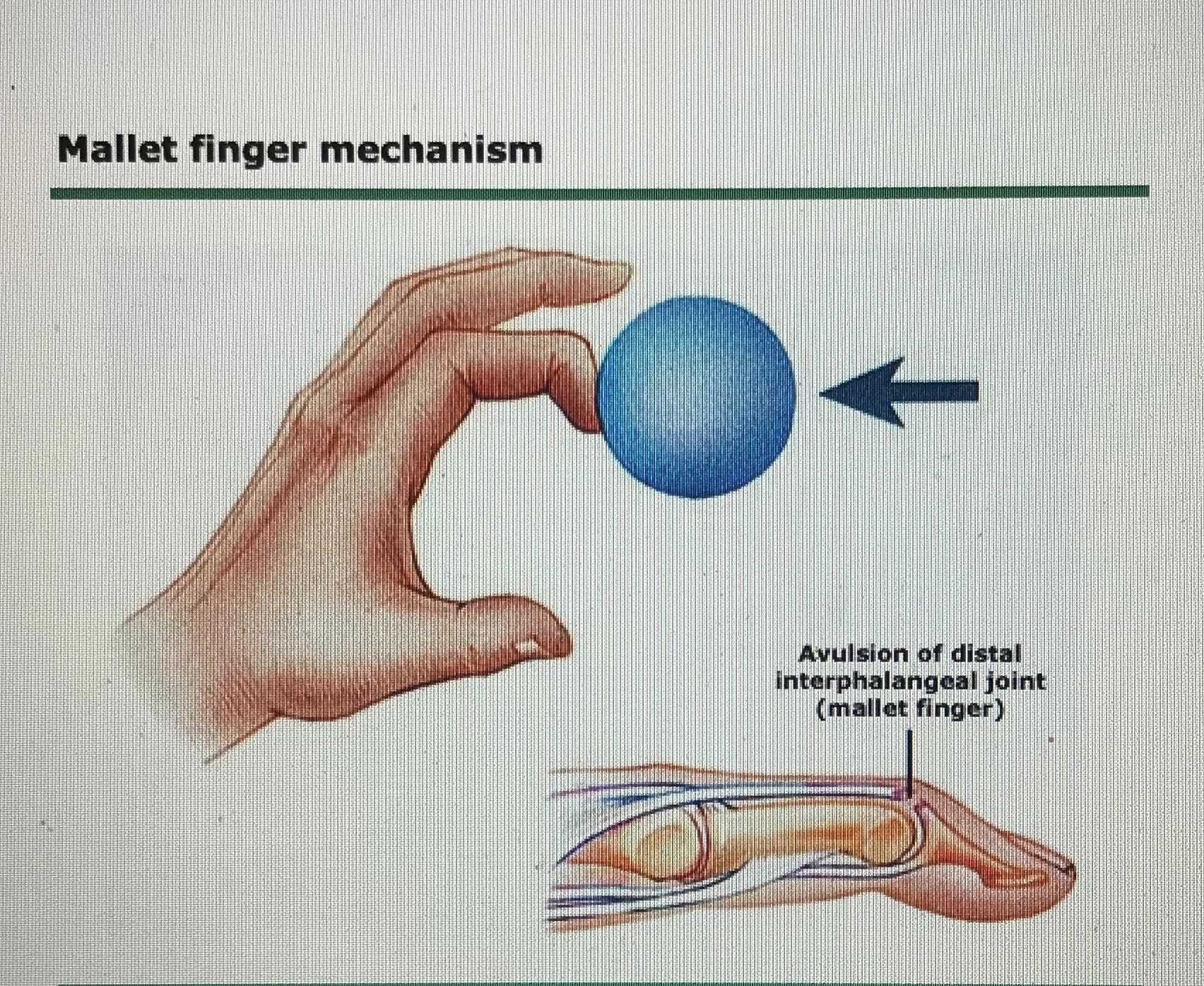
Traumatic Disruption of terminal Extensor tendon leading to inability to extend DIP. Common in baseball and football and resulted due to axial loading at fingertip
What is a Jersey finger hand deformity?
Rupture of flexor digitorum profundus
MOI: Forced passive extension during active finger flexion (grabbing a jersey). Common in ring finger.

What is Dupuytren’s Contracture hand deformity?
Multifactorial (ETOH, DM< TOBB, trauma). Thickening of the palmar fascia/skin leading to decreased finger extension, palpable nodules and secondary contractures of the MCP and PIP joints. More common in men than women.

What are other common hand deformities as a result of nerve injury?
Claw Hand (Ulnar): loss of interossei and hypothenar eminence.
Bishop’s: Ulnar palsy and loss of interossei, 2 interossei and hypothenar eminence
Ape Hand: Median nerve palsy and thenar eminence wasting

What are the collateral ligament injuries of the hand?
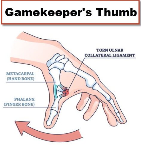
Gamekeeper’s (Skier’s) Thumb: UCL sprain or Stener’s lesion resulting in complete UCL disruption w/ retraction requiring surgical fixation.
MCP collaterals will be taught in flexion.
DIP collaterals will be tensioned through flex/extension
PIP collaterals taught in extension
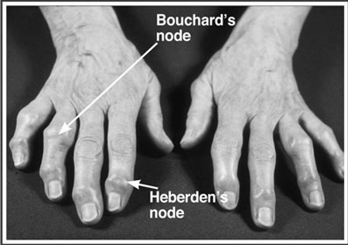
What are Herbeden’s and Bouchard’s Nodes?
They are bony swelling as a result of OA in the DIP/PIP
DIP: Hebeden’s
PIP: Bouchard’s
What are phalanx fractures?
They account for 45% of wrist and hand fractures and the most commonly fractured bone in the entire body is the distal phalanx. Is divided into the base, shaft, neck, and head
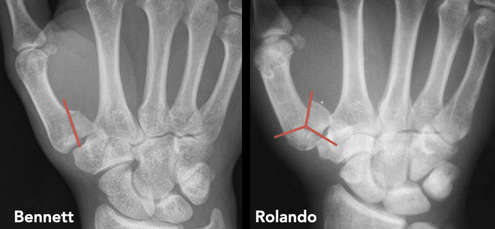
What are thumb metacarpal fractures?
Bennett Fracture: fracture/dislocation of the CMC
Rolando Fracture: >1 fractures involving articular surface
What is Complex Region Pain Syndrome and the Types?
CRPS Type I (RSD): pain syndrome triggered by a noxious event and not limited to the single peripheral nerve. Typically seen in PT
Causes: Fractures (Colle’s), Peripheral nerve injury, or no injury
Treatment: Avoid increased pain, CKC exercise, sensory techniques.
CRPS Type II (causalgia): pain syndrome involving direct partial or complete injury to a nerve or nerve branch
Signs and Symptoms: pain, edema, stiffness, skin temp, sweating.
What are ganglion cysts and boss carpals?
Ganglion Cyst: Thin-walled cyst containing mucoid hyaluronic acid that develop spontaneously over joint or tendon. May become painful w/ growth or nerve involvement.
Treatment: Needle aspiration or excison
Boss Carpal: Bony prominence typically at the base of the 2nd or 3rd metacarpal caused by forced wrist extension. Involves the ECRL and B
Treatment: Wrist splint w/ 10-15 deg ext

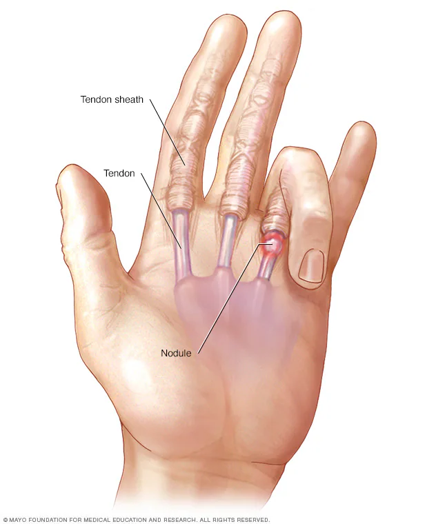
What is Trigger finger?
Painful or non-painful catching of flexor tendon at A1 pulley. Causes stenosing tenovaginitis and presence of distal palmar crease. Palpable and tender nodule.
Conservative: Modalities, orthosis, ROM, tendon gliding
Cortisone or surgical resection
What is intrinsic plus?
The intrinsic muscles put the hand into a protective/functional posture. Imbalance of spastic intrinsics and weak extrinsic (FDS, FDP, EDC).
Position: MCP flexion, PIP extension, DIP extension
Cause: trauma (direct/indirect, vascular injury, compartment syndrome), RA, neurologic injury
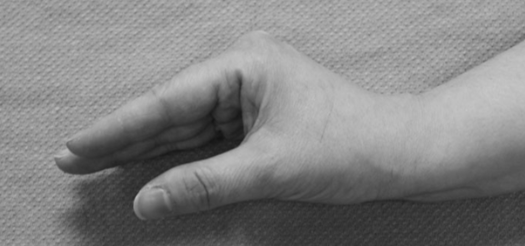
What is intrinsic minus?
Imbalance between strong extrinsic and weak intrinsics.
Position: MCP hyperextension, PIP flexion, DIP flexion
Cause: ulnar neuropathy, Charcot-Marie-Tooth (motor-sensory neuropathy), compartment syndrome

What are the types of splinting?
-Static
-Dynamic
-Functional Position