A&P II Chapter 20 blood vessels
 Call Kai
 Learn
Learn Practice Test
 Spaced Repetition
 Match
Match Flashcards
 Knowt Play
1/232

There's no tags or description
Looks like no tags are added yet.
Name | Mastery | Learn | Test | Matching | Spaced | Call with Kai | Chat |
|---|
No analytics yet
Send a link to your students to track their progress
233 Terms
Blood vessels functions?
Transport and distribute blood through the body.
Deliver oxygen, nutrients, and hormones to cells and carry away carbon dioxide and wastes
Return blood to the heart
How many layers do most blood vessels have?
Most have 3 layers, except for the smallest vessels.
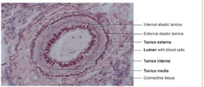
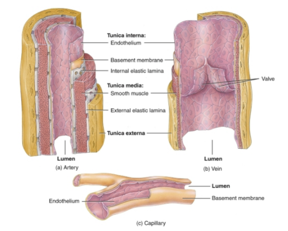
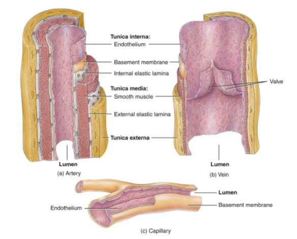
What are the three layers of blood vessels
The Tunica interna(innermost layer)
Tunica media(Middle layer)
Tunica externa(Outermost layer)
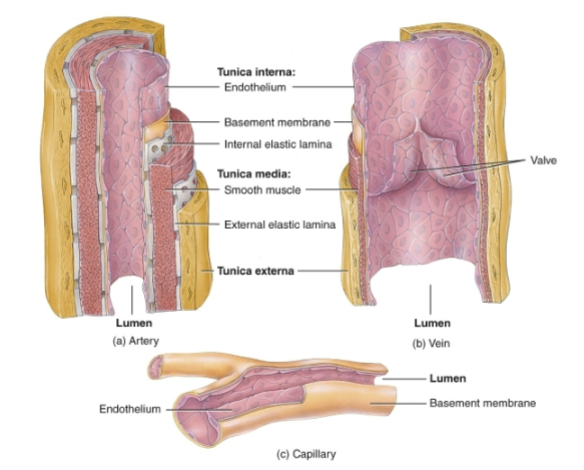

What makes up the Tunica interna and what does it do?
The endothelium, basement membrane and internal elastic lamina. Surrounds lumen, in direct contact with blood.


What makes up the Tunica media and what does it do?
Smooth muscle and external elastic lamina. Stretch and recoil, and changes diameter of lumen through vasoconstriction and vasodilation.
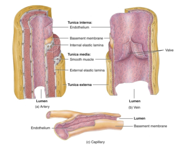

What makes up the tunica externa and what does it do?
Elastic and collagen fibers. Anchors vessels to surrounding tissue.

Function of arteries?
Carry oxygenated blood away from the heart.
Elastic arteries
Close to the heart. Have highly elastic walls to accommodate the surge of blood and project blood onward when ventricles are relaxing.
Muscular arteries
Have lots of smooth muscle in the tunica media for greater vasoconstriction and dilation to adjust blood flow to various regions of the body
Anastomoses
Form alternate route of blood flow to body part through collateral circulation
Abundant microscopic vessels are?
From small arteries branching from muscular arteries. They deliver blood to capillaries.
Tunics change
Thin tunica interna with fenestrated(small pores) internal elastic lamina.
Tunica media with one or two layers of smooth muscle

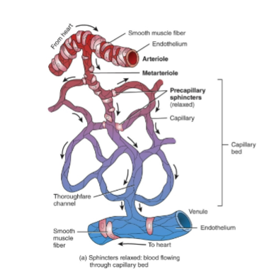
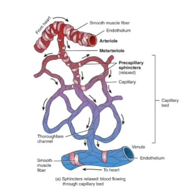
Metarteriole
Tapers toward junction with capillary.
Precapillary Sphincter(Single muscle fiber) at junction regulates blood flow through capillary bed by regulating resistance
Also affects blood pressure
Capillaries are?
Networks of microscopic vessels. They connect outflow of the heart through arteries and return to heart through veins.
Capillary bed
10 to 100 capillaries come from a single metarteriole
Blood flow is controlled by the precapillary sphincters in metarteriole.
Throughfare channel is?
The distal end of capillary bed with no smooth muscle. Provides a direct route to the venule.
Autoregulation?
Tissues automatically adjust blood flow to match its metabolic demands.
Types of capillaries?
Continuous
Fenestrated
Sinusoids

Continuous capillaries description
Endothelial cells form continuous tubes
intercellular clefts between adjacent cells allow only a few substances to move across, such as in blood-brain barrier.
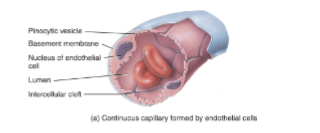
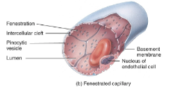
Fenestrated capillaries description
Endothelial cells have many fenestrations(small pores). Allows greater exchange of materials, such as in kidneys for filtration.


Sinusoid capillaries description
Endothelial cells with large fenestrations, incomplete basement membrane, and large intercellular clefts. Allows all components of blood to pass through.

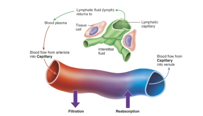
Capillary exchange
Movement of substances between blood and interstitial fluid.
Methods of Capillary exchange
Diffusion:
Across endothelial cell membranes - Gases and lipid soluble
through intercellular clefts or fenestrations - water soluble
Transcytosis: Pinocytic vesicles
What are the influences on capillary exchange
Filtration: capillary blood pressure pushes fluid out of capillary into interstitial fluid
reabsorption: Blood colloid osmotic pressure(due to plasma proteins) pulls fluid into capillary bed
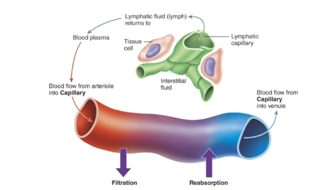
Where does filtration occur? Where does reabsorption occur?
Greater arteriole end
Greater venule end
Most capillaries reunite to form venules
Thin walls that do not maintain shape.
Merge to form veins.
-Close to capillary, very porous, function as exchange of nutrients and wastes and movement of white blood cells from blood stream into inflamed or infected tissue.
-Acquire thicker walls as enlarge, with no exchange.
In some parts of the body, blood passes from one capillary bed into another, instead of a venule.
-known as portal system
-portal vein connects the two capillary beds
(hepatic - Liver )(hypophyseal - Hypothalamus/pituitary)
Veins do what?
transport deoxygenated blood from tissues back to the heart.
Difference between walls of veins and walls of arteries?
Veins have thinner tunica interna and media than arteries. They lack elastic lamina.
(This leaves them unable to withstand high pressure)
what are valves in the vein and what do they do?
Flap like cusps of tunica interna.
They prevent backflow of blood segments in vein.
What is a Vascular sinus.
Vein with thin endothelial wall, such as coronary sinus.
Lacking smooth muscle so unable to alter its diameter.
Venous return
Volume of blood flowing back to the heart through systemic veins.
Limited pressure remaining from heart pump.
(Venous return) Skeletal muscle pump info?
Skeletal muscle compression pushes blood through valve closer to heart, “milking.”
Valves further away close as some blood is pushed back against it.
(Venous return) Respiratory pump info?
during inhalation the diaphragm moves downward, decreasing pressure in thoracic cavity and increasing pressure in abdominal cavity; blood flows into decompressed veins and into right atrium.
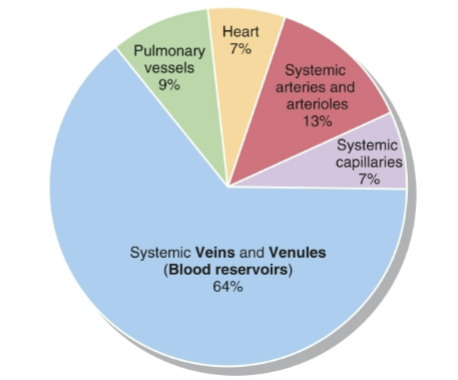
Blood distribution
At rest, 64% of blood volume is in systemic veins and venules.
13% systemic arteries and arterioles
9% pulmonary vessels
7% heart
7% systemic capillaries
Blood reservoir.
-Blood can be diverted quickly as need arises
-Brain stem sympathetic impulses can vasoconstrict veins, allowing greater blood volume to flow to skeletal muscles as needed during increased activity
-Similar mechanism during hemorrhage, to counteract blood loss and associated decrease in blood volume and pressure
Blood flow in any given tissues
-Varies with blood pressure gradient and resistance
-In all systemic vessels is equal to cardiac output
Blood pressure
Hydrostatic pressure exerted by blood on blood vessel walls.
Generated by ventricular contraction
Measured in mm Hg
Highest at aorta and falls progressively, with fluctuations disappearing at the capillaries
influenced by cardiac output, blood volume, and vascular resistance.
Systemic blood pressure
highest arterial pressure during systole(< The phase when the heart contracts to pump blood out)
Diastolic blood pressure
Lowest arterial pressure during diastole.(< the heart muscle relaxes and blood flows in and refills chambers)
Vascular resistance
Opposition to blood flow due to friction between blood and walls of blood vessel
An increase decreases blood flow and increases blood pressure
Factors affecting vascular resistance
Size of lumen - Higher resistance as vasoconstrict
Blood viscosity - higher resistance as ratio of blood cells and proteins increase
total blood vessel length - higher resistance if longer
Systemic vascular resistance(SVR)
total peripheral resistance from all systemic vessels
slight change in arteriole diameter has large effect

BP regulation
Cardiovascular center in medulla oblongata
In addition to regulating stroke volume(SV) and heart rate(HR), also regulates blood pressure and blood flow to specific tissues
Autonomic sympathetic and parasympathetic
neural regulation
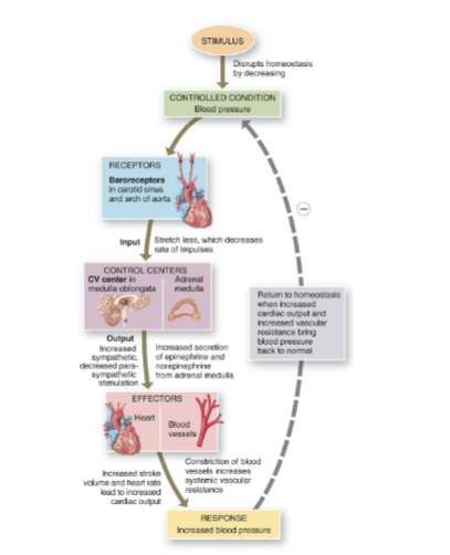
baroreceptor reflex - baroreceptors monitor blood pressure
Chemoreceptor reflex - chemoreceptors monitor chemical composition of blood
Hormones
Alter cardiac output, systemic vascular resistance, and total blood volume
Autonomic regulation of BP
Input from sensory receptors, cerebral cortex, limbic system and hypothalamus
output regulates heart rate, contractility and blood vessel diameter
Autonomic sympathetic regulation of BP
Cardiac accelerator nerves increase rate and force of heat contraction, that increases BP
vasomotor nerves maintain resting smooth muscle tone in blood vessel walls and stimulation of veins results in constriction and movement from venous blood reservoirs, that increases BP
Autonomic parasympathetic regulation of BP
Vagus nerves decrease heart rate, that decreases BP

(Neural regulation of BP)Baroreceptor reflexes
Carotid sinus receptors - initiate reflex to regulate BP in brain
aortic arch receptors - initiate reflex to regulate systemic BP
BP fall - receptor fewer impulses to cardiovascular center, decreased parasympathetic and increased sympathetic impulses increase BP
(Neural regulation of BP) Chemoreceptor reflexes
Carotid bodies and aortic bodies
detect drop in O2, increase in CO2, or increase in H+(acidic)
increased sympathetic impulses increase BP to drive gas exchange through the lungs
(Hormone reg of BP) Renin-angiotensin-aldosterone system
Stimulates increased systemic vascular resistance and increased reabsorption of sodium and water to increase blood volume, thus increase BP
(Hormone reg of BP) Epinephrine and norephinphrine
Stimulates increased rate of force of heart contraction and vasoconstriction of some arterioles and veins to increase BP, but vasodilation of arterioles in skeletal muscles
(Hormone reg of BP) Antidiuretic hormone
Stimulates vasoconstriction to increase BP
(Hormone reg of BP)Atrial natriuretic peptide
Stimulates vasodilation and loss of salt and water in urine to decrease BP.
Pulse is?
Alternate expansion and recoil of an artery wall after each contraction of the left ventricle.
Tachycardia
rapid heart rate
Bradycardia
slow heart rate
Pulse pressure is?
The difference between systolic pressure and diastolic pressure.
(Circ Routes) How many routes are blood vessels organized in?
Two
(Circ routes)Pulmonary circulation
Carries oxygen poor blood from right ventricle to the air sacs of the lungs
Returns oxygen rich blood to the left atrium
(CIrc routes) Systemic circulation
Carries oxygen rich blood from the left ventricle to systemic capillaries throughout the body(except to air sacs of lungs)
returns oxygen poor blood to the right atrium

Major systemic arteries
Ascending aorta
Right and left coronary arteries
Arch of the aorta
Brachiocephalic trunk
left common carotid artery
left subclavian artery
Thoracic aorta
Branches to thoracic aorta, passing through diaphragm
Abdominal aorta
Branches to abdominal organs
common iliac arteries supply pelvis and lower limbs
Major systemic Veins
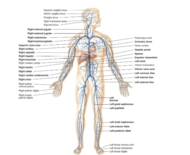
Superior vena cava
from head: dural venous sinus, internal jugular, external jugular, and vertebral veins
Upper limbs: superficial and deep veins
Thorax: azygos system
Inferior vena cava
Hepatic portal vein: Collects from splenic and superior mesenteric veins
Hepatic vein
Lower limbs: superficial and deep veins

Hepatic portal circulation
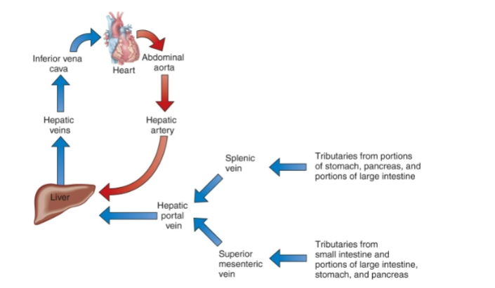
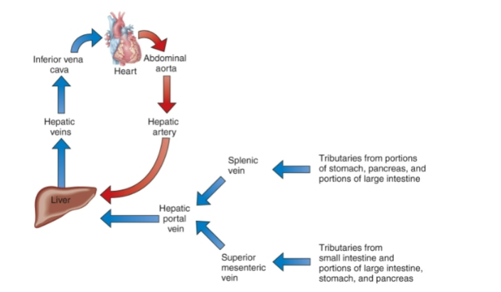
Directs venous blood from gastrointestinal organs and spleen into the hepatic portal vein of liver before it returns to the heart
enables the liver to utilize nutrients and detoxify harmful substances in the blood
Splenic vein
Drains the spleen stomach and large intestines
Superior mesenteric vein
drains the small intestine, large intestine, stomach, and pancreas

Hepatic veins
Drain the liver

What layer of an artery contains the lumen?
Tunica interna

What layer of an artery contains smooth muscle?
Tunica media
What layer anchors a blood vessel to surrounding tissue?
Tunica externa

What structure surrounds the lumen and contacts the blood?
Endothelium
What type of blood vessel has only an endothelial layer?
Capillary

Where are precapillary sphincters located?
At capillary entrances

What vessel provides a direct route from a metarteriole to a venule?
Thoroughfare channel
Where is the thoroughfare channel located relative to the capillary bed?
At the distal end

What type of capillary has large openings and an incomplete basement membrane?
Sinusoid

Where does filtration occur more strongly in the capillary bed?
Arteriole end
Where does reabsorption occur more strongly in the capillary bed?
Venule end

Where does filtered fluid move during capillary filtration?
Into interstitial fluid
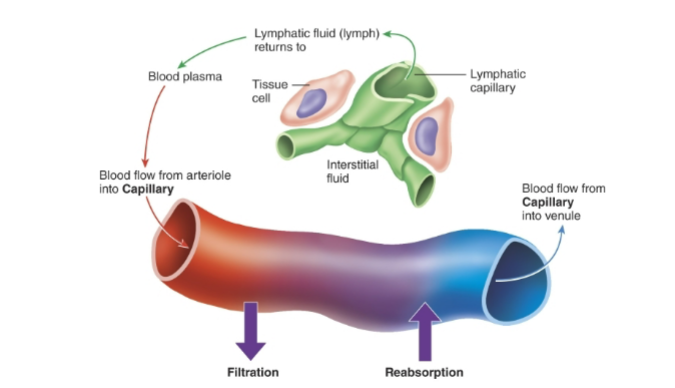
Where does reabsorbed fluid move during capillary reabsorption?
Into the capillary
During inhalation, where does blood move from decompressed veins?
Right atrium