cardiology
 Call Kai
 Learn
Learn Practice Test
 Spaced Repetition
 Match
Match Flashcards
 Knowt Play
1/35

There's no tags or description
Looks like no tags are added yet.
Name | Mastery | Learn | Test | Matching | Spaced | Call with Kai | Chat |
|---|
No analytics yet
Send a link to your students to track their progress
36 Terms
Pretest probability of CAD
If low risk then DC
If high risk go for further evaluation
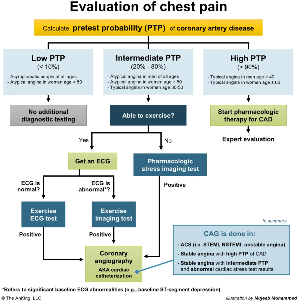
Factors that determine high and low risk in chest pain evaluation
Sex
Age
Risk factors
Symptoms (cardiac: relived by GTN, cardiac characteristics, exacerbated by exercise)
Right ventriculer MI always remember
No furosemide
No GTN
give iv fluide if indicated
Never give GTN to
RVMI
Phosphodiesarase
Hypotention
In case of sus. RVMI with nonspecific ECG changes
Do right-sided precordial ECG which may show ST elevation and T wave inversion
ECG may be normal in unstable angina what matters is
Clinical Pic + risk factors
Question trick
In some questions the question may be built up which means for example the patient may had an MI then undergo catheterization you must be able to identify whether the complication is related to Cath or MI
When you are suspecting STEMI, the Next step in management is always
PCI, the question is mentioning the surgery or open heart surgery you have to know that it is done later after identification of anatomy. Even if the patient is having a global ischemia
During catheterization after MI, cardiac catheterization, arterial wall is penetrated superior to right inguinal ligament. Which of the following is the most likely location of blood collection?
Retroperitonial space
Puncture above inguinal ligament
Exp:
Vascular access during cardiac catheterization is typically obtained through either the common femoral artery or radial artery. The common femoral artery is the continuation of the external iliac artery as it crosses the inguinal ligament. Arterial puncture above the inguinal ligament increases the risk of retroperitoneal hemorrhage, as this portion of the vessel lies directly inferior to the peritoneum. Accidental puncture of the posterior wall can cause blood to track along the loose connective tissue surrounding the vessel and accumulate within the interfascial planes of the retroperitoneum.
Bleeding in the retroperitoneal space cannot be controlled with external compression and can lead to life-threatening hemorrhage. Affected patients typically develop hemodynamic instability with significant hypotension, a drop in hemoglobin, and ipsilateral flank pain.
(Choice A) Bleeding into the pelvic cavity is frequently due to trauma (eg, pelvic fracture) or gynecologic hemorrhage (eg, ruptured ectopic pregnancy). Injury to the common femoral artery is less likely to cause intraperitoneal bleeding as the external iliac artery courses underneath the peritoneum.
(Choice C) The right paracolic gutter is a peritoneal recess found between the ascending colon and abdominal wall. Fluid (bile, pus, or blood) typically accumulates there from pathology involving the gastrointestinal organs, particularly the appendix and gall bladder.
(Choice D) Subcutaneous tissue hemorrhage from an arterial source typically presents with a large area of ecchymosis and a palpable hematoma. Development of significant hypotension is less likely with subcutaneous bleeding, as the potential space for blood accumulation is smaller than with peritoneal or retroperitoneal bleeding.
(Choice E) Hemorrhage into the thigh muscles would present with significant pain and edema in the thigh and possibly lead to compartment syndrome (severe pain, absent distal pulses).
Mechanism of action of nitroglycerin
At standard clinical doses, nitroglycerin causes predominantly venodilation (reducing venous return, preload, and myocardial oxygen demand) over arterial dilation.
decrease in left ventricular end diastolic volume
Mechanism of atheroma formation
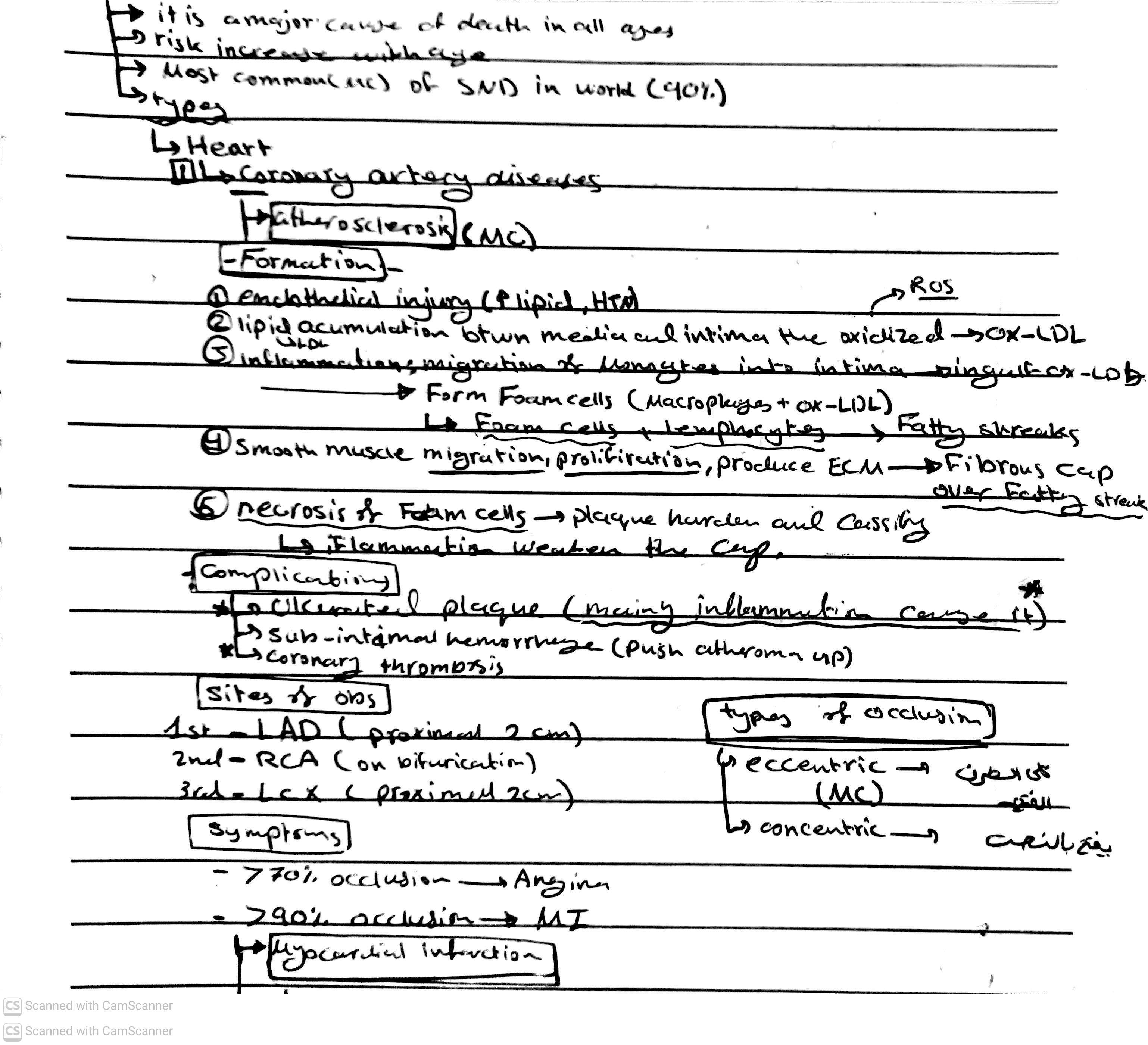
The most susceptible vascular beds to atherosclerosis are:
Coronary arteries and lower abdominal aorta
Exp:
Key Takeaways: Atherosclerosis Pathophysiology
* Pathogenesis & Progression:
* Initiated early in life (childhood) by risk factors like hypertension, diabetes, and hyperlipidemia.
* Begins as fatty streaks (intimal thickening from lipid-laden macrophages and extracellular matrix).
* Evolves over time into fibrous plaques / fibrous cap atheromas, eventually forming advanced lesions with necrotic lipid cores and calcification.
* Hemodynamic Factors:
* Vessel bends and branch points are most vulnerable due to turbulent blood flow.
* Turbulence causes endothelial dysfunction, damages wall integrity, and increases contact time between cholesterol particles and the endothelium.
* Most Susceptible Locations:
* The lower abdominal aorta and coronary arteries are the most prone to atherosclerosis due to local hemodynamics.
* Lesions can appear in these specific locations as early as the second decade of life.
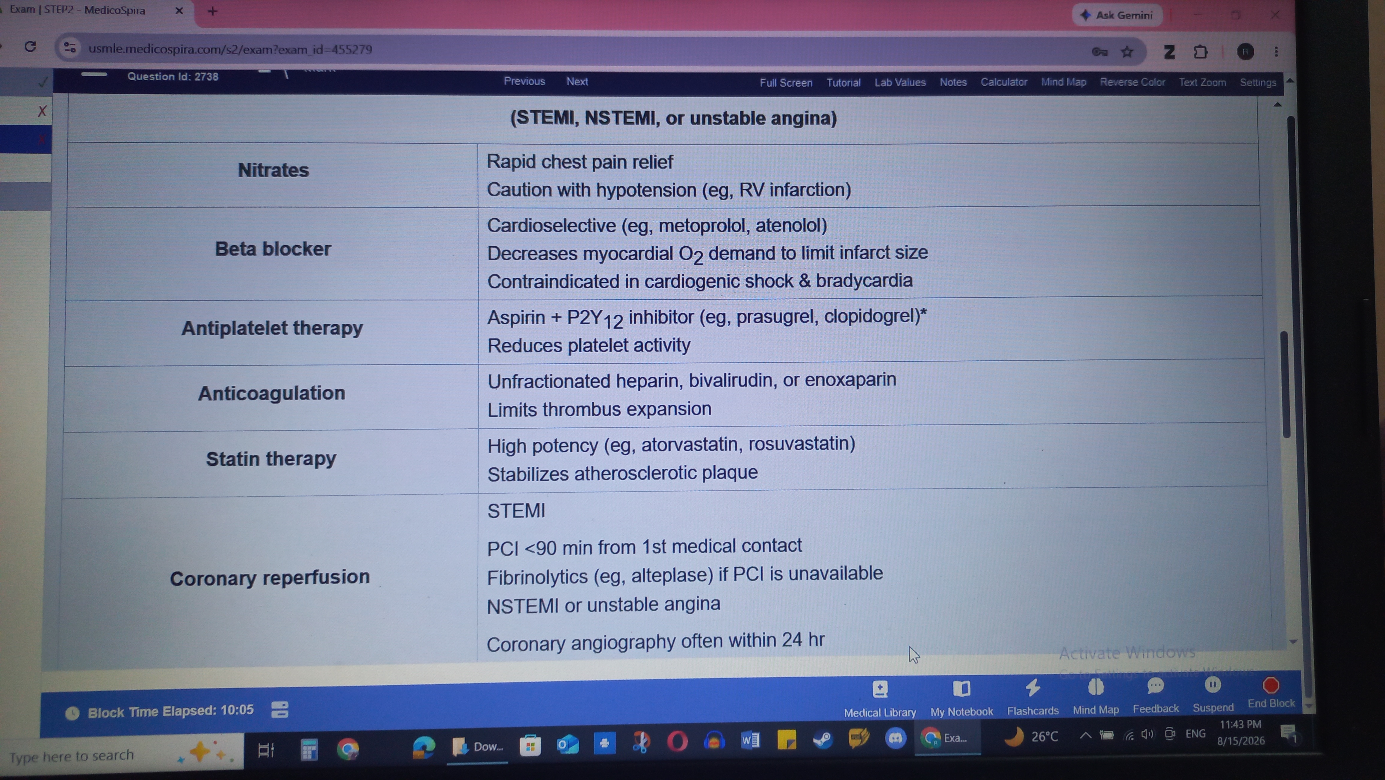
The cause or rapid reperfusion after acute MI mnangment is
Alteplase (fibrenolytic)
Cholesterol embolism clinical presentations
Atherombolism is most frequently observed after cardiac catheterization or other procedures involving a vascular manipulation
Onset acute or delayed sometimes 30 days or more after the getting event
Affected organs includes intestinal ischemia pancreatitis gastrointestinal bleeding acute kidney injury
Clinical picture includes blue toe syndrome which is cyanotic digit despite palpable pulse la vida reticularis ulcers and gangrene retinal examination might reveal hollenhorst plaques which are bright refractile cholesterol emboli within the retinal arteries
Management of athero embolism
Supportive. Renal impairment from Atheroembolism May persist for several weeks
Complications of cardiac catheterization
Complications
1. Vascular/access-site complications — most common
Hematoma
Bleeding
Femoral/radial artery pseudoaneurysm
Arteriovenous fistula
Arterial thrombosis/occlusion → limb ischemia
Retroperitoneal hemorrhage, particularly with femoral access
Infection
2. Contrast-related
Allergic/hypersensitivity reaction
Contrast-associated acute kidney injury
Rarely, severe anaphylaxis
3. Cardiac complications
Arrhythmias: VT/VF, bradycardia, AV block
Coronary artery dissection
Coronary perforation → hemopericardium/tamponade
Coronary thrombosis
Myocardial infarction
Acute heart failure/pulmonary edema
Cardiac tamponade
4. Cerebrovascular
Stroke/TIA, usually from embolization of thrombus or aortic/catheter debris.
5. Other rare complications
Aortic dissection
Cholesterol embolization
Air embolism
Vasovagal reaction/hypotension
Radiation injury with prolonged/interventional procedures
Death
Clinical presentations of complications of cardiac catheterization
Atrioventricular fistula: minimum symptoms with localized tenderness and slight swelling can develop plus a continuous bruit have to focus on a continuous bruit associated with palpable thrill. The Continuous bruit is resulted from a higher arterial pressure compared to Venus pressure and you have to note that the AV fistula is resulted from a accidental puncture
Femoral artery pseudo aneurysm present as a tender pencil Mass with a systolic bruitlike the continuous bruit of avf
Retro peritoneal Hemorrhage is life-threatening manifest within 24 hours present with severe flank pain and hemodynamic instability
Managment of ACS
Patient develop severe chest and sudden on such as pain and hypertension I mean he's in a shock within the first 24 hours after stent placement how would we think about this okay and and he had a ECG shows the elevation from V1 to V4 you have to think about the cause

Cocaine induced MI
First you have to remember it's a combination of three when a young patient has MI Plus symptoms or signs of sympathetic Overdrive and patient is young then think about toxicology rather than premature MI
The first priority for cocaine intoxication is benzo
Medications decreases myocardial remodeling after MI event
ACEI
Beta Blocker
Aldosteron antagonists
Complications of MI
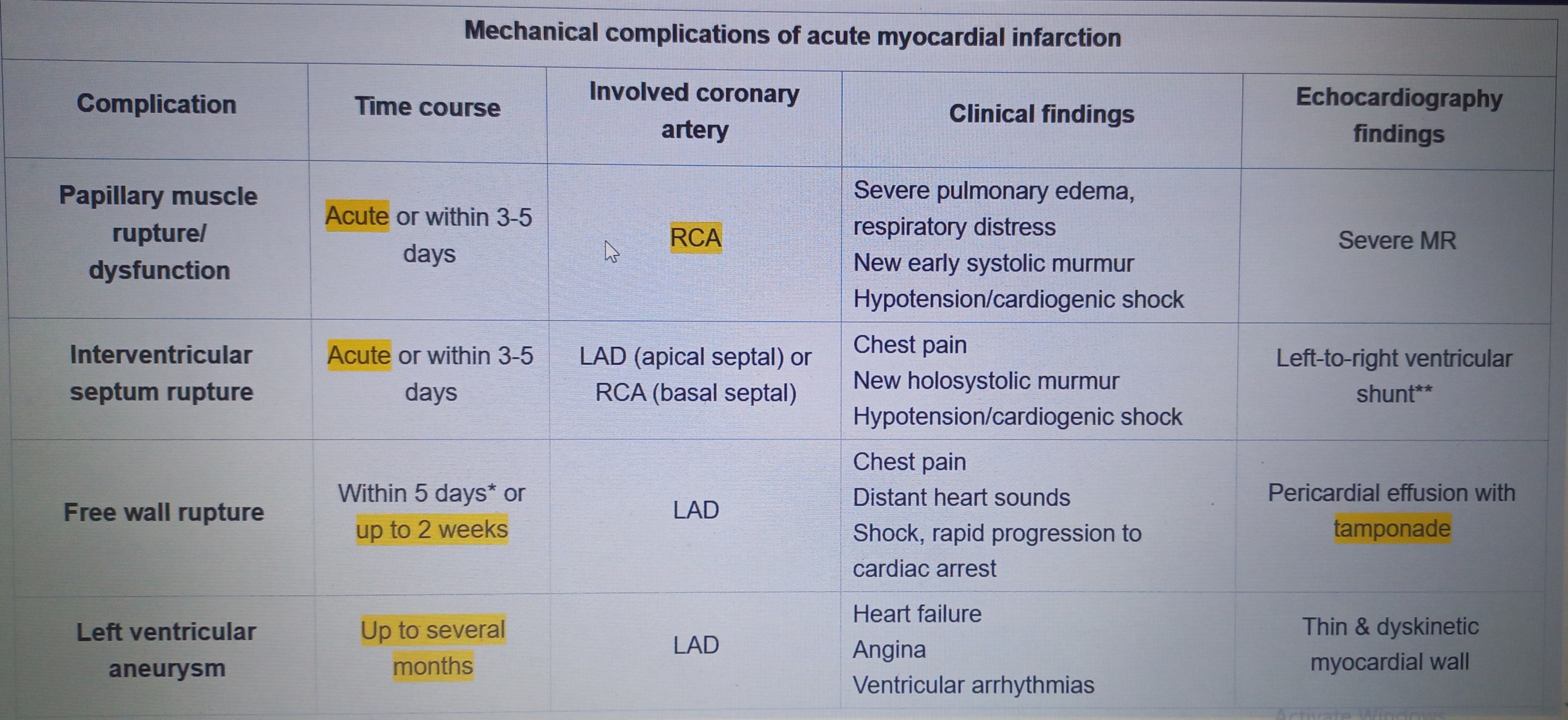
LV aneurysm is more associated with
Anterolateral MI
Presentations of LV wall aneurysm
Persistent ST segment elevation plus deep Q wave in the same leads
Symptoms of heart failure intractable enjoy now ventricular arrhythmias symptoms of mitral regurgitation including murmurs also stroke resulted from thrombo embolism formed in the aneurysm
Send and disconnect of the left ventricular wall corresponding to the prior MI
To distinguish between complications of MI ( free wall rupture vs interventriculer septum rupture)
Free wall rupture: the key distinguishing feature is absence of pul. Edema
Severe sudden chest pain, obstructive shock, pulseless electrical activity which means organized Rhythm without palpable pulse
Interseptal rupture: pul. Edema, holosystolic murmur and heart failure
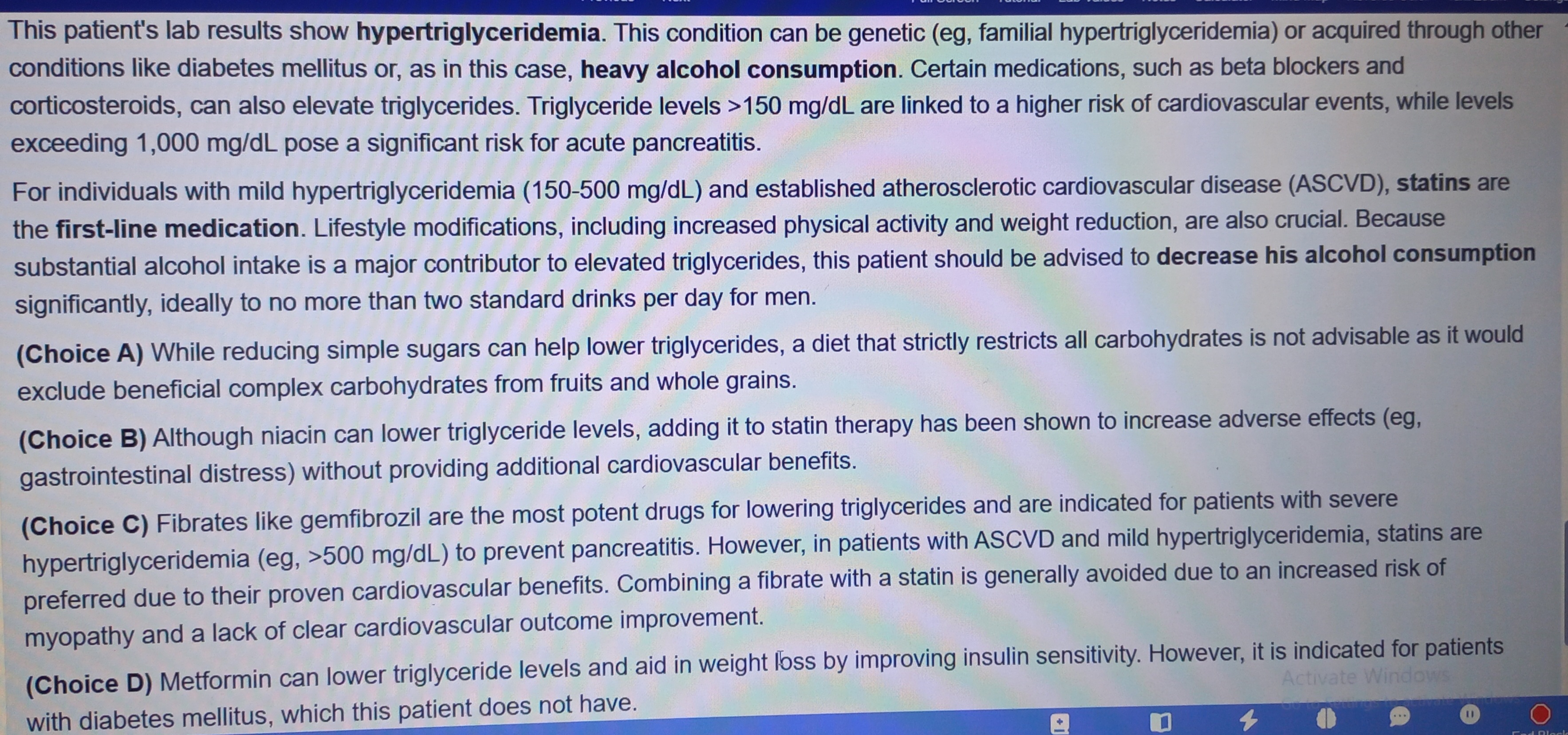
Best managment option for someone have a high cardiovascular risk and hyperlipidemia
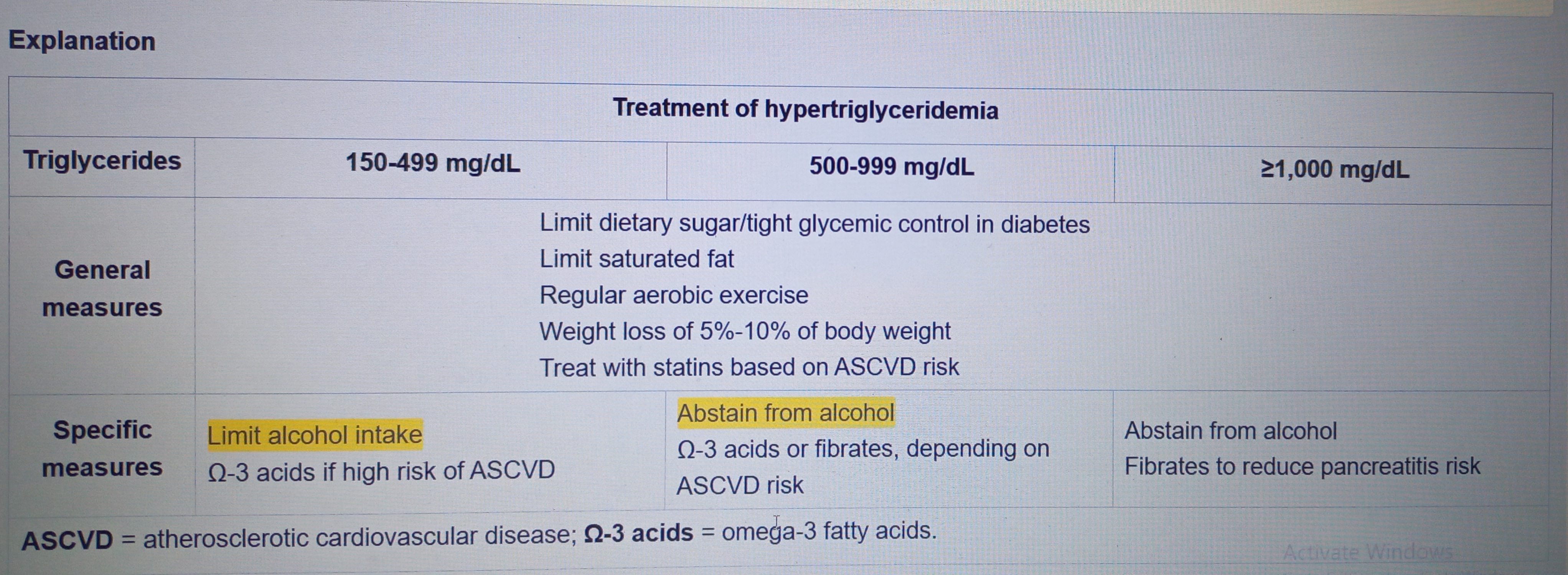
Acute mediastinitis after CABG
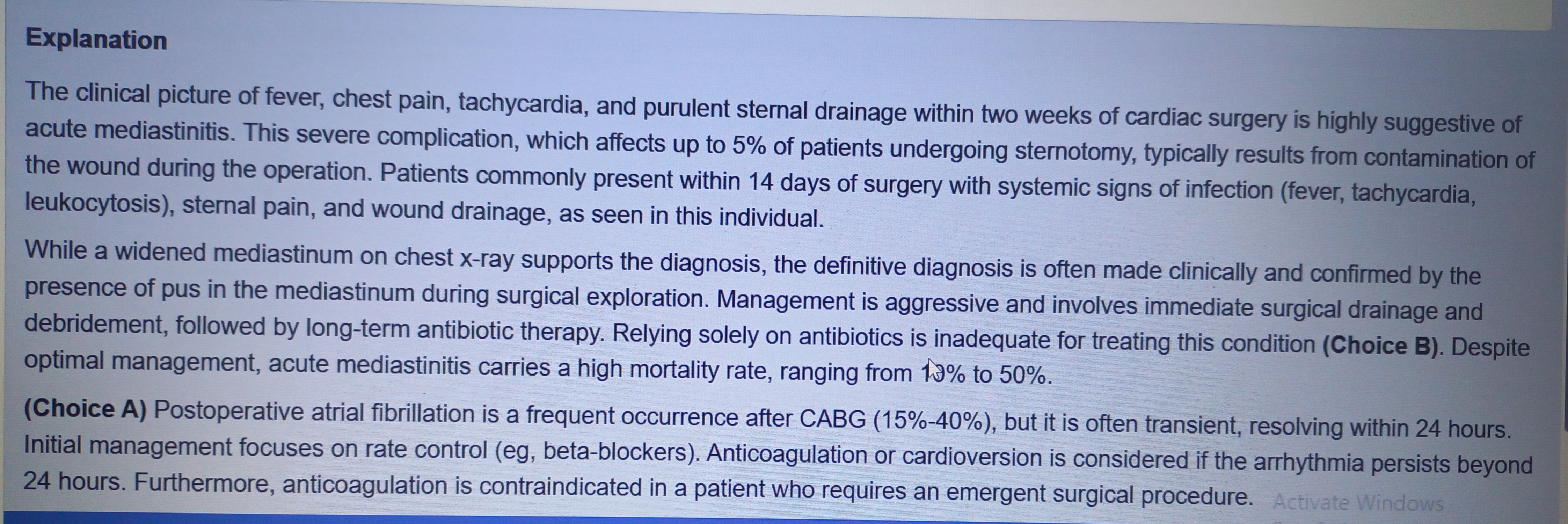
Complications of CABG
⭐ High-yield complications to remember
CABG →
Bleeding + MI + AF + stroke + AKI + pulmonary complications + mediastinitis
And if you're asked for the most common arrhythmia after CABG → atrial fibrillation.
Mechanical Complications increases by
Delayed or absent reperfusion therapy
Pleural effusion after CABG

Primary prevention of ASCVD guidelines to start statins
They said high intensity is superior bcs of effect

Evaluation of chest pain in ER
Don’t let previous hx of panic and anxiety trick you, when there is a risk for ACS approach for it
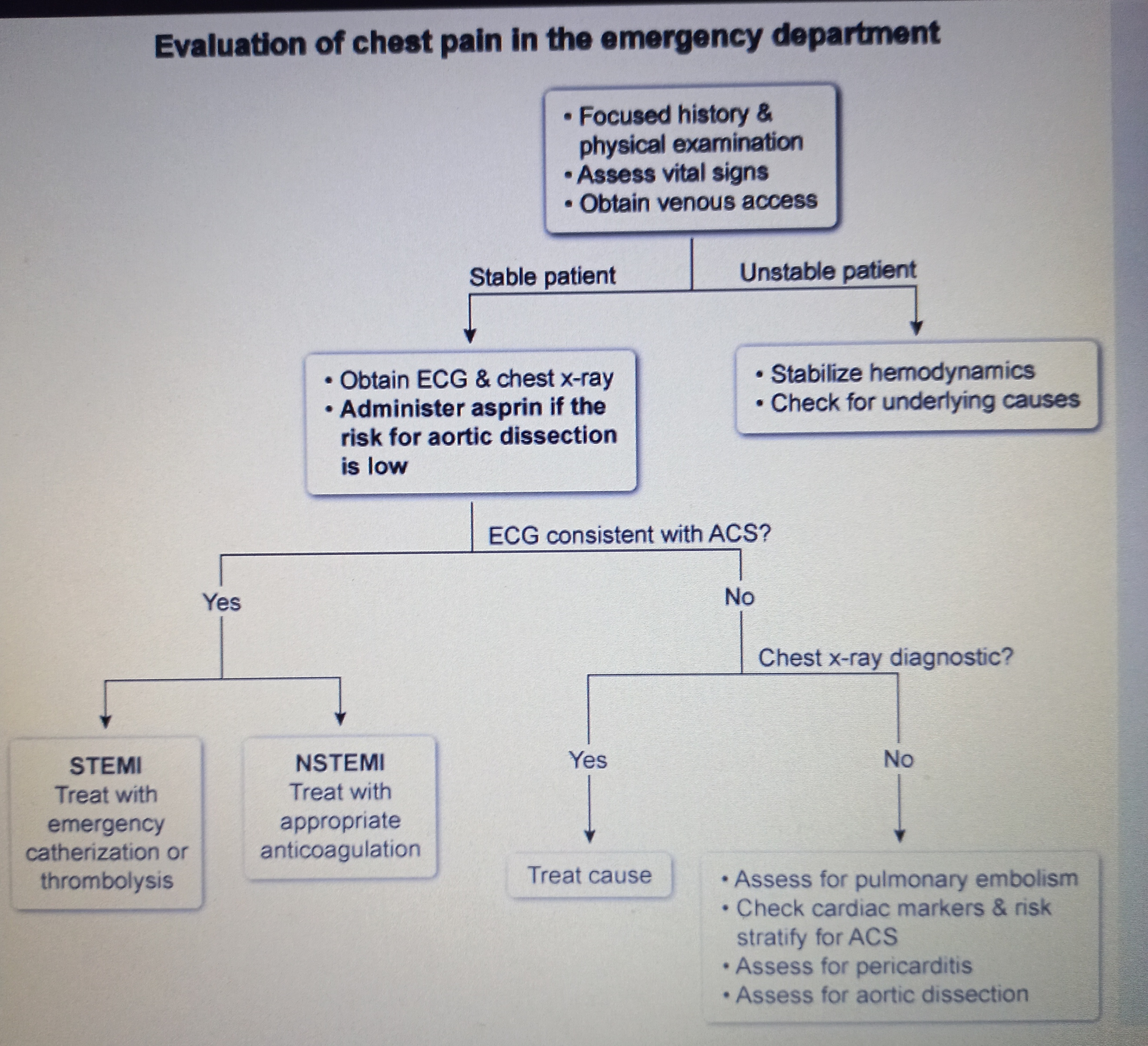
Why Aspirin first then heparin
Heparin is given for confirmed MI, while Aspirin is given for suspected patients
Cardiac tamponade
Present with hypotension and clear lungs
The single most important factor for improving patient's long-term survival after St elevation myocardial infarction is
To limit the amount of myocardial necrosis this can only be achieved by promptly reopening the included are three and restoring blood flow which is reperfusion by PCI or if it's not available we do the fibrinolytic therapy
Retroperitonial hematoma after CABG with femoral access
Within 12 hrs think about ( av fistula, retroperitoneal hematoma, pseudoanuerysm)
Non contrast CT abdominal and pelvis is diagnostic of choice
Presentations include hemodynamic instability suddenly with epsilateral flank pain or back pain often with external signs at the puncture site