Lab 1: Anatomical Terms, Histology, Integumentary System
 Call Kai
 Learn
Learn Practice Test
 Spaced Repetition
 Match
Match Flashcards
 Knowt Play
1/102

There's no tags or description
Looks like no tags are added yet.
Name | Mastery | Learn | Test | Matching | Spaced | Call with Kai | Chat |
|---|
No analytics yet
Send a link to your students to track their progress
103 Terms
Anatomical position
the body is upright, directly facing the observer, feet flat and directed forward. The upper limbs are at the body's sides with the palms facing forward.
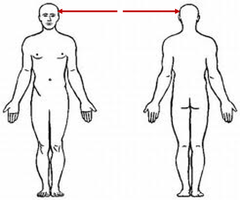
Cephalic region
head area
Cervical region
neck area

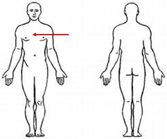
Thoracic region
chest area
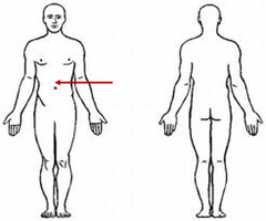
Abdominal region
stomach area
Pelvic region
groin area

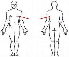
Axillary region
armpit area
Brachial region
arm area
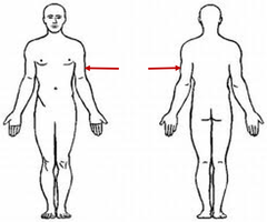
Lumber region
Back area (lower back)

Dorsal region
back side
Transverse sectional plane
divides the body into superior and inferior portions

Frontal sectional plane
divides the body into anterior and posterior portions
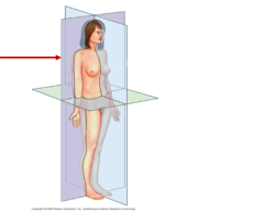
Sagittal sectional plane
divides the body into right and left portions

Histology
the study of cells and tissues
Four basic types of tissues
epithelial, connective, muscle, neural
Epithelial tissue
- Protective layer covering surfaces in contact with the outside world
- Lines tubular structures
- Glands are formed by this
- Has apical and basal surfaces
Epithelial tissue function
protection, absorption, secretion
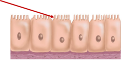
Apical surface
exposed surface of cells
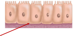
Basal surface
attachment point of cells
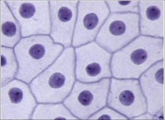
Simple squamous epithelium
single layer of flat cells
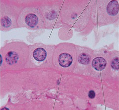
simple cuboidal epithelium
single layer of cube shaped cells
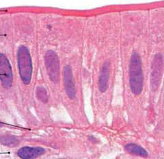
simple columnar epithelium
Made up of a single layer of tall cells that fit closely together
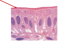
villi
Name this structure
stratified squamous epithelium
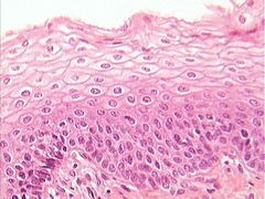
Stratified cuboidal epithelium
tissue that consists of two or more layers of cube-shaped cells, found in some ducts

stratified columnar epithelium

Transitional epithelium non-stretched
Protection and added distensibility
EX: urinary bladder and ureters
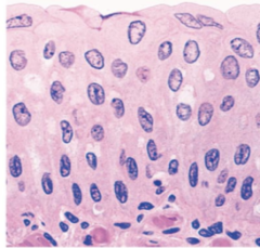
Transitional epithelium stretched
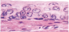
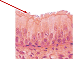
Pseudostratified ciliated columnar epithelium
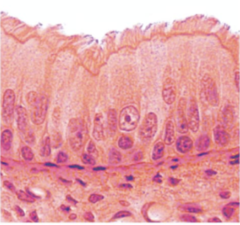
Cillia
Name this structure
Endocrine glands
release products directly into interstitial fluid or bloodstream
Exocrine glands
release products through ducts directly onto epithelial apical surfaces
Loose connective tissues (3)
areolar, adipose, reticular
dense connective tissue (3)
dense regular, dense irregular, elastic
Areolar connective tissue
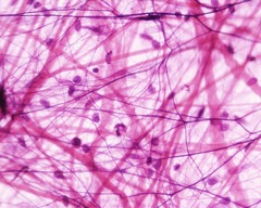
adipose connective tissue
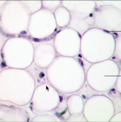
reticular connective tissue
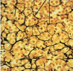
dense regular connective tissue

elastic connective tissue
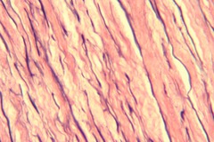
dense irregular connective tissue

Plasma
the watery matrix of blood made up of blood cells and cell fragments
formed elements
erythrocytes, leukocytes, platelets
Lymph
interstitial fluid that enters the lymphatic vessels
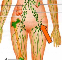
Supporting connective tissue
bone and cartilage
Chondrocytes
cells in the cartilage matrix

Hyaline cartilage

Elastic cartilage
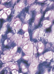
Fibrocartilage cartilage
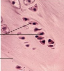
Bone
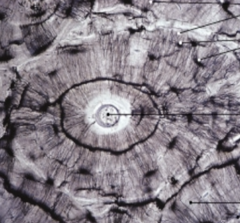
Osteocytes
bone cells
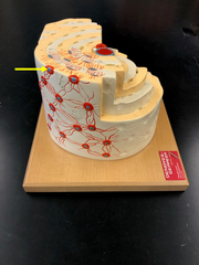
canaliculi
Passageways connecting osteocytes together
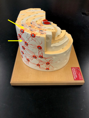
Osteoblasts
bone forming cells
Osteoclasts
Bone-destroying cells
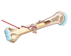
Periosteum
A dense fibrous membrane covering the surface of bones (except at their extremities) and serving as an attachment for tendons and muscles.
Name this layer
Skeletal muscle tissue
- striated
- voluntary

Cardiac muscle tissue
- striated
- involuntary
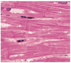
smooth muscle tissue
- non-striated
- involuntary

Neuron
cells in the nervous system that act as interconnected information processors, which are essential for all of the tasks of the nervous system
Cell body of a neruon
Name this structure
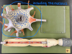
nucleus of neuron
Name this structure (red)

perikaryon
cytoplasm surrounding the nucleus
* Name this fluid*

dendrite
branching projections from cell body that receive stimuli
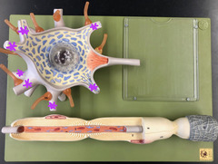
axon
A threadlike extension of a neuron that carries nerve impulses away from the cell body.

myelin sheath
covers the axon of some neurons and helps speed neural impulses
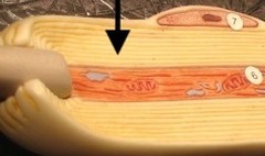
internode
region between nodes
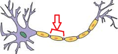
Nodes of Ranvier
gaps in the myelin sheath
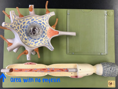
axon hillock
Cone shaped region of an axon where it joins the cell body.
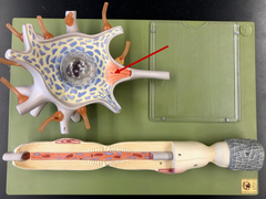
telodendria
fine terminal extensions at the end of the axon
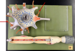
synaptic knob
rounded areas on the end of the axon terminals

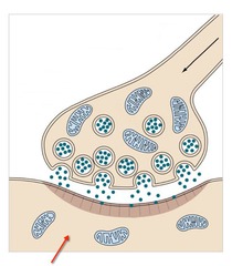
synaptic cleft
a gap into which neurotransmitters are released from the axon terminal
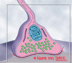
postsynaptic cell
cell that receives message
glial cells
cells in the nervous system that support, nourish, and protect neurons
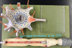
Oligiodendrocytes
glial cells in CNS
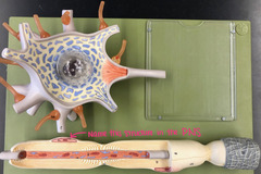
Schwann cells
glial cells in the PNS
stratum corneum
outermost layer of epidermis
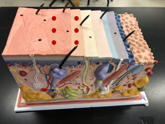
stratum lucidum (epidermis)

stratum granulosum (epidermis)
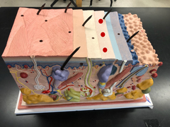
Stratum spinosum
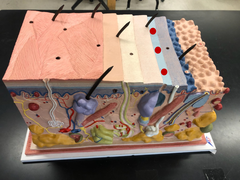
stratum germinativum (stratum basale)
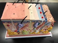
Basal lamina
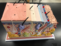
dermal papillae
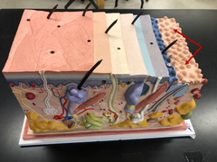
Papillary plexus

tactile corpuscle
"tomatoes"
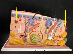
lamellated corpuscle
"watermelon"
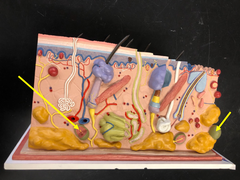
Rufinni corpuscles
"rufus" (from Kim Possible)
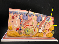
Pore of sweat gland
Name this opening

Apocrine sweat glands
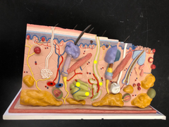
merocrine sweat glands

sebaceous glands
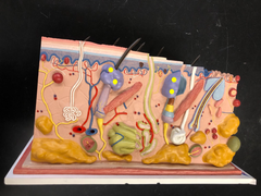
root hair plexus
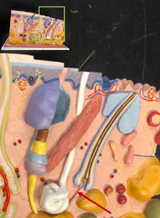
Arrector pilli muscle
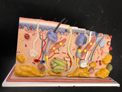
Hair root
everything below the sebaceous gland
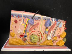
Hair shaft
everything above the sebaceous gland- part that is outside of skin
"Name this structure"
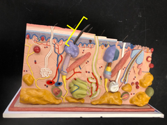
Hair follicle
organ that produces hair
hair bulb
The rounded, club-shaped part of hair located at the end of the hair root

Hair papilla
Name this indentation
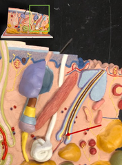
Medulla
White innermost layer of the hair

Cortex
Dark brown 2nd layer from the middle

Cuticle
"Name this layer"
The clear covering layer on the hair shaft
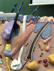
Internal root sheath
Tan layer 3rd from the middle
