BV 2 Disparity Notes
 Call Kai
 Learn
Learn Practice Test
 Spaced Repetition
 Match
Match Flashcards
 Knowt Play
1/77

There's no tags or description
Looks like no tags are added yet.
Name | Mastery | Learn | Test | Matching | Spaced | Call with Kai | Chat |
|---|
No analytics yet
Send a link to your students to track their progress
78 Terms
What is the Vieth-Müller circle, and what does it represent in binocular vision?
The Vieth-Müller circle is the circle that passes through the fixation point and the two entrance pupils.
It represents the locations in space where objects create equal oculocentric angles in the two eyes, meaning: βL=βR
Objects on this circle are seen with zero retinal disparity.
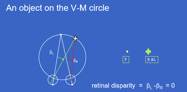
Why do objects on the Vieth-Müller circle have zero retinal disparity?
Objects on the Vieth-Müller circle project to corresponding retinal locations because the left-eye and right-eye oculocentric angles are equal, so, the visual system receives matching angular information from both eyes.
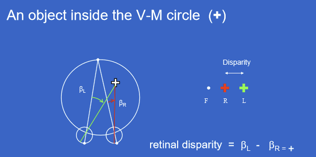
What type of retinal disparity occurs when an object is inside the Vieth-Müller circle?
An object inside the Vieth-Müller circle produces positive crossed retinal disparity. This means the left-eye and right-eye images are displaced in a crossed pattern relative to fixation.
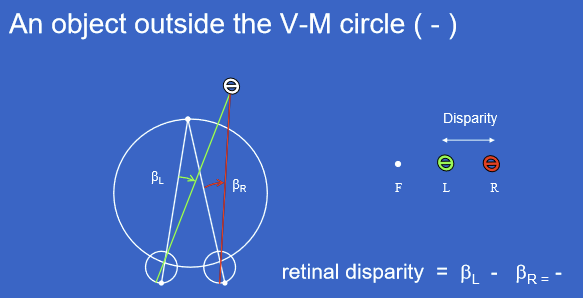
What type of retinal disparity occurs when an object is outside the Vieth-Müller circle?
An object outside the Vieth-Müller circle produces negative uncrossed retinal disparity. This means the left-eye and right-eye images are displaced in an uncrossed pattern relative to fixation.
In right esotropia, why does the object of regard produce uncrossed diplopia?
In right esotropia, the deviating right eye turns inward, so the object of regard is not fixated by the right eye.
The object produces uncrossed/negative disparity because: retinal disparity=βL−βR; and βL=0,βR=+. So: βL−βR=0−(+)=−
In strabismus, what determines the Vieth-Müller circle: the fixation point or the object of regard?
The Vieth-Müller circle is defined by the fixation point, not necessarily the object of regard.
In strabismus, the deviating eye may not fixate the object, so the object of regard can fall off the V-M circle, creating retinal disparity and diplopia.
What is relative disparity?
Relative disparity, also called η or eta, is the difference in absolute retinal disparity between two objects.
Why is fixation position unimportant for relative disparity?
Relative disparity depends on the difference between disparities of two objects, so the fixation-related terms cancel out. Because relative disparity uses the difference in binocular parallax angles, the absolute fixation point does not matter.
η=ϕL−ϕR
ϕL = angular separation between the two objects in the left eye
ϕR = angular separation between the two objects in the right eye
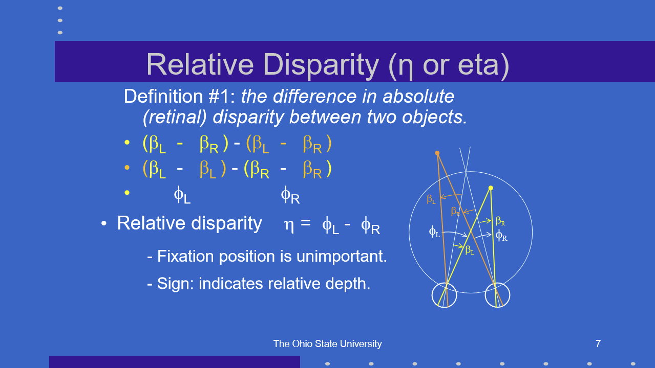
What does the sign of relative disparity indicate?
The sign of relative disparity indicates relative depth between two objects.
Positive vs. negative relative disparity tells whether one object is relatively nearer or farther than another.
Relative disparity is therefore important for judging depth relationships, not just whether one object is on or off the V-M circle.
What equation should you use to calculate relative disparity in radians?
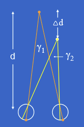
Relative disparity η\etaη in radians is:
η (rad)= (PD⋅Δd) / d²
Where:
PD = interpupillary distance / pupillary distance
Δd= depth separation between the two objects
d = viewing distance from the observer
How do you convert relative disparity from radians to seconds of arc?
Convert radians to seconds of arc using: η (sec)=η (rad)×206,000
How does viewing distance affect relative disparity?
Relative disparity is inversely proportional to the square of viewing distance. So as viewing distance increases, relative disparity gets much smaller.
What is the difference between dioptic and dichoptic stimulation?
Dioptic stimulation means the same target is seen simultaneously by both eyes.
Dichoptic stimulation means separate targets are presented one to each eye.
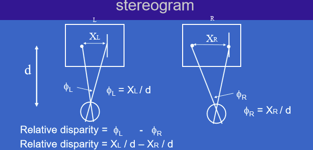
In a stereogram, how is relative disparity calculated from the left-eye and right-eye image separations?
In a stereogram relative disparity is: η=ϕL−ϕR, where ϕL=XL/d and ϕR=XR/d.
Or η= (XL−XR)/d
What do XL, XR, and d represent when calculating relative disparity in a stereogram?
XL = image separation in the left-eye image
XR = image separation in the right-eye image
d = viewing distance
What is an anaglyphic stereogram?
An anaglyphic stereogram presents separate left-eye and right-eye images, often using different colors or filters, to create binocular depth.
How does fixation choice affect the sign of disparity in a stereogram
Disparity is measured relative to the fixated target.
An object can appear further/behind the fixated target if it has an uncrossed/negative disparity
An object can appear nearer/closer the fixated target if ti has a crossed/positive disparity
This is true regardless of the fixation target/point
What are the possible perceptual outcomes when disparate images are presented to the two eyes?
When the two eyes receive disparate images, the visual system may produce:
Physiological diplopia: normal double vision of nonfixated objects due to retinal disparity
Fusion: combining the two monocular images into one percept
Rivalry: alternating or competing percepts when the two monocular images are not suitably related
What is the difference between motor fusion and sensory fusion?
Motor fusion is an eye movement response that adjusts eye position so the object of regard is imaged on both foveas.
Sensory fusion is a cortical process that combines images from the two eyes into a single perceptual whole.
What condition is required for sensory fusion to occur?
Sensory fusion requires the two monocular sensations to be suitably related to each other.
If the two images are too different or incompatible, the visual system cannot fuse them and may produce binocular rivalry instead.
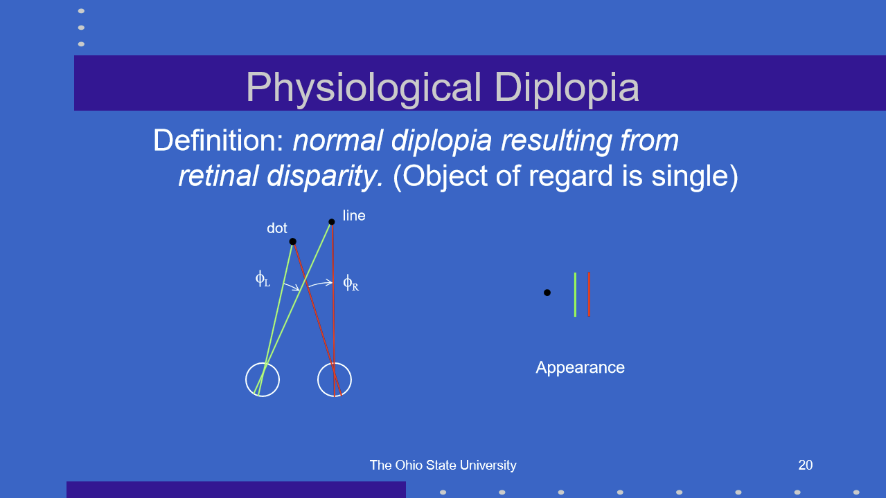
What is physiological diplopia, and how is the object of regard perceived?
Physiological diplopia is normal double vision caused by retinal disparity of objects that are not being fixated.
The object of regard is single because fixation places that object on corresponding retinal points, usually both foveas.
Nearby or farther nonfixated objects can appear double because they fall on noncorresponding retinal points.
Why do small retinal disparities usually not cause diplopia?
Small retinal disparities can be eliminated by a normal physiological fusion process.
Instead of perceiving double vision, the visual system fuses slightly different retinal images into a single binocular percept.
What is the diplopia threshold?
The diplopia threshold is the critical amount of retinal disparity at which double vision begins.
Below this threshold, disparity can usually be fused.
Above this threshold, the object is perceived as double.
What is Panum’s area, and why is it important for binocular vision?
Panum’s area is an area on the retina of one eye that can be stimulated simultaneously with a point on the retina of the other eye and still produce single binocular vision.
It allows slightly noncorresponding retinal points to be fused.
How does Panum’s area allow single vision even when disparity is present?
Panum’s area provides a tolerance zone for binocular fusion.
If the retinal disparity falls within Panum’s area:
The images are fused
Single binocular vision occurs
Depth can still be perceived
If the disparity exceeds Panum’s area:
Fusion fails
Diplopia may occur
Why can all 3 circles on the Randot test appear single even though disparity is present?
The disparities in the Randot targets are small enough to fall within Panum’s area, so the visual system can fuse them into single percepts.
This allows single vision despite retinal disparity.
What is stereopsis, and what visual stimulus produces it?
Stereopsis is the specific sensation of depth that arises from relative disparity between the two eyes’ images.
The stimulus for stereopsis is lateral disparity.
What are the key developmental and functional features of stereopsis?
Stereopsis is:
Innate
Matures at about 4 months
Provides relative depth information
Driven by lateral disparity
What is stereothreshold?
Stereothreshold is the minimum relative disparity required to produce stereopsis.
It represents the smallest disparity difference the visual system can detect as depth.
How is stereothreshold related to relative disparity and stereoacuity?
Stereothreshold is based on relative disparity:
Relative disparity=ϕL−ϕR
It can also be calculated using:
ηrad= (PD⋅Δd)/d²
How are images presented to the two eyes in clinic using dioptic versus dichoptic methods?
Dioptic presentation means the same target is seen by both eyes. Examples:
Standard visual acuity charts
Near point cards
Dichoptic presentation means different images are presented separately to each eye. Examples:
Stereoscope
Red-green anaglyphs
Polaroid methods
Alternating shutters
What devices or methods can be used for dichoptic image presentation in clinic?
Dichoptic presentation separates images so each eye receives different visual information.
Common methods include:
Stereoscope
Mirror stereoscope
Lens stereoscope
Anaglyphs, such as red-green filters
Polaroid filters
Alternating shutter systems
How does a typical red-green anaglyph present dichoptic targets?
A red-green anaglyph presents colored targets on a white background, with filters used to separate what each eye sees.
What is the clinical purpose of the Hess Lancaster screen?
The Hess Lancaster screen is used clinically to subjectively determine ocular deviation in multiple positions of gaze.
Basic setup:
White screen
Patient wears red-green glasses
Doctor shines a red light in nine positions of gaze
Patient uses a green light to overlay the red light
How does a red-green anaglyph on a black background create dichoptic presentation?
oth are forms of dichoptic presentation, but the background changes how the colored targets are presented.
White background: colored targets are seen by the eye behind the opposite filter color
Black background: colored targets are seen by the eye behind the same filter color
In a Wheatstone stereoscope, how is relative disparity calculated from image separations?
Relative disparity: η = -(XL-XR)/d
Where:
XL = separation in the left-eye image
XR = separation in the right-eye image
d = target distance from the eyes
In a Brewster stereoscope, how is relative disparity calculated?
Relative disparity: η= (XL-XR)/(Target distance from lens)
Where:
XL = separation/position in the left-eye image
XR = separation/position in the right-eye image
Target distance is measured from the lens, not directly from the eyes
How is convergence calculated in a Brewster stereoscope?
Convergence is calculated as:
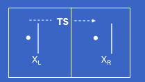
Convergence= (OS-TS)/(target distance from lens)
Where:
OS = optical separation
TS = target separation
Target distance is measured from the lens
Why is stereothreshold not considered one fixed value of relative disparity?
Stereothreshold depends on multiple stimulus characteristics, so it is not a single universal value.
Factors such as target design, viewing conditions, contrast, separation, and other stimulus properties can affect the smallest disparity detected.
Under the best conditions, how does stereothreshold compare with vernier acuity threshold?
Under optimal conditions, stereothreshold is slightly larger, meaning poorer, than the vernier acuity threshold.
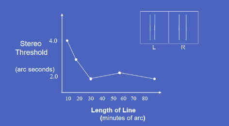
How does the length of vertical line targets affect stereothreshold?
Stereothreshold depends on the length of the vertical line targets.
Very short lines have poorer stereothresholds around 4 arcsec
Increasing line length improves stereothreshold to about 2 arcsec
After a certain length, improvement levels off
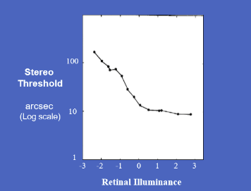
What is the relationship between target luminance/light adaptation and stereothreshold?
As retinal illuminance increases, stereothreshold generally improves, meaning the threshold decreases.
Low retinal illuminance → high/poor stereothreshold, around 100 arcsec
Higher retinal illuminance → lower/better stereothreshold, near 10 arcsec
Improvement eventually levels off
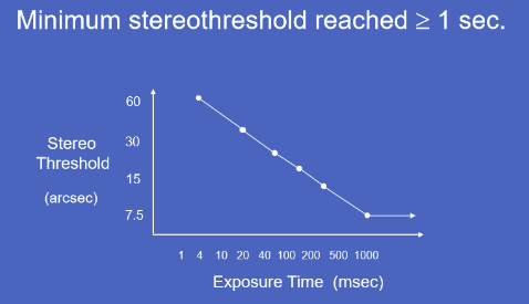
How does exposure duration affect stereothreshold?
Longer exposure duration improves stereothreshold until about 1 second.
Very brief exposure → poor stereothreshold, around 60 arcsec
Increasing exposure time progressively lowers the threshold
Minimum stereothreshold is reached at about ≥ 1 second
How does increasing blur affect stereo threshold and visual acuity threshold?
Increasing blur raises both:
Stereo threshold
Visual acuity threshold
Meaning performance gets worse as blur increases.
However, stereo threshold worsens more steeply than visual acuity threshold.
Which has a greater effect on stereothreshold: monocular blur or binocular blur?
Monocular blur raises stereothreshold more than binocular blur.
Why is stereothreshold clinically useful for detecting amblyopia, refractive error, and anisometropia?
Stereothreshold is very sensitive to blur and unequal image quality between the two eyes.
Because monocular blur greatly worsens stereopsis, poor stereoacuity can suggest problems such as:
Amblyopia
Uncorrected refractive error
Anisometropia
How does fixation position affect stereothreshold?
Stereothreshold is lowest when fixation is accurate and near the target.
If fixation is displaced:
In front of or behind the target, stereothreshold increases
Left or right of the target, stereothreshold also increases
The farther the fixation position is from the ideal target location, the poorer stereo sensitivity becomes
How does fixation disparity or small-angle tropia affect stereothreshold?
Fixation disparity or small-angle tropia raises stereothreshold because the eyes are not accurately aligned on the object of regard.
Lowest stereothreshold occurs around 0 degrees absolute disparity
As absolute disparity increases in either direction, stereothreshold rises on a log scale
Clinical note: Stereothreshold testing can help reveal micro-strabismus.
Why can stereothreshold testing reveal micro-strabismus?
Micro-strabismus may involve subtle fixation misalignment that is not obvious on routine observation.
Because stereopsis requires precise binocular alignment, even small fixation errors can raise the stereothreshold.
How does retinal eccentricity affect stereothreshold?
The foveal region has the lowest stereothreshold, meaning the best stereoacuity.
As the target moves farther from the fovea into peripheral retina:
Stereoscopic threshold increases
Stereoacuity gets worse
What clinical conditions can increase stereothreshold by forcing eccentric viewing or poor fixation?
Conditions that reduce central fixation or require eccentric viewing can increase stereothreshold.
Examples:
Central visual field loss
Eccentric viewing
Poor fixation
What types of patients are expected to have poor stereopsis due to impaired bifoveal fixation?
Patients who cannot fixate bifoveally will have poor stereopsis because precise binocular alignment is required for fine depth perception.
Examples:
Microtropia
Large fixation disparity
What visual conditions reduce stereopsis by preventing normal vision in both eyes?
Stereopsis requires usable, balanced visual input from both eyes. Poor stereopsis can occur when normal vision is lacking in one or both eyes due to:
Suppression
Amblyopia
Central scotoma
Anisometropia, especially because it creates monocular blur
What does the Helmholtz target demonstrate about complementary luminances?
The Helmholtz target can demonstrate multiple binocular phenomena, including:
Motor fusion
Stereopsis
Rivalry
Luster
The key point is that complementary luminance patterns can produce binocular interactions even when the two eyes receive different luminance information.
Is sensory fusion required for stereopsis?
No. Sensory fusion is not a prerequisite for stereopsis. Stereopsis can occur even when the two monocular images are not fully fused into a single sensory image.
Sensory fusion = combining two eye images into one perception
Stereopsis = depth sensation from binocular disparity
What is the stereoscopic depth interval?
The stereoscopic depth interval is the relative depth difference Δd that corresponds to a particular binocular disparity η\etaη.
The relationship is: η = (PD⋅Δd)/d²
Where:
η\etaη = relative disparity
PD = pupillary distance
Δd = relative depth interval
d = viewing distance
What is a telestereoscope, and how does it affect stereoscopic depth perception?
A telestereoscope is an optical device that effectively increases the observer’s pupillary distance/interpupillary distance (PD). Increasing effective PD changes the relationship between disparity and depth interval.
Why does increasing effective PD with a telestereoscope help at large viewing distances?
At large distances, normal PD produces very small binocular parallax/disparity cues.
A telestereoscope increases effective PD , which increases binocular separation and makes depth differences easier to detect.
At large distances, why should the simple approximation relative disparity equation be abandoned?
At large viewing distances, the approximation becomes less appropriate because disparity is better calculated using the difference in binocular parallax angles, or the difference in vergence demands.
What is the difference between fine stereopsis and coarse stereopsis?
Fine stereopsis occurs when depth sensation is linearly proportional to disparity.
Coarse stereopsis occurs when depth sensation has a non-monotonic relationship to disparity, meaning increasing disparity does not produce a simple proportional increase in perceived depth.
What is the relationship between physiological diplopia and coarse stereopsis?
Physiological diplopia is present over much of the broad region of perceived depth, especially when disparities are large enough that perfect single vision is not maintained.
Even when objects are physiologically diplopic, the visual system can still extract coarse depth information.
What is stereopsis?
Stereopsis is the specific sensation of depth arising from retinal disparity.
It provides relative depth information, meaning it tells the visual system which objects are closer or farther relative to each other.
What is the visual stimulus for stereopsis?
The stimulus for stereopsis is lateral disparity, meaning a horizontal positional difference between the images seen by the two eyes.
This retinal disparity allows the brain to compute relative depth.
What is stereoanomaly, and what type of stereopsis is affected?
Stereoanomaly is a common deficiency of stereopsis, affecting about 30% of the population, in which the deficit is limited to either crossed or uncrossed disparity.
It is mainly a defect of coarse stereopsis.
Why may stereoanomaly not be obvious on routine clinical stereopsis testing?
Stereoanomaly may not be detected clinically because many tests use conditions that do not isolate the defect well.
Also, patients with stereoanomaly can have a normal stereothreshold, so standard stereoacuity tests may appear normal.
What are the six classes of cortical disparity detectors?
The six classes of cortical disparity detectors are:
Far
Near
Tuned zero
Tuned inhibitory
Tuned far
Tuned near
How do fine and coarse stereopsis differ in neural pathway and response type?
Fine stereopsis:
Sustained response
Parvocellular units
“Tuned” detectors
Feature selective
Small disparity range
Linear response
About 3% defects
Coarse stereopsis:
Transient response
Magnocellular units
“Far and near” detectors
Non-feature selective
Large disparity range
Non-monotonic response
About 30% defects
Which type of stereopsis is more commonly defective: fine or coarse?
Coarse stereopsis is more commonly defective.
Fine stereopsis defects: about 3%
Coarse stereopsis defects: about 30%
What is local stereopsis, and what type of stimulus is commonly used to test it?
Local stereopsis occurs when relative disparity is processed independently for each local target pair.
It is also called:
Contour stereopsis
Example stimulus:
Simple line targets
What is global stereopsis, and what type of stimulus is commonly used to test it?
Global stereopsis occurs when relative disparities are processed interactively over a large region of the binocular field.
Example stimulus:
Random dot stereogram
It is also important in highly structured visual environments, such as:
Forest or jungle
“Leaf room”
What is the key significance of random dot stereograms (RDS) for understanding stereopsis?
Random dot stereograms show that stereopsis does not require monocularly identifiable contours.
The observer cannot identify the shape using either eye alone. The depth percept only emerges when the two eyes’ images are combined binocularly.
Why are random dot stereograms considered a test of global stereopsis?
Random dot stereograms require the visual system to process many relative disparities interactively across a large binocular field.
There are no obvious monocular contours, so the brain must integrate the dot pattern globally to extract depth.
Why is stereopsis useful for revealing camouflage?
Stereopsis can reveal objects that are camouflaged in monocular images because binocular disparity can separate an object from its background in depth.
Even if monocular cues are weak or absent, disparity can make the hidden object stand out.
What is the key significance of random dot stereograms (RDS) for understanding stereopsis?
Random dot stereograms show that stereopsis does not require monocularly identifiable contours.
The observer cannot identify the shape using either eye alone. The depth percept only emerges when the two eyes’ images are combined binocularly.
Why are random dot stereograms considered a test of global stereopsis?
Random dot stereograms require the visual system to process many relative disparities interactively across a large binocular field.
There are no obvious monocular contours, so the brain must integrate the dot pattern globally to extract depth.
Why is stereopsis useful for revealing camouflage?
Stereopsis can reveal objects that are camouflaged in monocular images because binocular disparity can separate an object from its background in depth.
Even if monocular cues are weak or absent, disparity can make the hidden object stand out.
What is the clinical value of random dot stereograms?
Random dot stereograms are useful clinically because they detect the presence of stereopsis and provide an unambiguous response.
Because the target cannot be identified monocularly, a correct response strongly suggests true binocular stereopsis.