BSC-700 Histology Module 4
1/39
Earn XP
Description and Tags
Graded Quizzes
Name | Mastery | Learn | Test | Matching | Spaced |
|---|
No study sessions yet.
40 Terms
Which intestine segment has the highest density of goblet cells?
a) duodenum
b) jejunum
c) ileum
d) colon
Colon
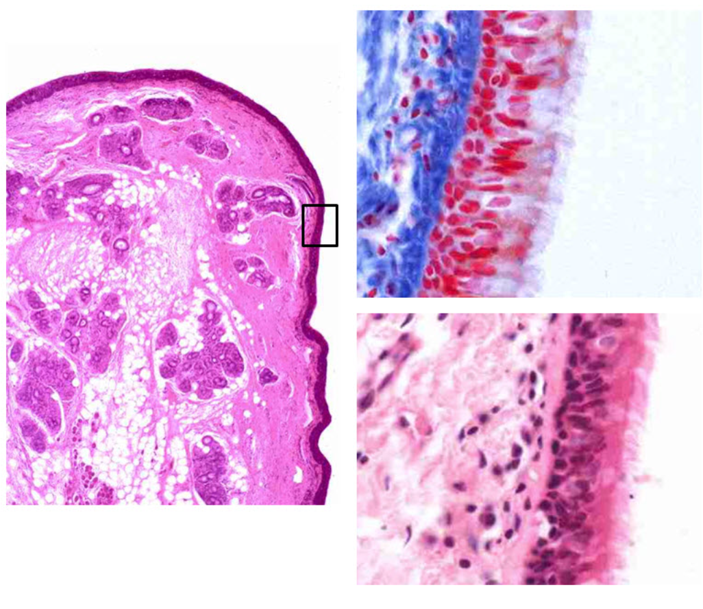
Different views of the same organ are displayed. Left image is stained with Hematoxlin and Eosin. Right image is stained with Azan Alcian Yellow (mucus is stained green). Identify the organ represented by this specimen.
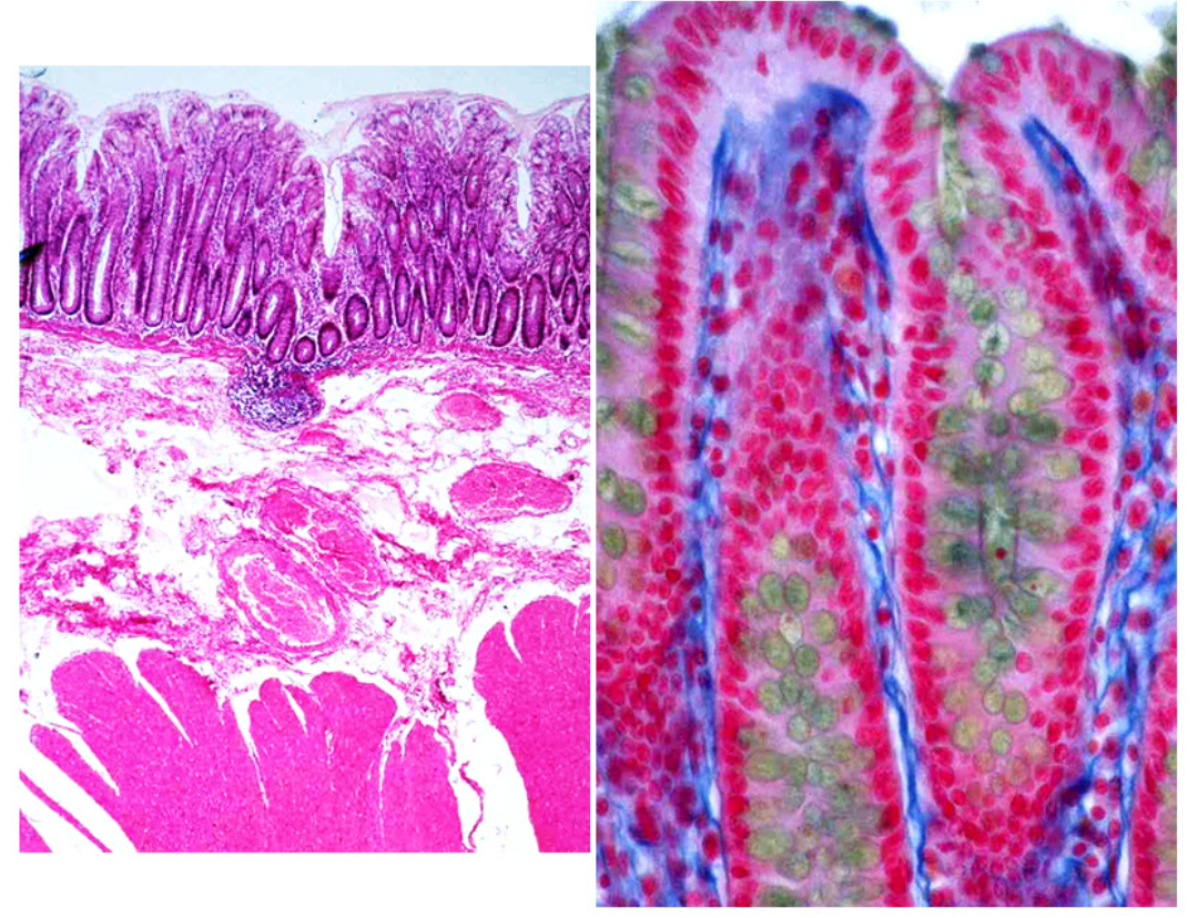
a) stomach
b) duodenum
c) esophagus
d) small intestine
e) colon
Colon
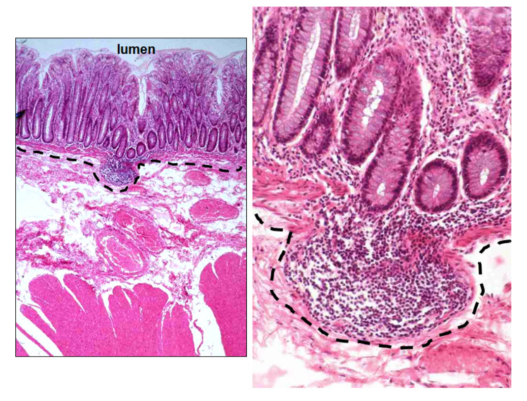
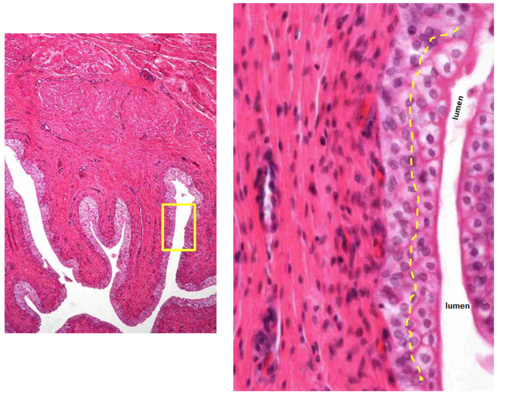
Identify the layer in this GI tract specimen that lies between the two yellow dashed lines.The right image is a higher power view of the rectangular boxed in area.
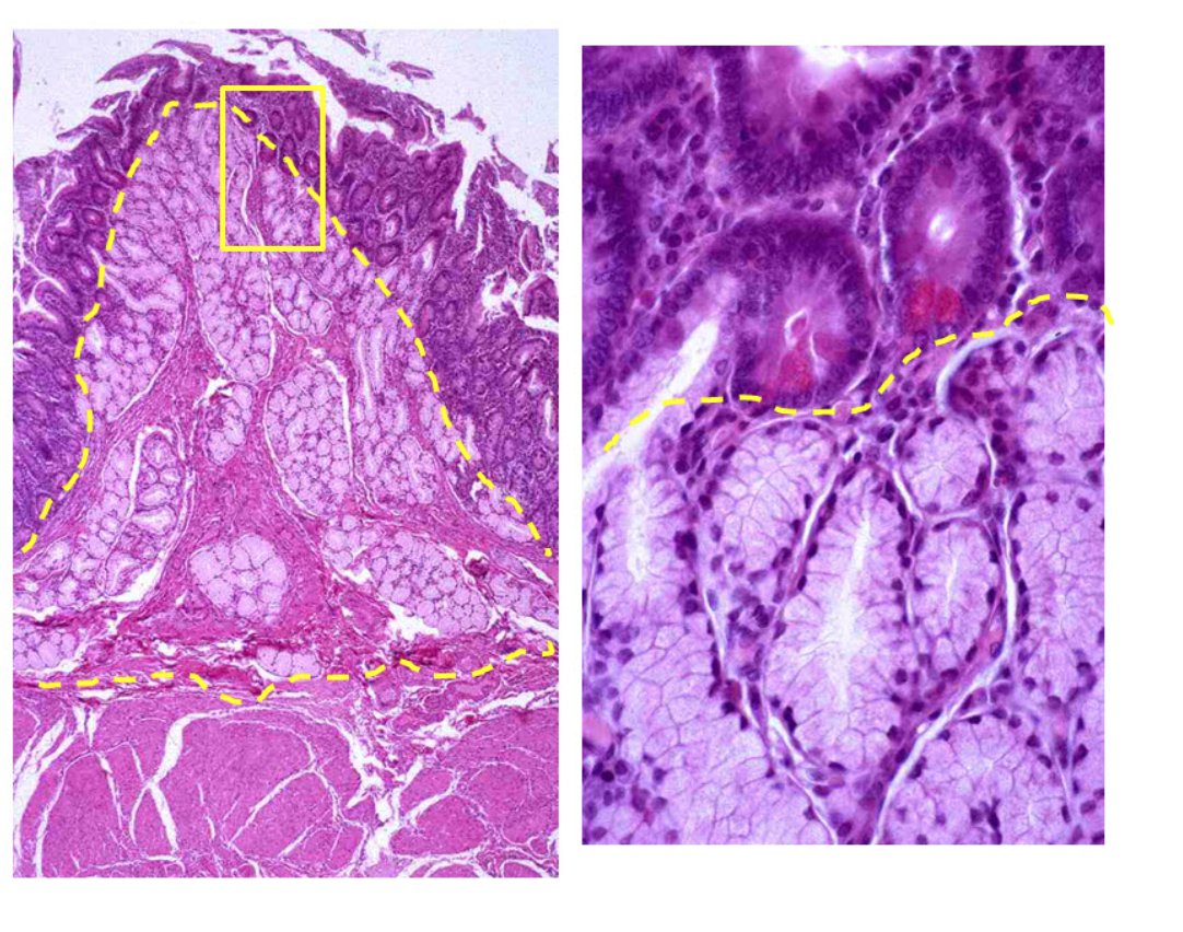
a) mucosa
b) submucosa
c) muscularis mucosa
d) muscularis externa
e) adventitia
Submucosa
Which of the following contains a muscularis externa where the upper 1/3 portion is striated skeletal muscle, the middle third 1/3 portion is a mix of striated skeletal muscle and smooth muscle, and the lower 1/3 portion is all smooth muscle?
a) trachea
b) esophagus
c) stomach
d) jejunum
e) duodenum
Esophagus
Examine the low and higher power view of this specimen.Identify the organ this specimen represents.
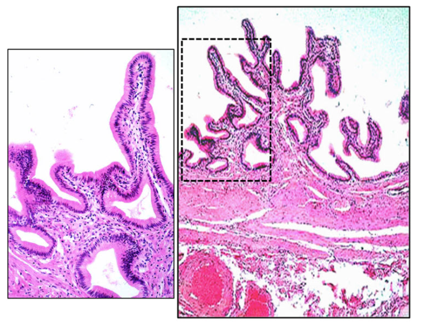
Gallbladder
A mucosa is found in all of the locations EXCEPT:
a) male reproductive tract
b) urinary tract
c) large artery
d) gastrointestinal tract
e) respiratory tract
Large Artery
Which is the location of stem cells in the intestine?
a) tip of villus
b) base of villus
c) intestine gland wall
d) intestine gland base
e) lamina propria
Intestinal gland wall
Which of the following describes folds of the mucosa with submucosa at the core and function to increase surface area?
a) Plica Circularis
b) Mesentary
c) Meissner’s Plexi
d) Peyer’s Patches
e) Villus
Plica circularis
Which nephron segment has simple squamous epithelium bordering its lumen?
a) proximal convoluted tubule
b) distal convoluted tubule
c) proximal descending tubule
e) distal straight descending tubule
e) thin segment of Henle’s loop
Thin segment of Henle’s Loop
The endothelial cellular component of the glomerular filtration membrane has slit pores that are covered with a thin diaphragm.
False
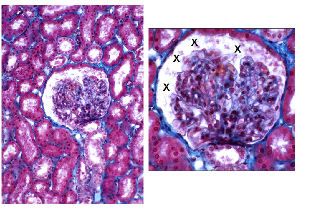
Identify the tubule that is marked by the letter X in several examples.
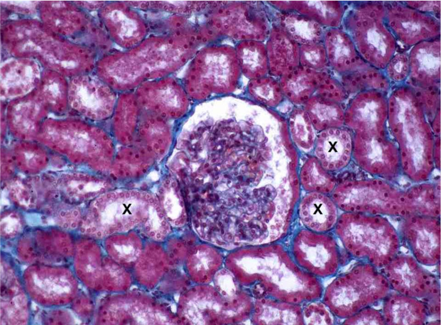
Distal convoluted tubule
Examine the low and higher power images of this specimen stained with Goldner Trichrome Stain. Identify the area in which the letter Xs are placed.
a) Medullary rays
b) Renal corpuscle
c) Bowman’s capsule
d) Distal convoluted tubule
e) Proximal convoluted tubule
f) Urinary space
g) Urinary pole
h) Vascular pole
Urinary space
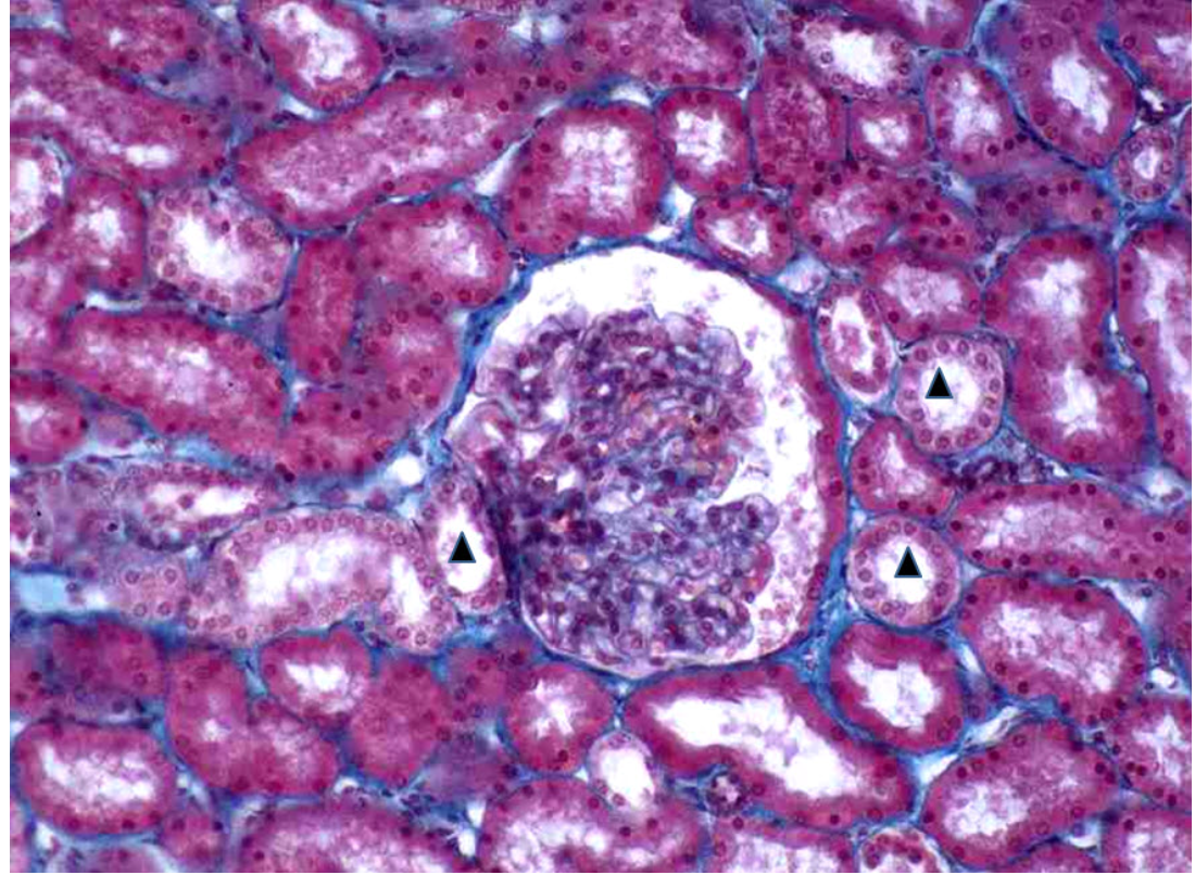
Identify the segment of the nephron indicated in two examples in this specimen with a black triangle in their lumens.
a) Medullary rays
b) Renal corpuscle
c) Bowman’s capsule
d) Distal convoluted tubule
e) Proximal convoluted tubule
f) Urinary space
g) Urinary pole
h) Vascular pole
Distal convoluted tubule
Which type of Epithelium lines the region of the Larynx where the vocal cords are found?
a) Stratified Squamous Non-keratinizing
b) Pseudostratified ciliated columnar
c) Simple squamous non-keratinizing
d) Transitional epithelium
e) none of the above
Stratified squamous non-keratinizing
Which denotes the usefulness of viewing liver microscopic architecture as liver acini?
a) flow of bile
b) flow of portal blood
c) oxygen delivery to hepatocytes
d) distribution of von Kupffer cells
e) distribution of Ito cells
Oxygen delivery to hepatocytes
Which is the location of fat storing cells of Ito in the liver?
a) portal area
b) lining central veins
c) lining bile canaliculi
d) lining sinusoids
e) space of Disse
Space of Disse
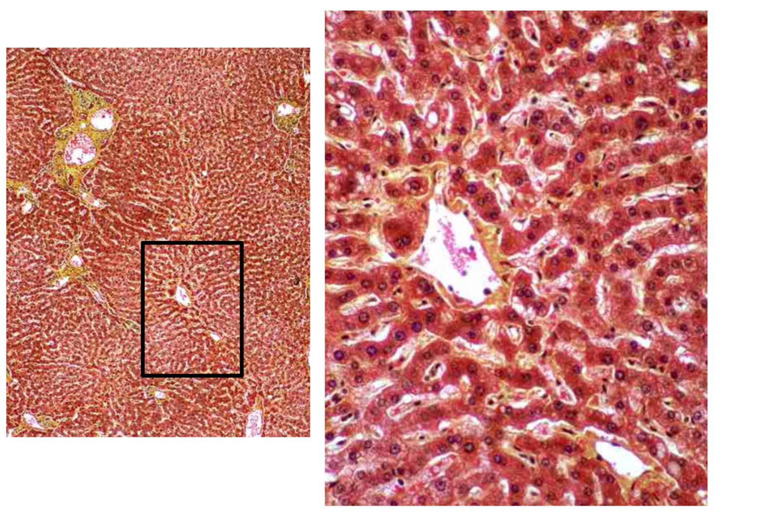
Examine the low (left image) and higher power (right image) and then identify the cell type in the higher power that is dominant and have rounded nuclei.
a) Hepatocytes
b) Parietal cells
c) Absorptive cells
d) Surface mucous cells
e) Paneth cells
f) Chief cells
g) Neuroendocrine cells
h) Goblet cells
Hepatocytes
The main function of the von Kupffer cell of the liver is to synthesize and secrete fibrinogen.
False
The structures that are located in the portal area of the liver are bile duct, hepatic artery, and central vein.
False
An alveolar septum includes all of the following EXCEPT:
a) macrophages
b) Clara cells
c) type II pneumocytes
d) type I pneumocytes
e) elastic fibers
f) capillaries
g) fibroblasts
Clara cells
Which of the following is the correct range of thickness of the air-blood barrier in the alveoli of the lung?
a) 0.1-1.5 centimeters
b) 0.1-1.5 millimeters
c) 0.1-1.5 micrometers
d) 0.1-1.5 nanometers
e) 0.1-1.5 angstroms
0.1-0.5 micrometers
Which of the following cells function in respiratory Epithelium to secrete a variety of peptides via small electron dense granules?
a) Neuroendocrine cells
b) Goblet cells
c) Brush cells
d) Ciliated cells
e) Paneth cells
Neuroendocrine cells
Which of the following consists of a wall of series of alveoli interrupted by thin bands of smooth muscle lined by squamous cells?
a) Alveolar ducts
b) Respiratory bronchioles
c) Alveolar sacs
d) Alveoli
e) Bronchus
Alveolar ducts
Alveolar macrophages clear foreign substances from the respiring airways.
True
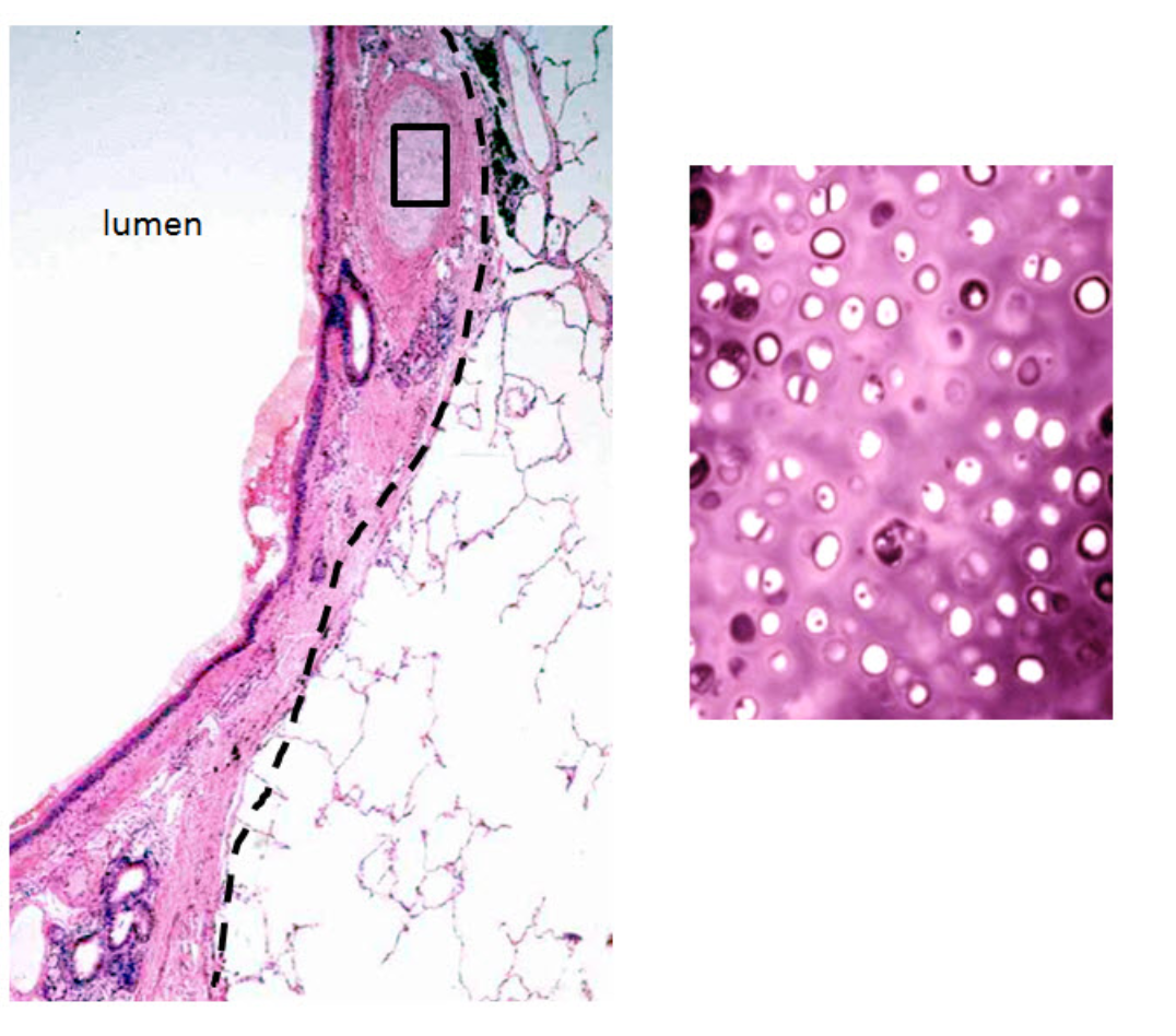
Identify the segment of the respiratory tract represented by the area from the dashed line to the lumen. The enlarged view on the right of the boxed in area should be carefully considered.
a) False volal fold (ventricular fold)
b) Alveolar duct
c) Bronchiole
d) Bronchus
e) Trachea
f) Alveoli
g) True vocal fold
Bronchus
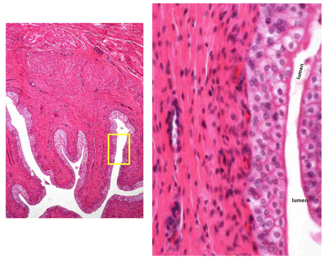
Identify the layer of this specimen between the dashed line and the lumen. Right image is a higher power view of the layer.
a) Mucosa
b) Submucosa
c) Muscularis mucosa
d) Muscularis externa
e) Adventitia
Mucosa
Examine the low power and the higher power view of the boxed in area. The upper higher power view is stained with a Trichrome Stain. The lower higher power view is stained with Hematoxylin and Eosin. What part of the Respiratory Tract is represented by this specimen. It is not the nasal or oral cavity.
a) True vocal fold
b) False vocal fold (ventricular fold)
c) Trachea
d) Bronchi
e) Bronchiole
f) Alveolar duct
g) Alveoli
False vocal cord (ventricular fold)
What organ does this histological pattern and cell types seen in this specimen represent?
a) submandibular gland
b) liver
c) sublingual gland
d) pancreas
e) parotid gland
Submandibular gland
Which of the following glands is composed of almost 100% serous cells (some text state there may be a few mucous cells) forming its acini?
a) labial gland
b) sublingual gland
c) submandibular gland
d) parotid gland
Parotid gland
Which intestine cell types function to absorb glucose, amino acids and lipids?
a) enterocyte
b) goblet cell
c) enteroendocrine cell
d) Paneth cell
Enterocyte
Which is an antigen presenting cell in the intestine?
a) enterocyte
b) absorptive cell
c) M-cell
d) T-cell
e) Paneth cell
M-cell
Which is the location of a Peyer's patch?
a) duodenum
b) jejunum
c) ileum
d) ascending colon
e) transverse colon
Ileum
Which function is associated with intracellular canaliculi?
a) pepsinogen synthesis
b) pepsin production
c) hydrochloric acid production
d) mucus production
Hydrochloric acid production
Examine the low and higher power views of this specimen. Name the circled structures in the specimen.
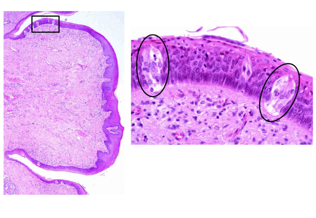
a) Filiform papilla
b) Fungiform papilla
c) Circumvallate papilla
d) Tase buds
e) Buccal glands
Taste buds
Which is the function of cytoplasmic vesicles in a facet cell of the urinary bladder?
a) stores urine
b) stores calcium
c) stores plasma membrane
d) contracts surface cells
e) removes sodium from urine
Stores plasma membrane
Choose the correct name of the layer of cells between the yellow dashed line and the lumen in the higher power view of this specimen.
a) Basal cell layer
b) Intermediate cell layer
c) Facet cell layer
d) Crusta cell layer
Facet cell layer
Examine the low power and the higher power view of the boxed in area. Identify the organ.
a) Either urinary bladder or ureter
b) Either jejunum or ileum
c) Either oral cavity or esophagus
d) Either gallbladder or colon
Either urinary bladder or ureter
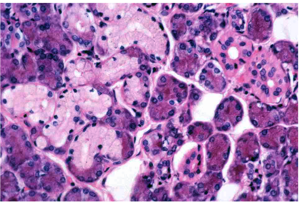
Examine the low and higher power views of this specimen. Identify the organ represented by this specimen.
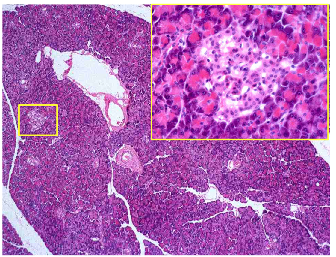
Pancreas
Identify the cell group indicated by the arrows whose cytoplasm is acidophilic containing granules.
Serous demilune
The centroacinar cell of the pancreas is a part of the structure of an islet of Langerhans.
False