4.1 - Structure of the Respiratory System
1/80
There's no tags or description
Looks like no tags are added yet.
Name | Mastery | Learn | Test | Matching | Spaced |
|---|
No study sessions yet.
81 Terms
Respiratory System
A biological system responsible for the exchange of gases between the internal and external environments of the body.
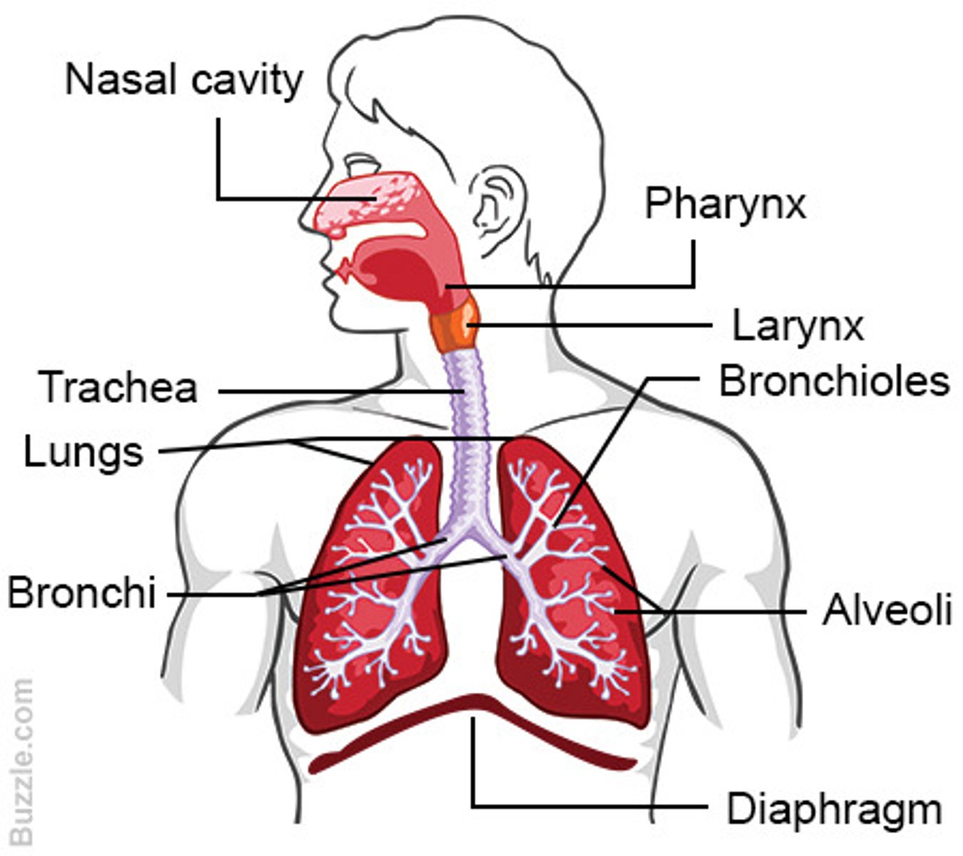
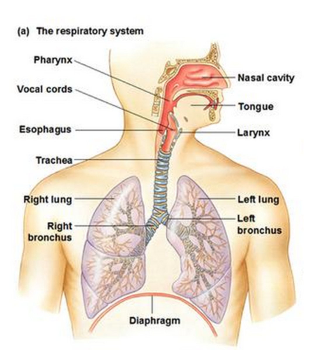
Structure of the Respiratory System
Nasal Cavity, Larynx, Pharynx, Epiglottis, Trachea, Ribs, Bronchi, Intercoastal Muscles, Bronchioles, Lungs, Diaphragm, Alveoli
Mouth + Nostrils
Structure:
- Opening to the outside air.
Function:
- Air enters the body during inspiration and exits during expiration.
Nasal Cavity
Structure:
1. Lining is convoluted and lined with mucous membranes.
2. Has hair and mucous lining.
3. Chamber
Function:
1. Increased surface area and air are warmed and humidified.
2. Traps debris. (Dust, pollen, microbes)
3. Resonant speech sound
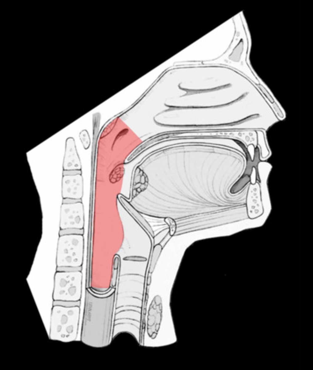
Pharynx
Structure:
- The region from the nasal cavity to the epiglottis.
Function:
- Food and air travel through it before separating into the larynx (air) and oesophagus (food and liquids)
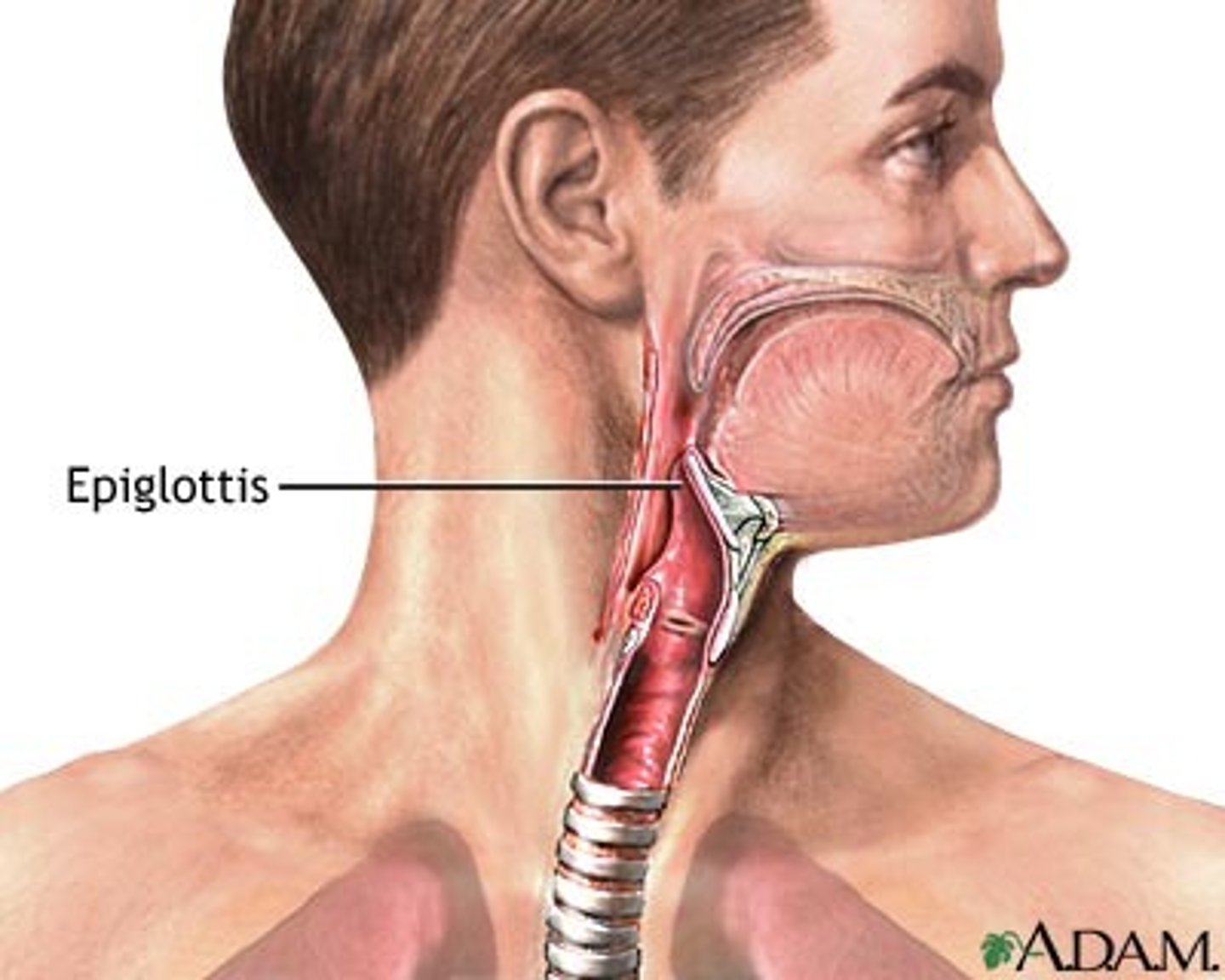
Epiglottis
Structure:
- Flap of elastic cartilage.
Function:
- During swallowing, it covers the trachea to prevent food or liquids from entering the lungs.
Larynx (voice box)
Structure:
1. Cartilage structure joining pharynx to trachea
2. Contains vocal cord; mucous membranes
Function:
1. Maintains an open passage way for air.
2. Vibrate as air passes, creating sound for speech.
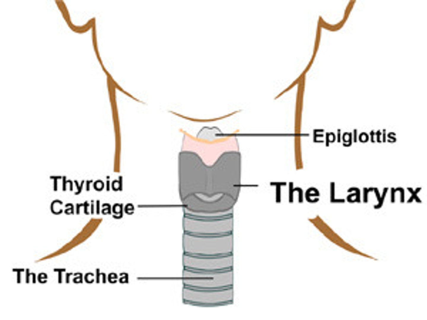
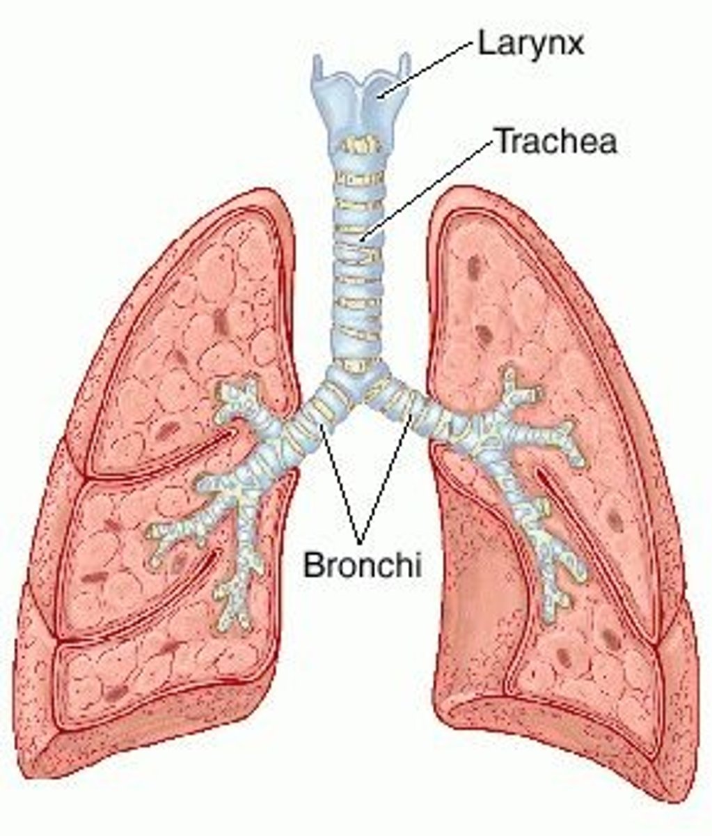
Trachea (windpipe)
Structure:
1. Joins larynx to two primary bronchi.
2. Wall have c-shaped cartilage rings.
Function:
1. Carries air into and out of the lungs.
2. Holds the airways open to maintain constant airflow.
Characteristics the Trachea, Bronchi and Bronchioles have are:
Structure:
1. A wall with an epithelial lining that produces mucus.
2. Epithelial cells have cilia, tiny hairlike projections.
Function:
1. Traps dust and debris, preventing it from entering the lungs and protecting them from contaminants.
2. Beat in a wave-like motion to move mucus with debris up to the pharynx to be swallowed and digested.
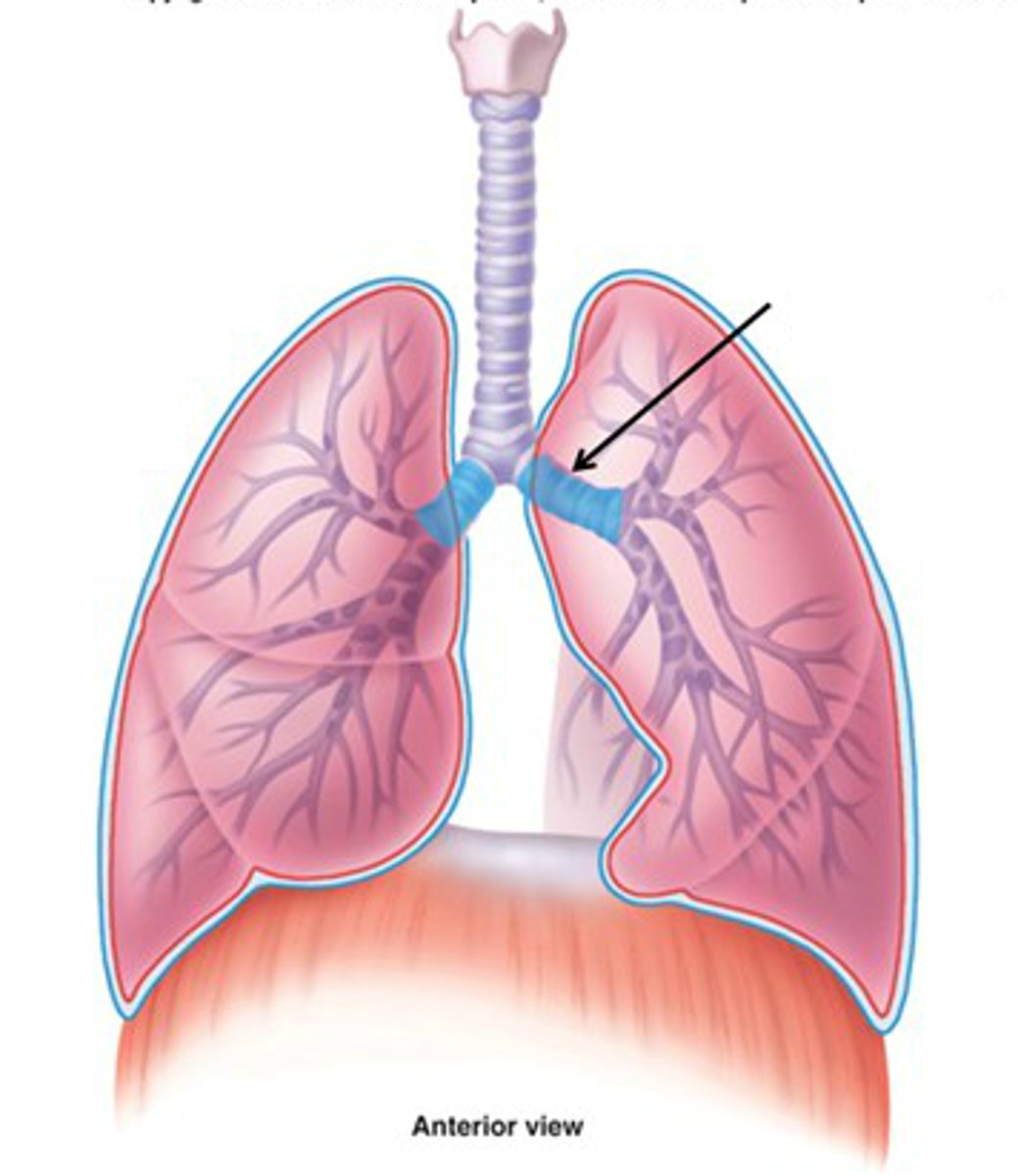
Primary Bronchi
Structure:
1. Two tubes: Left and right
2. Walls have C-shaped cartilage rings.
Function:
1. Connect the end of the trachea to the to lungs.
Left tube -> Left Lung, Right tube -> Right Lung.
2. Hold the airways open to maintain airflow.
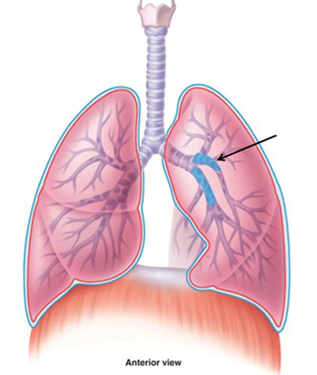
Secondary Bronchi
Structure:
1. Branches of narrower tubes.
2. Cartilage rings are more spread out.
Function:
1. Connects to the lobes of each lung.
2. Holds the airways open to maintain airflow.
Tertiary Bronchi
Structure:
1. Branches of narrower tubes.
2. Smooth muscle and elastin walls.
Function:
1. Connects to bronchioles.
2. Holds the airways open to maintain airflow.
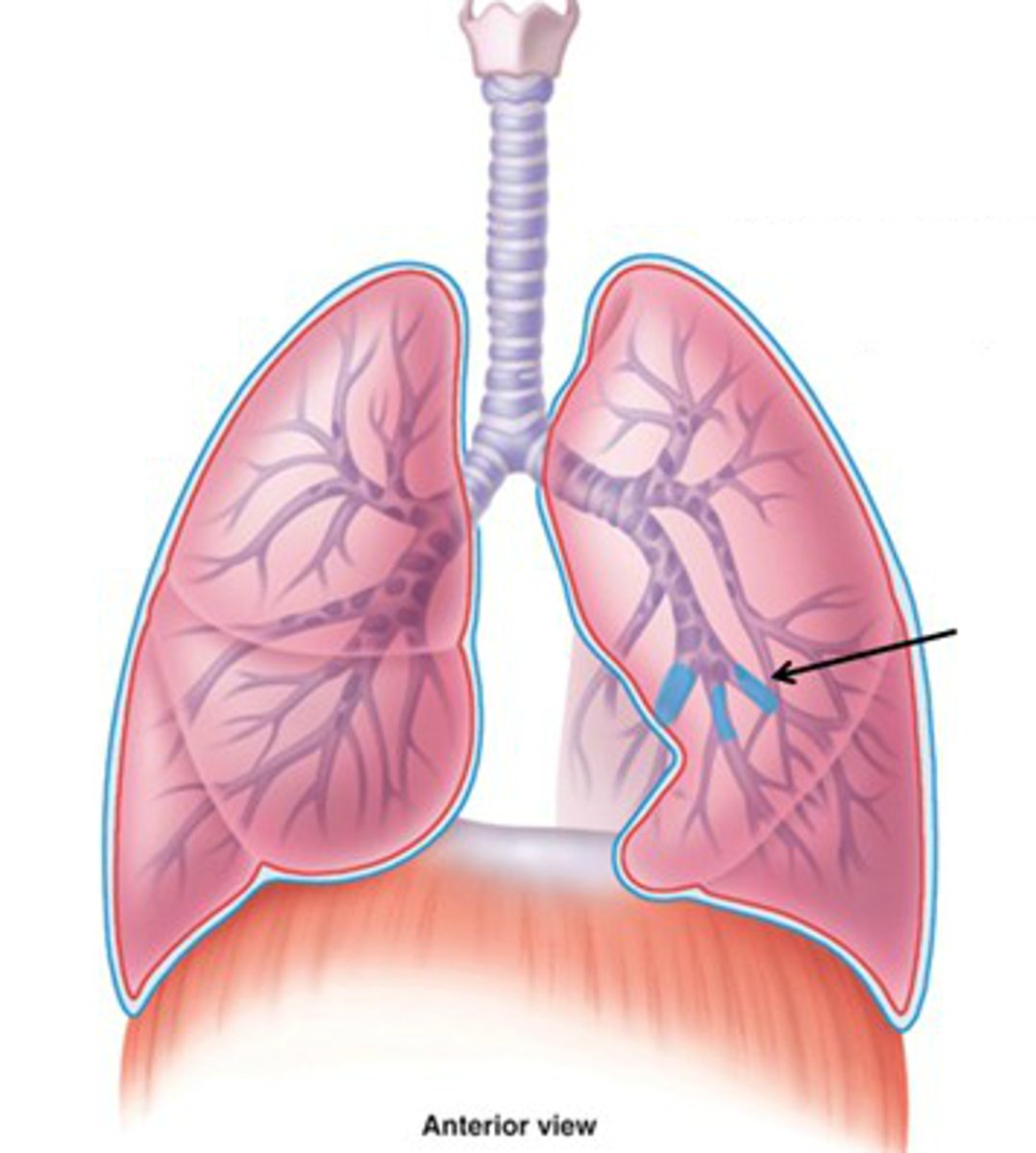
Bronchioles
Structure:
1. Branching of narrower tubes with terminal ends.
2. Smooth muscle and elastin walls. (no cartilage)
Function:
1. Alveoli at the ends.
2. Holds the airways open to maintain airflow and controls the flow of air -> Expanding when the body needs more O2.

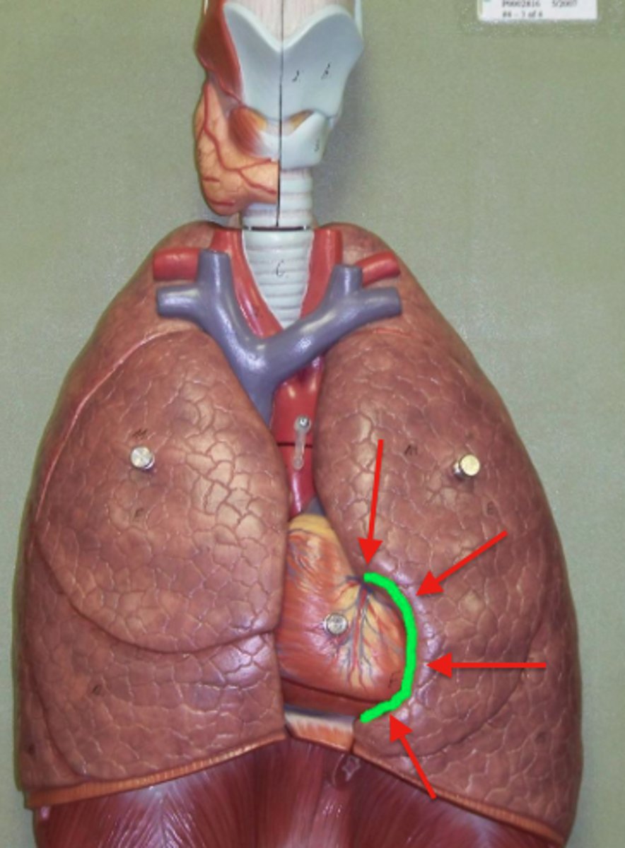
Caridac Notch
Where the heart lies on this medial concavity of the left lung, it leaves the appearance of a 'dent' in the left lung.
Alveoli
- Clusters of tiny air sacs at the end of the smallest terminal bronchioles.
- The site of gas exchange:
- O2 moves from the air sac to the blood.
- CO2 moves from the blood into the air sac.
- 1 Cell-thick walls and surrounded by capillaries.
Mediastinum
Structure:
- The space between the lungs.
Function:
- Contains the heart and blood vessels.
Pleural Sac:
1. Visceral Pleura
2. Pleural Cavity
3. Parietal Pleura
Structure:
1. A thin slippery membrane covering the lungs.
2. Contains a lubricating plural liquid.
3. Lines the thoracic cavity.
Function:
1. Reduces friction between the lungs and the thoracic cavity during respiration.
2. Cushions the lungs.
3. Acts as a barrier against microbes -> Prevents disease.
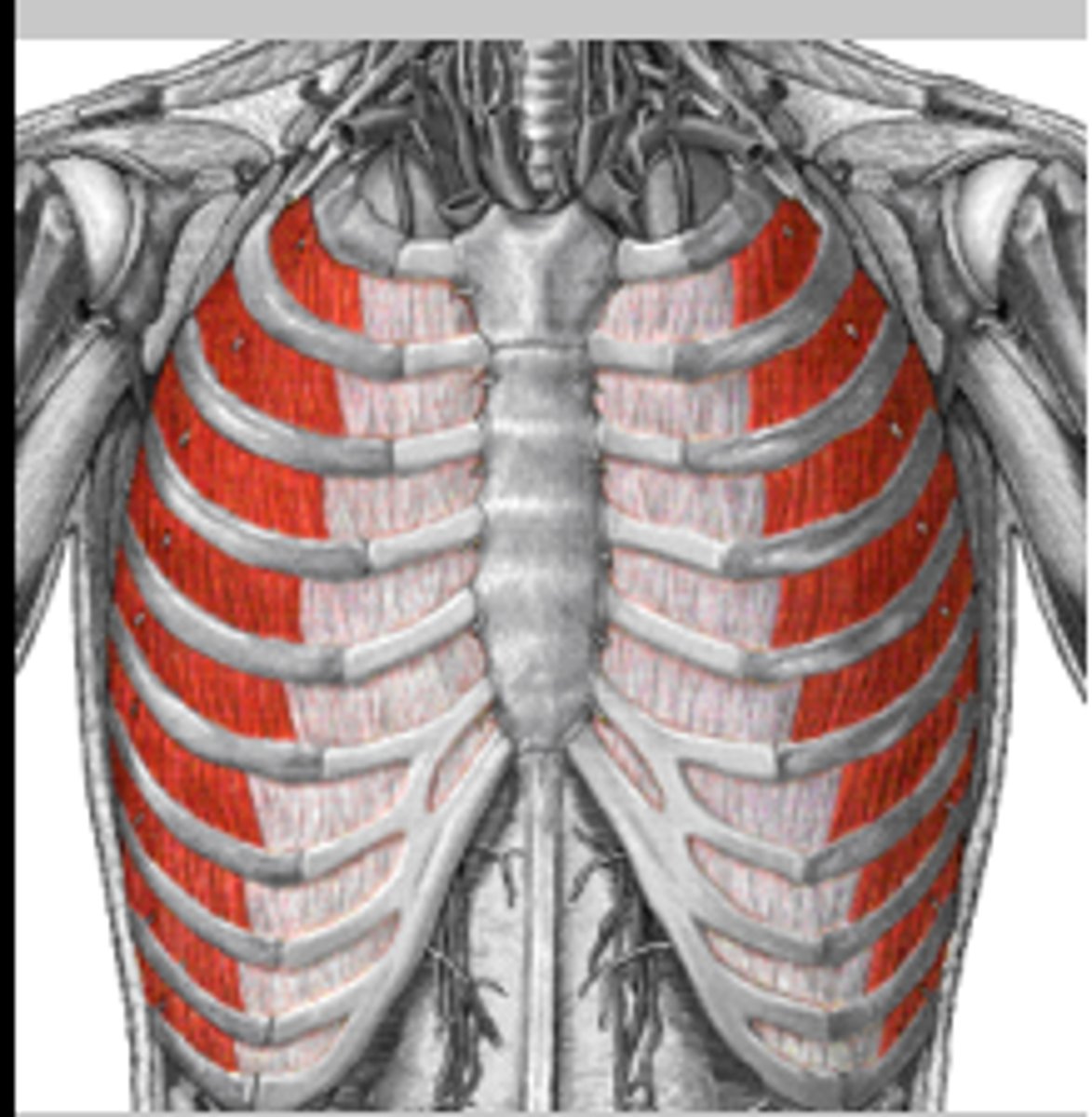
Ribs
Structure:
- Long curved bones wrapping the thoracic.
Function:
- Forms the framework of the chest.
- Protects heart and lungs.
- Moved up and down by intercoastal muscles through inspiration.
Intercoastal Muscles
Structure:
- Skeletal muscles between the ribs.
Function:
- Moves the ribs up and down, increasing the volume of the thoracic cavity for inspiration.
Diaphragm
Structure:
- A dome shaped skeletal muscles
Function:
- Separates the thoracic cavity from the abdomen.
- Flattens when it contracts. (Inhalation)

1. Maintain the concentration gradient between the O2 in the alveoli and in the blood
2. Effect for gas exchange.
3. Air must be constantly replaced.
- The process of moving air into and out of the lungs, due to differences in the air pressure.
- Manipulated by changing the volume of the thoracic cavtiy.
- Air moves from areas of higher pressure to lower.
- Taking air into the lungs.
- Pressure in the lungs is less than the atmospheric pressure
- Increase in volume of the thoracic cavity.
- Diaphragm position is flattened.
- Intercostal muscles pull the ribcage up + out.
- Moving air out of the lungs.
- Pressure in the lungs is more than the atmospheric pressure.
- Decrease in volume of the thoracic cavity.
- Diaphragm position is domed.
- Intercostal muscles pull the ribcage down + in.
1. The diaphragm contracts, moving down and becoming flatter due to the external intercostal muscles contracting and pulling the ribs up and out.
2. Volume of the thoracic cavity increases.
3. The pleura pulls on the lungs, making the lungs expand and increase in volume.
4. Air pressure in the lungs decreases below that of the outside.
5. Higher air pressure in the external environment -> air moves into the lungs until it becomes equal.
1. The diaphragm relaxes, moving up and becoming domed-shaped due to the external intercostal muscles relaxing and moving the rib cage down and in.
2. Volume of the thoracic cavity decreases.
3. The pleura pushes on the lungs, making the lungs deflate and decrease in volume.
4. Air pressure in the lungs increases above that of the outside.
5. Higher air pressure in the internal environment -> air moves out of the body until equal.
- Caused by long-term exposure to irritants entering the lungs. (eg. Smoke, Dust, Pollution)
- Alveoli become damaged from the particles, making them lose their elasticity, often becoming fibrous tissue and may down, which reduces the internal surface area of the alveoli.
Thus, two problems: Inadequate surface area for gas exchange and difficulty in ventilating the lungs, as it requires voluntary effort rather than a passive process.
- No cure, progression of the disease cannot be ceased.
- The development of a mass of cells that divides in an uncontrolled way, which is a tumour.
- Exposure to asbestos fibres, pollutants, and tobacco from smoking increases the risk for lung cancer
Begins in the walls of the air passages, usually the bronchi, and the inhaled particles constantly irritate the mucous membrane that lines the air passages.
This creates an excessive production of mucus, causing the cells at the base of the membrane to divide more rapidly. (The accumulation of mucus cannot be removed.)
- This results in 'smokers cough', and the trapped mucus causes the alveoli to rupture. -> Emphysema -> Cancer in the air passages can travel around the body.
- Pneumonia is an infection of the lungs caused by bacteria, viruses, fungi or other organisms.
- The inflammation caused by the infection causes secretion of fluid and mucus into the alveoli, decreasing the amount of air they can contain.
- Surface area for gas exchange is also reduced.
- Tuberculosis is an infection of the lungs by the bacterium, 'Mycobacterium tuberculosis.'
- It spreads through droplets by sneezing, coughing or spitting. So any droplets of moisture containing the virus can be inhaled by others.
- Asthma is a medical condition that causes difficulty breathing due to the narrowing of the airways. This occurs due to:
- The smooth muscles contract -> Narrowing the airways.
Inflammation causes the lining of the airways to thicken -> Narrowing its diameter.
Asthma can be caused by an allergic response or a non-allergic response.
- Triggers include respiratory infections, cigarette smoke and normal smoke, and exercise.
- During an attack, the muscles surrounding the bronchioles go into spasm, which causes involuntary contractions, the narrowing of the air passages, and thus difficulty breathing.
1. Ventilation brings in oxygen-rich air and removes carbon dioxide-rich air, keeping a high oxygen and low carbon dioxide concentration gradient in the alveoli.
2. Oxygen dissolves in the moisture lining of the alveoli and diffuses into the blood, where it binds to haemoglobin in erythrocytes, forming oxyhaemoglobin.
3. Carbon dioxide in the blood diffuses into the alveoli and is breathed out.
4. Blood flow constantly brings in low-oxygenated and high-carbon dioxide blood, which maintains the concentration gradient between the blood and the alveoli.
In the alveoli are active surfactants, which help with breaking the water tensions to enable O2 to dissolve.
Structural Features for efficient Gas Exchange:- Alveolus Wall
- 1 Cell thick -> Gas molecules have a short distance to diffuse.
- Exchange is faster/more efficient.
Structural Features for efficient Gas Exchange:- Blood Supply
- A huge network of capillaries closely surrounds each alveolus.
- Allows for a continuous flow of blood to maintain the concentration gradient.
- Delivers blood high in CO2
- Removes blood high in O2
Structural Features for efficient Gas Exchange:- Constant Ventiliation
- Air is constantly replaced to maintain concentration gradient.
- Delivers air high in O2
- Removes air high in CO2
Structural Features for efficient Gas Exchange:- Deep inside the body
- Reduces the loss of essential fluid by evaporation.
- Gas must dissolve before they can diffuse into the body.
Structural Features for efficient Gas Exchange:- Surface Area
- More sites for exchange -> Exchange becomes faster.
A system that extracts nutrients from the food we eat and absorbs them into the bloodstream for it to be used by cells for:Energy Growth Cell division
1. Ingestion.2. Mechanical Digestion 3. Chemical Digestion 4. Movement 5. Absorption 6. Elimination
1. Mastication:Chewing in the mouth by the teeth
2. Churning:The action in the stomach where three smooth muscle layers (circular, longitudinal, and transversal) breaks the chyme down further. (Chyme is food in stomach)
3. Emulsification:The gall bladder releases bile, produced by the liver, into the duodenum. (small intestine)The bile salts act as emulsifying agent for dividing fat into smaller droplets. (Dish soap washing off fat.)
4. Segmentation:is the contractions of circular smooth muscle in the duodenum.breaks up the bolus, mixes it with digestive juices, and reduces clumping. (Agitation)
Ribonucleic Acid (RNA),Nucleic Acids, Phosphate and ribose sugar backbone with nitrogenous base -> Pancreatic Ribnuclease (Duodenum) -> RNA Fragments are used to make RNA.
Deoxyribonucleic Acid (DNA)Phosphate and deoxyribose sugar backbone with nitrogenous bases. -> Pancreatic deoxyribonuclease (Duodenum) -> DNA fragments are used to make DNA.
The Different types of Molecules broken down in Chemical Digestion:Carbohydrates
Starch/ Glycogen -> Get broken down by Salivary Amylase -> Into Disacchrides Maltose, Sucrose, Lactose -> They get broken down by Maltase, Sucrase and Lactase -> into Monosacchrides.
Sucrose -> gets turned into 1 glucose and 1 fructose. Maltose -> gets turned into 2 glucose.Lactose -> gets turned into 1 glucose and 1 galactose.Used as a main source of energy.
The Different types of Molecules broken down in Chemical Digestion:Proteins
Proteins -> Get broken down by pepsin -> into large polypeptides (Many amino acids joined by peptide bonds.) Polypeptides -> Get broken down by Pancreatic protease/ Trypsin -> into short peptides -> Get broken down by Intestinal peptidase -> into Amino Acids (Glutamine)Used to make enzymes, hormones, and used for energy.
The Different types of Molecules broken down in Chemical Digestion:Lipids
Lipid-triglyceride -> Gets broken down by Pancreatic and Intestinal Lipase -> Into 3 fatty acids and 1 monoglyceride.
- Material stays in the large intestine therefore movement through the canal is reduced, and the food remains at a large size.
- Because it stays in the large intestine, water is constantly absorbed, and the faeces becomes drier and harder.
- Defecation à More difficult and more painful.
- The main cause is being of the lack of fibre. Roughage is cellulose, which is a major component found in plant-based foods. There are no enzymes to digest cellulose, its's very important as it stimulates movement.
- Lack of exercise
- Stress/emotional problems.
- Movement through the canal is increased, thus there will be frequent defection
- Because there is excess water (not absorbed into the body), the faeces will be watery.
- Defecation à More frequent and waterier.
If the diarrhoea is severe or happens over prolonged periods, it may lead to dehydration due to the faeces being pushed through the canal too quickly and not having enough time for water to be absorbed by the body.
-> Bacteria -> E. coli, Salmonella-> Virus -> Norovirus-> Cancer (Bowel Cancer)-> Lactose intolerance-> Coeliac disease
- Soluble and insoluble fibres are found only in foods that derive from plants.
- Soluble -> Dissolves into water-> Lower cholesterol-> Decreased cancer-> Benefit blood glucose levels-> Fibre holds onto and traps fats in the intestines, which helps prevent their absorption in the body.
- Sources-> Fruits-> Vegetables-> Bran-> Soy-> Barley
- Research suggests that it may be linked to diet, high alcohol consumption and smoking.-
> Diets high in red meats (especially processed meat - Salami, Ham) and low in fibre may increase the risk of developing bowel cancer.
-> Overweight/Obese-> Lack of exercise
- If you are genetically exposed to cancer or it runs in the family, the risk of bowel cancer increases due to the slight correlation.
- Caused by the consumption of foods that have gluten in them. Gluten is found in:
-> Wheat
-> Rye
-> Barley
- Eating foods that contain gluten will force the body to release an immune response by damaging the villi in the small intestine.
- Without healthy villi, nutrients cannot be absorbed à Malnourishment à despite the quantity of food consumed or whether you have a healthy diet.
- Muscle cramp + Pain in abdomen
- Pain -> tingling in joints and legs.
- 'No symptoms' -> Tiredness.
- Genetic -> Coeliac Disease is inherited. Thus, if you have the gene for the disease, it may be activated later in life. -> No cure -> Gluten-free diet
- Main excretory organ
- Responsible for maintaining a constant concentration of materials in the body fluids.
- Excretes urine, excess H2O, Urea, Uric Acid, Creatine, NaCl
- Removal of bile pigments in faeces from the breakdown of haemoglobin in erythrocytes.
NOTE! Bile pigments are a metabolic waste; however, we use the term elimination instead of excretion for faeces.
- Processing of many substances
- Produces urea from the breakdown of specific unrequired amino acids.
- Detoxifies alcohol and many drugs.
- Converts excess amino acids into useful energy.
- Detoxifies alcohol and other drugs.
- Converts excess amino acids into useful energy.
-> Breaks down proteins into amino acids.
-> Breaks down amino acids when needed for energy.
- Prepares materials for excretion
-> Breaks down haemoglobin in dead erythrocytes to produce bil pigments removed by proteins into amino acids.
- Deactivates hormones and converts them into a form that can be excreted.
- Excess protein cannot be stored, therefore it gets broken down into amino acids.
- Amino acids can be:
-> reused to make new proteins, cell structures, enzymes.
-> Lost through urine, skin, hair and fingernails.
-> Used for energy, Only when required
Amino Group (NH2)
- Enzymes are used to remove NH2.
- Liver cells convert it to Ammonia (NH3)
-> Extremely soluble in water and is highly toxic to cells.
- Rapidly converted into urea -> Less toxic.
- Transported in the blood to be eliminated from the body.
- Then excreted by the kidneys into urine.
- Then excreted by the kidneys into urine.
- Used for aerobic respiration, releasing: Energy, CO2 + H2O, Ketone bodies, Acetyl CoA.
- Formed from solid crystals that build up inside the kidneys.
- They usually form when urine is too concentrated.
- It believed that they are caused by insufficient fluids in the diet.
- Smaller stones, while painful, may pass with the aids of fluids.
- Larger stones may need to be broken up with sound waves or physically removed via surgery.
- When the kidneys lose their ability to excrete wastes and control the level of fluid in the body.
- Most kidney diseases affect the glomeruli -> reducing their ability to filter blood.
- Diabetes, high blood pressure or kidney diseases slowly destroy the nephrons. Until the only way to maintain life is by dialysis or a kidney transplant.
1. Peritoneal dialysis: A treatment for kidney failure that uses the lining of your abdomen (the peritoneum) to filter waste products from your blood.
- Works based on the principle of diffusion.
- A fluid called dialysate (which contains glucose and other substances but no waste) is placed into the abdominal cavity via a catheter.
- Because the concentration of waste is higher in the blood than in the fluid, waste diffuses out of the blood and into the fluid.
- Useful substances do not move because their concentrations are the same in both fluid and blood.
- Done daily
2. Haemodialysis: Involves passing the blood through an artificial kidney or dialysis machine.
- Blood is removed from the body and pumped through fine tubes with differentially permeable membranes.
- These tubes are bathed in fluid that is similar to blood in composition but contains no waste.
- Because the concentration of waste is higher in the blood, waste diffuses out into the fluid.
- Clean blood is returned to the body.
- Takes 4-5 hours, done 3 times/ week.
- Caused by infection, autoimmune problems, genetic disorders, cancer, excessive alcohol consumption and a fatty diet.
- Symptoms include:
-> Build up of toxins that would normally be eliminated. Which would cause a yellow tinge to the skin (Jaundice)
-> Abdominal pain and swelling
-> Dark urine
-> Pale or dark faeces colour.