Unit 6: Genetics
 Call Kai
 Learn
Learn Practice Test
 Spaced Repetition
 Match
Match Flashcards
 Knowt Play
1/114

There's no tags or description
Looks like no tags are added yet.
Name | Mastery | Learn | Test | Matching | Spaced | Call with Kai | Chat |
|---|
No analytics yet
Send a link to your students to track their progress
115 Terms
Advantages and Disadvantages of Sexual VS Asexual Reproduction
Asexual = Binary fission, mitosis, budding, fragmentation, vegetation reproduction, parthenogenesis
Produce genetically identical offspring by individuals that are adapted to an existing environment
Rapid population growth (Under stable conditions)
Energy efficient (Lack need for mate)
BUT…
Limited ability to adapt
Mass death once environment changes
Sexual
Produce offspring with gene combinations and thus variation needed for adaptation to a changed environment.
Eliminate harmful mutations over time
Provide defence against parasites and diseases
BUT…
Needs time & energy
Produce small populations
Role of Meiosis and Fusion of Gametes in Sexual Life Cycle
Meiosis = Breaks up parental combinations of alleles
Production of haploid gametes (‘n’)
Fusion of gametes (Fertilisation) = Produce new combinations
‘2n’ number of chromosomes
Male VS Female in Sexual Reproduction
Male Gamete = Travels to female gamete
Smaller (~5 μm), flagellum for movement
Less food reserves than egg (Since so many are being produced)
Increase chance of fertilisation through quantity
Female Gamete
Invest in quality and survival of offspring
More energy (Larger food resources)
More parental care after fertilization
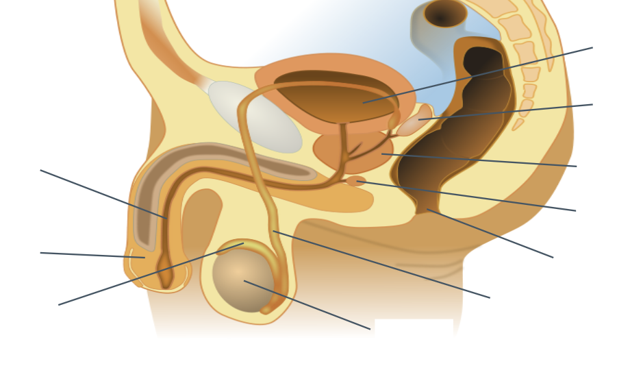
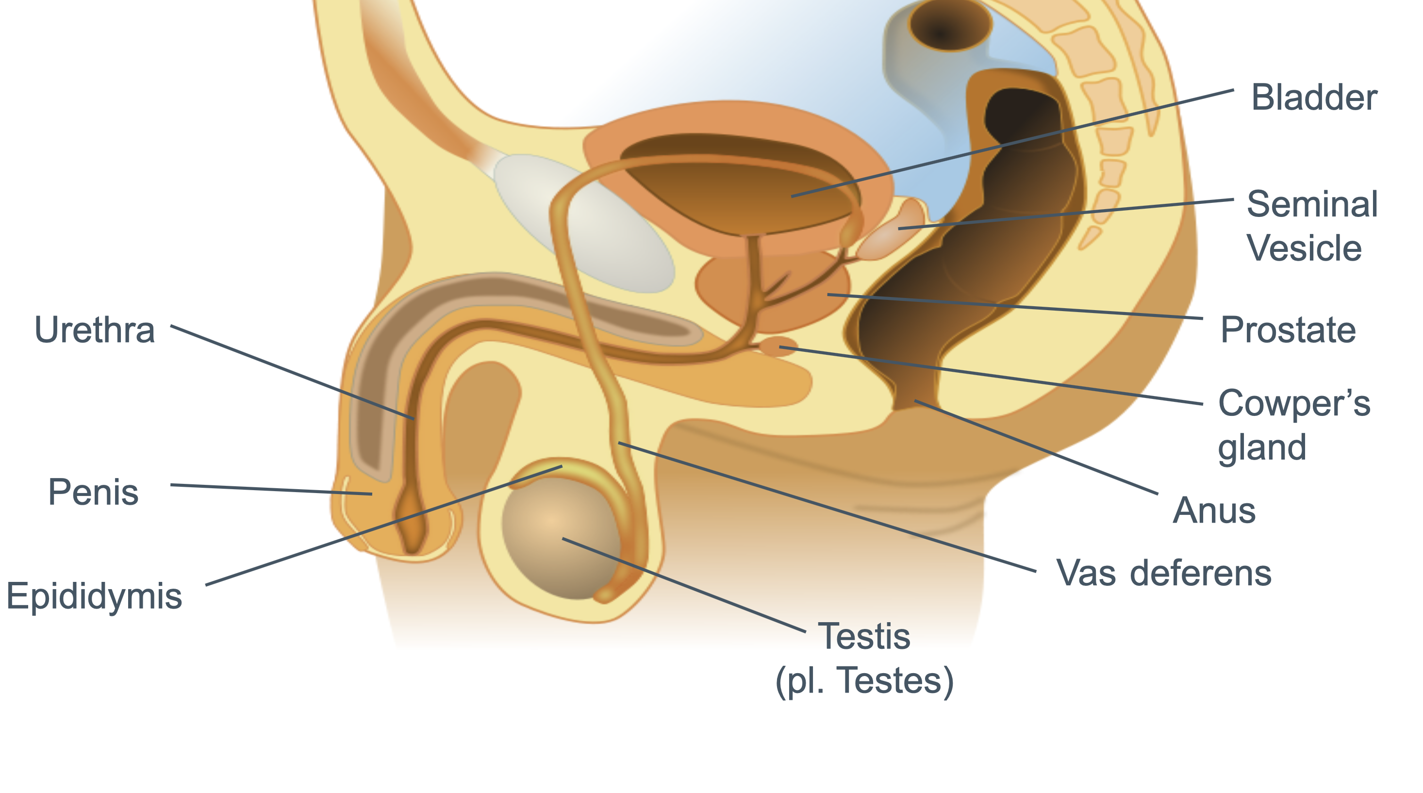
Anatomy of the Human Male Reproductive System + Function (HINT: 11 features)
Testes
Produce sperm + testosterone
Vas Deferens
Carry sperm from epididymis → junction with urethra
Rectum
Cowper’s Gland
Secrete mucus-rich fluids into semen → Protects sperm from acids in urethra
Prostate
Secrete alkaline buffer into semen → Protect sperm from acidic environment in vagina
Seminal Vesicle
Secretes fructose into semen → Provide energy for sperm
Bladder
Epididymis
Produce + Store sperm
Penis
Deposit sperm into vagina during ejaculation; Contains urethra
Urethra
Carries semen during ejaculation / urine from bladder to exterior
Scrotum
Protects testes + Thermoregulation
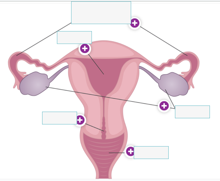
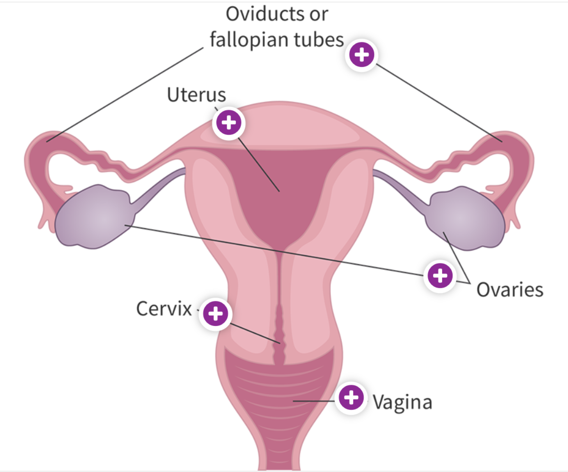
Anatomy of the Human Female Reproductive System + Function (HINT: 5 features)
Fallopian Tubes
Ducts that carry egg to uterus
Ovaries
Produce / release eggs (secondary oocytes) + Secrete oestradiol
Uterus
Muscular structure; Where embryo is implanted and develops
Endometrium = Inner lining of uterus
Cervix
Lower portion of uterus; Opening allows sperm to enter for fertilisation
Vagina
Muscular tube; Leads from external to cervix / Where semen is ejaculated
Changes During the Ovarian and Uterine Cycles (Hormonal Regulation)
Roles of estradiol, progesterone, luteinising hormone (LH), follicle-stimulating hormone (FSH) + Feedback Mechanism
Pituitary gland → FSH + LH / Ovaries → Oestradiol + progesterone
Ovarian Cycle = Production & release of egg / Release of oestradiol & progesterone
Follicular phase (Day 1-13)
FSH increase production and secretion of oestradiol → Increased density of blood vessels (vascularization) in endometrium
Rising oestradiol levels → Increase secretion of FSH and LH (Positive feedback loop)
FSH & LH result in Graafian follicles
Ovulation (Day 14) – Release of egg
Mostly LH & FSH surge → Triggers release of egg; Most mature egg cell burst from follicle through ovary wall
Empty follicle releases progesterone
Luteal phase (Day 15-28)
Empty follicle forms corpus luteum (Endocrine structure) → Produces MOSTLY progesterone and some oestrogen
Progesterone → Promotes endometrium thickening
High levels of progesterone & oestrogen → Inhibit GnRH → Prevent production of FSH & LH → Low LSH & FH → No further Graafian follicle → Corpus luteum break down → Progesterone & oestrogen decrease → Endometrium shed
Uterine Cycle = Preparation / Maintenance of endometrium to receive egg
Menstruation (Day 1-5)
Shedding of endometrium
Proliferative phase (Day 6-14)
Estradiol → Regeneration of endometrium
Secretory phase (Day 15-28)
Progesterone → Thickening and vascularisation of endometrium
The ovarian and uterine cycles together constitute the menstrual cycle (~28 days)
Feedback Mechanisms Involved with Ovarian / Uterine Cycles
Positive Feedback
Rising estradiol levels → Triggers LH surge for ovulation
Negative Feedback
High levels of estradiol and progesterone → Suppress FSH and LH
(So that eggs are not all released)
Fertilisation in Humans
Many sperms penetrate follicle cell layer and zone pellucid using hydrolytic enzymes stored in acrosomes
Fusion of sperm cell membrane with egg cell membrane
First sperm to reach perivitelline layer attaches to its cell membrane receptor
Entry of sperm nucleus into egg + Destruction of the tail and mitochondria; Dissolution of nuclear membrane of sperm and egg nucleus
Sperm nucleus (Haploid) in pronucleus structure, surrounded by membrane
Nuclear membrane of sperm and egg dissolves
Two pronuclei come together and fuses; Only sperm nucleus enters cell
Cortical granules fuse with the egg membrane such that the vitelline layer becomes impenetrable
All condensed chromosomes participate in a joint mitosis to produce two diploid nuclei (46 chromosomes)
Use of Hormones in IVF Treatment
Eggs are fertilised by sperm in lab (outside of body)
Normally, once egg matures per month and release from ovary during ovulation, controlled by natural hormones
In IVF:
Normal secretion of hormones is suspended
Prevent secretion of GnRH by hypothalamus → No FSH or LH by pituitary → No menstrual cycle
Artificial doses of hormones induce superovulation
Artificial FSH → Stimulates development of multiple eggs in follicle cells until appropriate size
Inject hCG → Triggers final cell maturation
hCG = Structurally similar to LH + Longer half life in body than LH → Release more eggs for collection
Control of Puberty by GnRH and Steroid Sex Hormones
Increased release of GnRH by hypothalamus in childhood → Trigger increased LH and FH release by pituitary gland
Hypothalamus releases GnRH in pulses, increasing frequency during puberty
LH and FSH trigger gonads to produce sex hormones (male: testes = testosterone // female: ovaries = estrogen + progesterone)
Increased sex hormone production → Changes in puberty
Growth spurt; Increase in height and body mass
Development of secondary sex characteristics
Growth of underarm / pubic / facial / chest hair
Acne
Ability to reproduce
(For females)
Development of breasts
Menstrual cycle begins
Bone structure of hips widens to prepare for possible childbirth
(For males)
Deeper voice
Testes and penis enlarges
Erections begin
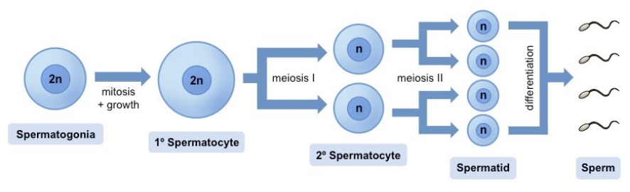
Spermatogenesis in Humans
Occurs in seminiferous tubules
Sperm = Made in testes; stored in epididymis → Carried in vas deferens / Seminal vesicle adds fluid → Leaves by urethra
Starts at germinal epithelium (outside tubules) → Move to lumen (inside tubules) during maturation
Mitosis → Cell growth → Two devisions of meiosis → Differentiation
Spermatogonia (2n) x1 —Mitosis
Spermatocytes (2n) —Meiosis I & II
Primary Spermatocyte (2n) x1 → (Meiosis I) → Secondary spermatocyte (n) x2
Secondary spermatocyte (n) x2 → (Meiosis II) →
Spermatids (n) x4 —Differentiation
Spermatozoa (n)
Receive nourishment from Sertoli cells
When mature, detach → Epididymis for storage
Oogenesis in Humans
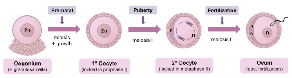
Mitosis → Cell growth → Two devisions of meiosis → Differentiation
Oogonia + Follicle cells → (Mitosis) → Many oogonia and follicle cells in ovaries
Oogonium (2n) → (Growth) → Primary oocytes (2n) surrounded by layer of primary follicle
Primary oocytes → (Begins meiosis; Arrested during Prophase I)
(Each menstrual cycle) Few primary follicle cells → (Meiosis I) → Secondary oocyte (n) → (Begins meiosis II; Arrested at Prophase) + First polar body (n) → Degeneration
Primary follicle layer surrounding the secondary oocyte divides forming two rings of follicle cells, separated by fluid filled space, forming a Graafian follicle
Fluid increases between layers → Ovum (Secondary oocyte + Inner follicle layer) is released from ovary to fallopian tubes at ovulation
At fertilisation, secondary oocyte completes Meiosis II → Fuses with spermatozoon to form zygote nucleus
Difference Between Gametogenesis in Humans
Gametogenesis results in different numbers of sperm and eggs + different amounts of cytoplasm
Numbers
Sperm = Millions daily → Increase chances of fertilisation
Eggs = One / month → Only one is fertilised in pregnancy
Cytoplasm
Sperm = Little → Small + mobile to reach egg
Egg = Lots → Provide nutrients for early embryo development
Mechanisms to Prevent Polyspermy
Polysemy = ≥1 sperm fertilizes an egg → Abnormal embryo development
Acrosome reaction allows a sperm to penetrate the zone pellucida
Enzymes in acrosome are released upon contact and breaks down egg’s outer layer
Cortical reaction prevents other sperm from passing through
First sperm fuses with egg membrane → Cortical granules inside egg fuse with egg plasma membrane → Releases cell contents
Zona pellucida hardens + Sperm receptors are altered/destroyed (No attachment sites) → Prevents additional sperm entry
Maintain correct chromosome number → Prevent genetic abnormalities + Ensure normal embryo developments → Increases chances of successful pregnancy
Compare & Contrast Spermatogenesis and Oogenesis (HINT: 8 VS 3)
Differences
Location
Testes = Testis
Ovary = Ovary
Production
Testes = Lifelong production
Ovary = Fixed amount
Gametes per parent cell
Testes = 4
Ovary = 1
Beginning of process
Testes = Puberty
Ovary = Fetal development
Timing of Gamete Formation
Testes = Continuous
Ovary = Once a month (during menstrual cycle)
End of process
Testes = Lifelong; Reduces
Ovary = Stops at menopause
Timing of gamete release
Testes = Anytime
Ovary = Monthly cycle
Meiotic divisions
Testes = Uninterrupted
Ovary = Arrested
Similarities
Processes in sexual reproduction
Produce haploid gametes
Both undergo meiosis and mitosis
Development of Blastocyte + Implementation in the Endometrium
Blastocyst = Ball of cells, gravitating in a certain direction, allowing a fluid-filled cavity to form in middle
Zygote undergoes rapid cell division as it travels toward uterus → Forms a blastocyst after 4-6 days
At Day 4: Enters uterus; Specialise into inner cell mass and outer layer (precursor to placenta when implanted)
At Day 6-10: Blastocyst attaches to endometrium; Outer layer invades endometrium and connects with mother’s blood supply (Placenta)
Pregnancy Testing by Detection of hCG Secretion
Production of hCG in embryo / developing placenta
Implantation → Production of hCG in embryo and cells of placenta
Levels rise rapidly in early pregnancy
hCG = Prevents more eggs developing in ovary
Appears in blood and urine of pregnant women
At Week 3: Embryo development reaches organ formation
Use of monoclonal antibodies that bind to hCG
Antibodies are attached to test strip
When urine containing hCG flows over strip, hCG bind to antibodies → Color change → Visible result
Made by injecting antigen into mice → Retrieving plasma cells + tumour cells → Hybridoma cells → Monoclonal antibody
Role of Placenta in Foetal Development Inside Uterus
Placental structure = Large SA of placental vili for exchange
Placenta = Temporary organ developed during pregnancy; Connects developing foetus to uterine wall
Allows for nutrient, gas, and waste exchange
Maternal and fetal blood do not mix directly
Exchange processes in placenta → Allow foetus to be retained in uterus til later stage of development than in mammals that do not develop a placenta
Types of exchanges
Nutrient transfer (Glucose, amino acid, fatty acids)
Gas exchange (Oxygen → foetus / CO2 → mother)
Waste removal (Urea, etc.)
Some hormone transfer
Allows for longer gestation period / Fetus can develop to more advanced stage before birth (More complex brain development)
Mammals without placenta (e.g. Marsupials) → Shorter gestation period → Foetus is born in less developed stage
Hormonal Control of Pregnancy and Childbirth
Continuity of pregnancy is maintained by progesterone secretion; From corpus luteum → placenta
Initial secreted by corpus luteum in ovary after ovulation
Later produced by placenta at Week 8-12
Functions include:
Prevent uterine contractions
Support foetal development
Prepare breast form milk production
Changes on childbirth are triggered by decrease in progesterone levels → Allows increase in oxytocin secretion due to positive feedback
At childbirth, progesterone levels decrease / oestrogen levels increase → Increased sensitivity of uterus to oxytocin / Increased production of prostaglandins (in uterine wall) → Widens cervix
Oxytocin = Primary hormone for uterine contractions; Progesterone levels drop → Oxytocin more effective
Positive feedback loop for increasing oxytocin → More frequent contractions
Baby head pushes against cervix → Trigger nerve impulse so hypothalamus → Oxytocin release from pituitary gland → Stimulates uterine contractions → Pushes baby’s head harder against cervix
Contractions continue after birth to eject placenta
Stretch receptor no longer stimulated → Oxytocin levels fall → Contractions stop
Hormone Replacement Therapy & Risk of Coronary Heart Disease
HRT replaces declining hormones during menopause e.g. oestrogen and sometimes progesterone → Relieve menopausal symptoms (anxiety, mood change, skin condition, feelings of loss of self, sleeping difficulties, etc.)
Early epidemiological studies: Women undergoing HRT → Reduced CHD incidence (Cause-effect relationship?)
HRT patients = Higher socioeconomic status → Lower HCD risk
Later randomised controlled trials: HRT → Small increases in CHD risk
Sexual Reproduction in Flowering Plants (Production of Gametes, Pollination, & Fertilisation)
Sexual even if plant species is hermaphroditic
Male = Stamens → Pollen
Female = Carpels → Ovules
Sexual Reproduction:
Production of gamete inside ovules and pollen grains
Male gametes produced inside pollen grains by meiosis; Pollen forms in anthers of stamens
Female gametes produced inside ovules by meiosis; Ovules forms within ovary of carpels
Pollination
Transfer of pollen from anther to stigma
Can self-pollinate or cross-pollinate but must be same species
Fertilisation
Once pollen lands on stigma, pollen tube grows down the style towards the ovary → Carry male gametes to ovule
2 haploids gametes fuses to produce a diploid zygote at ovule
Features of an Insect-pollinated Flower
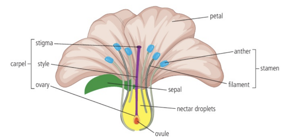
Function of flower parts:
Sepal
Protecting the developing flower while it is inside the bud
Petal
Colourful → Attract pollinators
Anther
Part of stamen (male parts) that produces pollen (male gametes)
Pollen
Contain male nuclei for fertilisation
Filament
Stalk of stamen that hold up the anther
Stigma
Sticky top of carpel (female parts); Where pollen lands
Style
Part of carpel that supports the stigma
Ovary
Base of carpel; Contains 1+ ovules
Ovule
Chamber in ovary where female nuclei develops
Methods of Promoting Cross-Pollination
Promotes genetic diversity and stronger offspring
Different maturation times for pollen & stigma
Prevents self pollination
E.g. Sunflower anthers mature and shed pollen inwards before stigma lobes elongate and become receptive
Separate male and female flowers / plants
At different heights
E.g. Corn has male flowers in tassels at top of plant + female flowers on the ears of corn at lower parts of stalk
Role of animals of wind in transferring pollen between plants
Through wind, insects / birds / animals, water, mechanical means
Adaptations to attract specific pollinators
Scent and color to attract pollinator + Reward of nectar and pollen (Diet)
Self Incompatibility Mechanisms to Increase Genetic Variation
Self pollination leads to inbreeding → Decreased genetic diversity and vigour
Decreased ability to adapt to environmental changes
Plants being smaller in size, making fewer seeds, increased susceptibility to disease
Genetic mechanisms in many plant species ensure male and female gametes fuse during fertilisation are from different plants
Self incompatibility = Plant recognises genetics of pollen from same plant and reject as “self” pollen
Specific genes create proteins on pollen → Proteins are detected by stigma when pollen lands → If proteins match, pollen tube growth is inhibited → Pollen does not reach ovule
E.g. Roses
Dispersal and Germination of Seeds
Distinguish seed dispersal from pollination
Seed dispersal = Spreading of seeds away parent plant; Occurs after fertilisation and seeds are produced
Seed dispersal agents include…
Wind (light seeds with wings)
Animal (consumption and excretion or stick to fur / feathers)
Water (buoyant seeds for aquatic plants)
Explosive (forceful ejection of seeds from fruit)
Pollination = Transfer of pollen from an anther to stigma
Growth and development of embryo + Mobilisation of food reserves
Seed structure:
Seed coat → Protective outer layer
Embryo → Undeveloped plant
Endosperm → Food storage tissue (Starch for energy, proteins fro enzyme production and growth, lipids as concentrated energy storage)
Steps (IAESC):
Imbibtion = Seed absorbs water and swells
Activation = Enzyme activates → Breaks down food reserves (Cell respiration + protein synthesis increases)
Emergence = Radicle (embryonic root) emerges first; Grows down into soil due to gravity
Shoot Development = Hypocotyl (below cotyledons) lifts seed above ground → Early shoot straightens; Orients growing plant perpendicular to ground + Epicotyl pushes the plumule (embryonic shoot; top of epicotyl) above earth and grows upwards, forms first true leaves
Cotyledon Function = (As endosperm becomes depleted) Becomes leaves to photosynthesise and transfer nutrients to growing parts
Root structure development
DNA Replication
Production of exact copies of DNA with identical base sequences
Required for reproduction, growth, and tissue replacement in multicellular organisms
Role of Complementary Base Pairing
Allows genetic information to be replicated and expressed accurately and quickly
When two DNA strands split open, they act as templates for synthesis of new strands at high accuracy
Complementarity is based on hydrogen bonding
A-T (2 H-bonds)
G-C (3 H-bonds)
Semi-conservative Model of Replication
Semi-conservative replication = Each strain in DNA double helix acts as template for synthesis of new complementary strand
Each daughter helix = Old strand from parental DNA + New strand
Complementary base pairing and retention of one original strand of DNA allows high degree of accuracy in copying base sequences
Using the unzipped single DNA strand, complementary nucleotides could be used to synthesise the new strand → Ensures two identical copies of DNA are produced from the original strand
Role of Helicase and DNA Polymerase in DNA Replication
Helicase = Hexomer; Only binds to one DNA strand on either side to split hydrogen bonds as they go in opposite directions (E.g. Pulling open__ )
Unwinds and breaks hydrogen bonds between DNA strands
DNA Polymerase
Adds free nucleotides to the leading and lagging strands in a 5’ to 3’ direction
Directionality of DNA Polymerases
Difference between 5’ and 3’ terminals of strands of nucleotides
5’ end = Phosphate group attached to 5’ carbon of deoxyribose
3’ end = Hydroxyl group on 3’ carbon of deoxyribose
DNA Polymerases can only add the 5’ end of a DNA nucleotide to the 3’ end of existing nucleotide
DNA chain must be built in 5’ to 3’ direction → Creates alternating sugar-phosphate backbone of DNA
Replication on the Leading Strand VS Lagging Strand
Due to the specific direction of Polymerase III from 5’ to 3’…
Synthesis of leading strand = Relatively fast; Continuous
Lagging strand = Discontinuous; Synthesised in Okazaki fragments
Requires DNA ligase to bind the fragments together
Replication has to be initiated with RNA primer only once on leading strand (because it is continuously formed) but repeatedly on lagging strand
Functions of Molecules in DNA Replication (In Prokaryotic System)
DNA Primase
Synthesises and attaches RNA primers to start of sequence; Signals DNA polymerase I where to begin synthesis
DNA Polymerase III
Removes RNA primers and replaces the sequence with DNA nucleotides
DNA Polymerase I
Synthesise new strand of DNA based on complementary bases in the direction of 5’ to 3’
DNA Ligase
Joins Okazaki fragments together to form a single chain of DNA
DNA Proofreading
DNA Polymerase III removes any nucleotide from the 3’ terminal with a a mismatched base, and replaces it with a correctly matched nucleotide
An incorrect base may be present in growing DNA chain
DNA Polymerase III excises the incorrect base and replace with correct base before proceeding with replication (Stalling)
DNA Replication Process (FULL)
Topoisomerase uncoils DNA
Helicase unwinds and splits H bonds between the DNA strands, creating a replication fork
Primase forms RNA primer and it attaches itself to site of synthesis
DNA polymerase III adds nucleotides to new Okazaki fragment only at 3’ end and continues until it meets the primer of the pervious fragment
OR
DNA polymerase III adds nucleotides to synthesise leading strand continuously
DNA polymerase I hydrolyses the primer and replaces it with DNA
DNA ligase catalyses the formation of phosphodiester linkage, joining the Okazaki fragments together
Polymerase Chain Reaction
PCR = Amplifies amount of DNA sample; Each cycle doubles the # of copies of target gene → Production of many copies quickly; In thermocycler…
Source DNA
DNA primers = Short DNA strands complementary to regions with sequence of interest on either strand; Used to isolate a gene
Free nucleotides (dNTPs)
Taq polymerase = From T. aquaticus; Bacteria found in hot springs → Enzymes have high optimum temperatures
Good for PCR since utilises temp. > normal body temp. + Much more efficient. than human DNA polymerase
Reaction buffer
Temperature cycles are…
Denaturing (94-95ºC) = High temp. → DNA strands to separate
Annealing (50-56ºC) = Lower temp. → Primers bind to DNA
Extending (72ºC) = Optimal temp. for Taq → Effective replication
Gel Electrophoresis
Since DNA is inherently -ve charged, will move through the gel from negative electrode to positive electrode.
Gel electrophoresis = Separation of DNA fragments based on size
Small DNA fragments → Travel further
Large DNA fragments → Travel less
Applications of PCR and Gel Electrophoresis
DNA profiling for paternity
Compare common sequences
Forensic investigations
Small amount of DNA from crime scenes can be amplified and searched against murder suspects’ DNA
Human genome studies for evolutionary biology
Historical
Clone DNA from extinct species
Medical
Detect mutations by comparing DNA; Predict risk of disease and cancers
NOTE: Increasing number of markers used reduces probability of false match in DNA profiling
Generation of New Cells by Cell Division
In all living organisms, a parent cell (AKA mother cell) divides to produce two daughter cells
Cytokinesis
Splitting of cytoplasm in parent cell between daughter cells
Occurs once cell reaches certain size
In an animal cell, a ring of contractile actin and myosin proteins pinches a cell membrane together to split the cytoplasm
Actin and myosin filaments contract to form cleavage furrow
Furrow deepens (Inward pinching) until pinched into two separate cells
Ensures equal share of cytoplasm in daughter cell
In a plant cell, vesicles assemble sections of membrane and cell wall to achieve splitting
Vesicles which make up new cell membrane and walls assemble to form a cell plate
Vesicles expand outwards and fuse with cell membrane → Divides cell into two
Equal and Unequal Cytokinesis
Division of cytoplasm is usually, but not always, equal; Both daughter cells must receive at least one mitochondrion and any other organelle that can only be made by dividing a pre-existing structure
Identical daughter cells → Ensures new cell contains necessary organelles to survive and function (i.e. 1 mitochondrion / chloroplast in each cell)
Examples of unequal cytokinesis:
Oogenesis in humans
4 haploid cells are produced (Each containing 23 chromosomes)
One cell retains most cytoplasm → Ovum (Can be fertilised to make zygote)
Other cells form smaller polar bodies which degenerate
Budding in yeast
Small bud receives part of cytoplasm and a nucleus → Grows and eventually detaches to become daughter cell
Role of Mitosis and Meiosis in Eukaryotes
Nuclear division is needed before cell division to avoid production of anucleate cells
Nuclear division must occur before cells division to ensure each new cell contains a nucleus
Anucleate cells cannot survive or function properly
Mitosis maintains the chromosome number and genome of cells
Mitosis results in two identical daughter cells
Essential for growth, repair in somatic cells + Asexual reproduction
Meiosis halve the chromosome number and generate genetic diversity
Meiosis results in FOUR daughter cells with only HALF the DNA
Used to make gametes; Each daughter cell receives different combination of genes
For sexual reproduction; Combination of genetic material from two parents at fertilisation → Genetic diversity (Beneficial to species survival and evolution)
DNA Replication as a Prerequisite for Both Mitosis and Meiosis
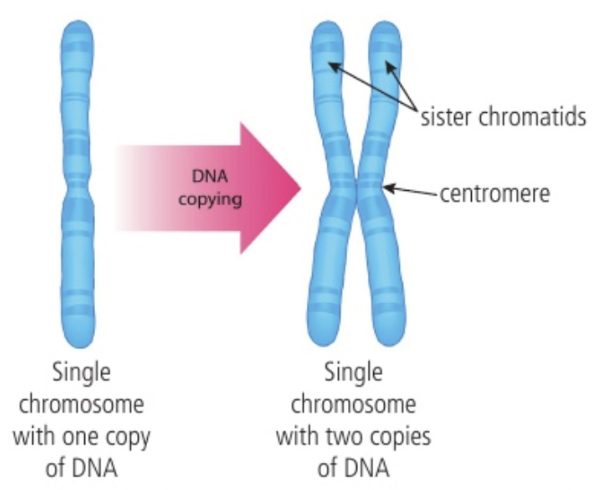
After replication, each chromosome consists of two chromatids held together until anaphase
DNA replication must occur before cell division → Ensure each new cell contains complete set of DNA
Chromatids = Attached a centromere; Two sisters → Chromosome
Condensation and Movement of Chromosomes
Shared features of mitosis and meiosis
Role of histones in condensation of DNA by supercoiling
DNA is usually spread out in long chains inside nucleus; Must be condensed by supercoiling before replication to prevent breaking
Condensed bt wrapping around histones → Nucleosomes
Nucleosomes further coil into → Chromatin
Chromatin further condense → Fully supercoiled Chromosome
Use of microtubules and microtubule motors to move chromosomes
Centrosome = Organelle allowing microtubule spindle fibres to move chromosome
Microtubules = Constructed / Disabled as needed; Act as highways guiding chromosomes to poles of cell
Motor proteins = Push / pull objects around cell
Dynein and kinesis can transport chromosomes by walking along microtubules
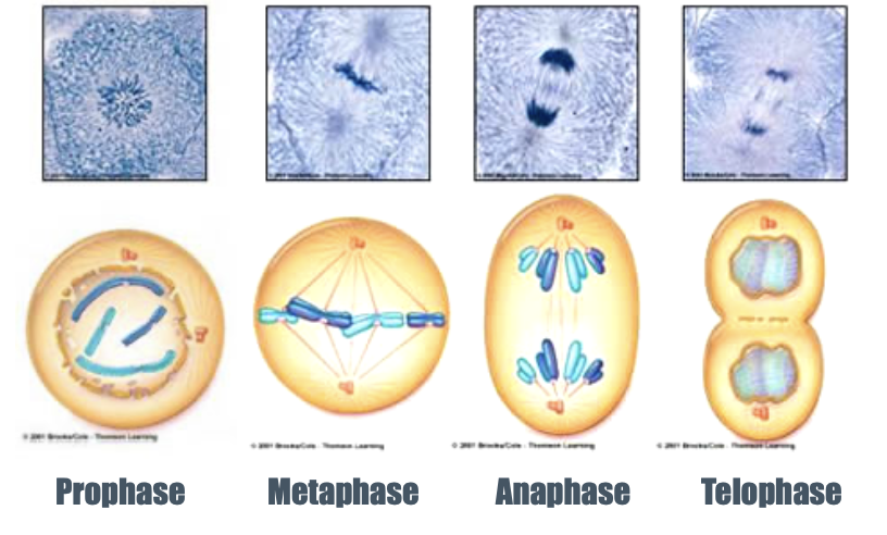
Phases of Mitosis
INTERPHASE (NOT A PHASE)
Cells spend most time here; Perform general functions (Protein synthesis, respiration, growing larger, etc.)
Enters mitosis when ready to divide
PROPHASE
Chromatin condense by supercoiling
Nuclear membrane breaks down; Nucleolus disappears
Mitotic spindle forms (New microtubules built from centrosome)
Kinetochores attach to spindle (Region in centromere)
Microtubules lengthen → Centrosomes move towards opposite poles
METAPHASE
Metaphase plate is formed; Chromosomes move to equator of cell
Centromeres align on plate
Spindle acts to move chromosomes
Centrosomes at opposite poles
ANAPHASE (Shortest phase)
Chromatids move towards opposite poles of cell due to motor proteins pushing microtubule in opposing directions → Each pole has complete, identical set of chromosomes
Centromeres move towards pole first (Point of contact with spindles)
TELOPHASE
Nuclear membrane begins to reform
Nucleoli reappear; Chromosomes elongate
Spindles disappears
Cell is elongates → Prepares for cytokinesis
Meiosis as a Reduction Division
A diploid cell undergoes two rounds of cell division to produce four haploid gametes
Diploid = 2n; 46 chromosomes
Haploid = n; 23 chromosomes
Two divisions of meiosis produces four haploid nuclei from one diploid nucleus; Need for meiosis in sexual life cycle
At fertilisation, a full number of chromosome is achieved
Two rounds of segregation in meiosis
MEIOSIS I (PMAT) → Separates chromosome pairs
Each chromosome makes an identical copy of itself which stays together
Chromosomes line up in pairs; As cell divides in two, one chromosome from each pair goes into new cell
MEIOSIS II (PMAT) → Separates chromatids
New cells divide again; Each chromosome is split into two → Haploid cells
Compare and Contrast Meiosis and Mitosis
Down syndrome and Non-disjunction
Error in mitosis = Extra or fewer chromosomes in gamete
Gamete with extra chromosome fertilising a normal gamete = Zygote with three copies of chromosomes (trisomy)
E.g. Down syndrome
Extra chromosome 21 (Total 47 chromosomes)
Causes developmental delays, physical growth limitations, distinctive facial features
Due to non-disjunction during formation of mother’s egg cells
Occurs when homologous chromosomes or sister chromatids fail to separate properly
Can occur during Meiosis I or II
Meiosis as a Source of Variation
Meiosis generates genetic diversity by random orientation of bivalents and by crossing over
Produce 4 cells with different chromosomes
Due to crossing over when genetic material swaps between non-sister chromatids → Creates recombinant chromatids; Increase variety among offspring
Random orientation during crossing over occurs because chromosomes can line up in different ways in Metaphase I
Chiasma = Point / Locus of crossing over
Cell Proliferation for Growth, Cell Replacement, and Tissue Repair
Cell proliferation = Cell division and multiplication; For growth, maintenance, and tissue repair
For example…
Growth within plant meristems
Meristems = Area where plant cells divide rapidly; Undifferentiated cells will differentiate into specialised cells
Apical meristem tissue = Root tips and branch tips; Undergoes mitosis to lengthen plant
Lateral meristematic tissue = Stems; Allow plant to grow wider (Responsible for continuous growth)
Early stage animal embryos
After fertilisation, embryonic stem cells in zygote divide by mitosis to produce cells which self-organise into layers → Embryo
Skin
Occurs during routine cell replacement and wound healing
Skin cells die off and are replaced every 28 days by mitosis (New cells are made in bottom layer, dead cells flake off at top layer)
When broken or wounded, cells in surrounding area divides, fills gap, and repair tissue → Closes wound
Phases of Cell Cycle
Cell cycle = Allows for cell proliferation; Include G1, S, and G2 as stages of interphase, followed by mitosis, and then cytokinesis
Interphase (Growing)
G0 (Stalled growth)
G1 (Growth)
Cell grows in size; Proteins and organelles are synthesised
Intense cellular activity as cell prepares for DNA replication
S (Synthesis)
DNA replication
G2 (Growth)
Cell completes growth, prepares for mitosis
Checks DNA for errors and repairs
Produce organelles and proteins for cell division
Dividing (Mitosis)
Nucleus and cell divides
Cell Growth During Interphase
Interphase = Metabolically active period; Longest phase
Growth involves biosynthesis of cell components i.e. DNA and proteins
Ribosomes are active as they translate mRNA into proteins
# of mitochondria and chloroplast are increased by growth and division of organelles
Growth and division via binary fission
Control of Cell Cycle Using Cyclins
Cyclins = Proteins that regulate progression of cells throughout cell cycle
Cyclin and cyclin-dependent-kinase (CDK) forms a complex which binds to a target protein and modifies it by phosphorylation
Phosphorylated protein triggers event in cell cycle
Cyclin is then destroyed
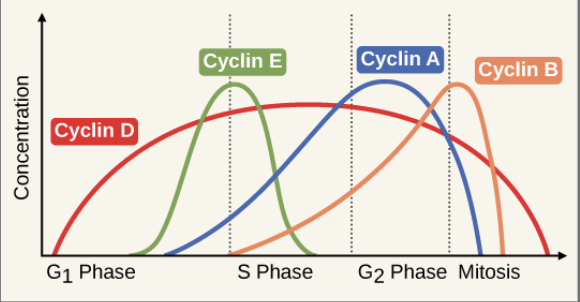
Concentration of different cyclins increasing and decreasing during cell cycle
Cyclin D – G1 Phase
Cyclin E → Starts S phase
Cyclin A → Starts G2 phase
Cyclin B → Starts Mitosis
Threshold level of a specific cyclin is necessary to pass each checkpoint
Overproduction or underproduction of cyclins lead to uncontrolled cell division → Cancer
Consequences of Mutations in Genes that Control Cell Cycle
Mutations in proto-oncogenes that convert them to oncogenes and mutations in tumour suppressor genes result in uncontrolled cell division
Proto-oncogenes = Help cells grow
When mutated, becomes oncogenes; Allow cells to grow uncontrollably into tumours
Tumour suppressor genes = Prevent uncontrolled cell division; Can slow down cell division, repair DNA mistakes, or trigger apoptosis
When mutated or silenced, cells grow unregulated; Damaged cells are allowed to continue dividing
Can be caused by environmental exposure or genetic inheritance
Can be inherited (germ-line) or acquired during lifetime (somatic)
Difference Between Tumours for Metastasis
Tumour = Abnormal growth of tissue that forms a mass
Benign = Non-cancerous
Low rate of cell division; Grows slowly
No capacity for metastasis; Do not spread to other parts of body
Malignant = Cancerous
Grows rapidly; Higher mitotic index; Greater rate of cell division
Capable for metastasis and invade neighbouring tissue
Whether benign or malignant can be determined by biopsy, medical imaging, or blood tests
Primary tumour = Occurs at original site
Secondary tumour = A tumour that has spread elsewhere via metastasis
Mitotic Index
(Number of cells undergoing mitosis) / (Total number of cells)
High mitotic index = Rapid growth
If mitotic index >0.75, cancerous
Polyploidy (Diploids & Haploids)
Production of haploid gametes in parents and their fusion to form a diploid zygote as a means of inheritance
Gametogenesis = Production of haploid gametes via meiosis
Diploid cell undergoes two rounds of cell division to produce four haploid gametes. Number of chromosome is halved and creates four genetically unique haploid cells.
Fertilization = Fusion of gametes
Fusion of haploid egg cell and haploid sperm forms a diploid zygote which undergoes mitotic cell division and develop into multicellular organism.
Diploid cell = Contains two copies of each autosomal gene
Pattern of inheritance is common to all eukaryotes with a sexual life cycle
Pollination and Fertilisation in Plants
Methods for conducting genetic crosses (fertilisation) in flowering plants
Pollination = Transfer of pollen to the stigma; Required for fertilisation to occur and produce zygote / Carry out genetic cross
Pollen contains male gametes
Female gametes are located in the ovary
Plants such as peas produce both male and female gametes on the same plant, allowing for self-pollination and therefore self-fertilisation
Results in lack of genetic diversity
Cross-pollination is when pollen is transferred between different individual plants
Genetic crosses are widely used to breed new varieties of crop or ornamental plants
Plant breeders help fertilisation by using brushes to transfer pollen grains to stigma of other plants → Increase chance of desirable traits for general produce or ornamental plants
Genetic Cross Terminology
P generation = Parental generation
F1 generation = First filial generation
F2 generation = Second filial generation
Genes and Alleles
Genotype = Combination of alleles inherited by an organism
Homozygous = Same alleles
Heterozygous = Different alleles
Hemizygous = Only one allele
Occurs only in males (Different sex chromosomes)
Genes VS Alleles
Genes = Inherited features encoded by specific DNA sequences; Located at specific positions on chromosome (gene locus)
Every gene has a specific locus on a chromosome
Gene is found in same position on both homologous chromosomes (one paternal and one maternal)
Alleles = Variations of a specific feature; Alternate forms of genes
Dominant and Recessive Alleles
Effect of dominant and recessive alleles on phenotype
Recessive allele is only expressed when in homozygous state
Both a homozygous-dominant genotype and a heterozygous genotype for a particular trait will produce the same phenotype
Dominant allele can fully express trait regardless of the state of the other allele
Phenotype + Example of Traits
Phenotype = Observable traits of an organism resulting from genotype and environmental factors
Examples of traits in humans due to…
Genotype ONLY
Eye colour
Blood type
Environment ONLY
Accent or speech patterns
Interaction between genotype AND environment
Height (Nutrition and health)
Susceptibility to certain diseases (Lifestyle choices e.g. diet and exercise + Environment factors e.g. stress and pollution)
Phenylketonuria (PKU)
Phenylketonuria = Recessive genetic condition caused by mutation in autosomal gene that codes for enzyme needed to convert phenylalanine to tyrosine
Deficiency / lack of enzyme → Accumulation of phenylalanine in body → Developmental issues such as intellectual disability
Example of a human disease due to a recessive allele
Single-Nucleotide Polymorphisms (SNPs) + Example
Single-nucleotide polymorphisms and multiple alleles in gene pools
SNP = Single-base differences in DNA sequences that occur at specific loci
Any number of alleles of a gene can exist in the gene pool but an individual only inherits two
More than two variants of the gene can exist within a gene pool → Contributes to genotypic diversity → Leads to broad range of phenotypic expression for the trait associated with that gene
Provide genetic variation that enables organisms to adapts to changing environments
For example…PTC Gene and Bitter Taste Perception
tt = Non-taster / Tt = Moderate Taster / TT = Supertaster
Co-dominance + ABO Blood group as an Example
Co-dominance = Multiple alleles presented at once; E.g. ABO blood groups
IA and IB are both co-dominant alleles
Can both be expressed individuals; E.g. AB blood type
i is a recessive allele
To be expressed, must be homozygous recessive; E.g. O blood type (No antigens are expressed)
ABO blood group is inherited in an autosomal co-dominant manner
Co-Dominance VS Incomplete Dominance
Difference between the patterns of inheritance of incomplete dominance and co-dominance at phenotypic level
In co-dominance…
Heterozygotes have dual phenotype
Heterozygous phenotype is a combination of the two homozygous phenotype, with both fully expressed simultaneously
E.g. AB blood type (IAIB)
In incomplete dominance…
Phenotype of heterozygous individual is intermediate between (in the middle) phenotypes of two homozygous individuals; Dominant allele does not completely mask the effects of a recessive allele, resulting in a new phenotype
E.g. Four o’clock flower or marvel of Peru (Mirabilis Jalapa)
R1R1 = Red / R2R2 = White / R1R2 = Pink (Intermediate between red and white)
Phenotypic Plasticity
Phenotypic plasticity = Capacity to develop traits suited to environment experience by an organism by varying patterns of gene expression
NOT due to genotype (No change in genotype); Occurs by regulation of gene expression
Different environmental cues can trigger the activation or suppression of specific genes
Changes in traits may be reversible during a lifetime
Organisms can revert to their original phenotype when environmental conditions change back to previous state
Sex Determination in Humans
Sex determination in humans and inheritance of genes on sex chromosomes; Far more genes are carried by the X chromosomes than the Y chromosome
Sex-linkage = Genes found on sex chromosome
Females = XX // Male = XY
Sex-linked condition are usually X-linked (Few genes exist on Y chromosomes; ~2000 VS 78 genes)
Exhibits different patterns of inheritance; Females cannot express Y-linked traits / Males cannot mask X-linked recessive traits → More commonly expressed in males (E.g. haemophilia)
Sex chromosome in sperm determines whether a zygote develops certain male-typical or female-typical physical characteristics
Y chromosome contains SRY gene which produces TDF (testis-determining factor) → Induces medulla of embryonic gonads to develop into testes → Produced testosterone; Initiate development of male sexual characteristics
Absence of SRY gene and TDF → Cortex of embryonic gonads develop into ovaries → Embryo develops female characteristics
Example of a Sex-Linked Genetic Disorder
Haemophilia = Caused by recessive allele on X chromosome; Leads to uncontrolled bleeding
Normal clotting factor allele = XH // Faulty clotting factor allele = Xh
Since females can be…
XHXH or XHXh or XhXh
And males can be…
XHY or XhY
Carrier females (XHXh) have 50% chance of passing Xh to offspring
XHXh + XhY → 2*(XhY) + 2*(XHXh)
Affected males (XhY) can inly pass Xh to daughter
XhY + XHXH → 2*(XHY) + 2*(XHXh)
Pedigree Charts
Pedigree Charts = Used to deduce patterns of inheritance of genetic disorders; Maps genetic history of family over several generations
Autosomal recessive: Traits can skip generations; Affect males and females equally
Neither parent is affected → Offspring is affected
Autosomal dominant: Traits do not normally skip generations; Affect males and females equally; Unaffected members are homozygous recessive
Both parents are affected → Offspring are unaffected
X-linked dominant
100% incidence of affected daughters + 0% incidence of affected sons from an affected father
X-linked recessive
0% incidence of affected daughters + 100% incidence of affected sons from an affected mother
Genetic basis for the prohibition of marriage between close relatives in many societies
Consanguine marriage = Marriage between individuals who are closely related → Increased risk of recessive genetic disorders in offspring of close relatives
Polygenic Inheritance
Continuous variation due to polygenic inheritance and/or environmental factors
Polygenic trait = Influenced by combined effects of multiple genes
For example…skin colour in humans
Each gene contributes small effects to the overall skin colour → Continuous range of skin tones
Environ. Factor: Exposure to UV radiation → Skin pigmentation (Amount of melanin produced in response to sun exposure)
Discussing Skin Tone and ABO Blood Group (Continuous VS Discrete Variables, Measures of Central Tendency)
Continuous variables = Infinite number of possible values within a range
For polygenic traits, bell curve is produced (y = frequency)
Ends of curve = Rare cases = Extreme phenotypes
Middle of curve = Common cases = Average phenotype
Discrete variables = Limited set of possible values
Measures of Central Tendency
Mean, Median, Mode
Relationship between these measures → Reveal distribution of traits within gene pool
Negatively skewed (Right)
Normal / No skew (Symmetrical)
Positively skewed (Left)
For example…skin tone
Continuous variable
To find measures of central tendency, must convert into discrete, numerical data first
Use Fitzpatrick scale
For example…ABO blood group
Discrete variable
Mode → Most common blood group // Median or Mean → Distribution of blood group frequency (?)
Mendelian Ratio and Test Crosses
Punnet grids = For predicting genotypic and phenotypic ratios in dihybrid crosses involving pairs of unlinked autosomal genes
MENDEL’S SECOND LAW (Law of Independent Assortment) = Alleles of one gene sort into the gametes independently of the alleles of another gene
Two dual heterozygous parents → 9:3:3:1 phenotypic ratio
Heterozygous + Homozygous recessive parents → 1:1:1:1 ratio
(Law only applies if genes are on different chromosomes or are far apart enough on one chromosome so that recombination rate reaches 50%)
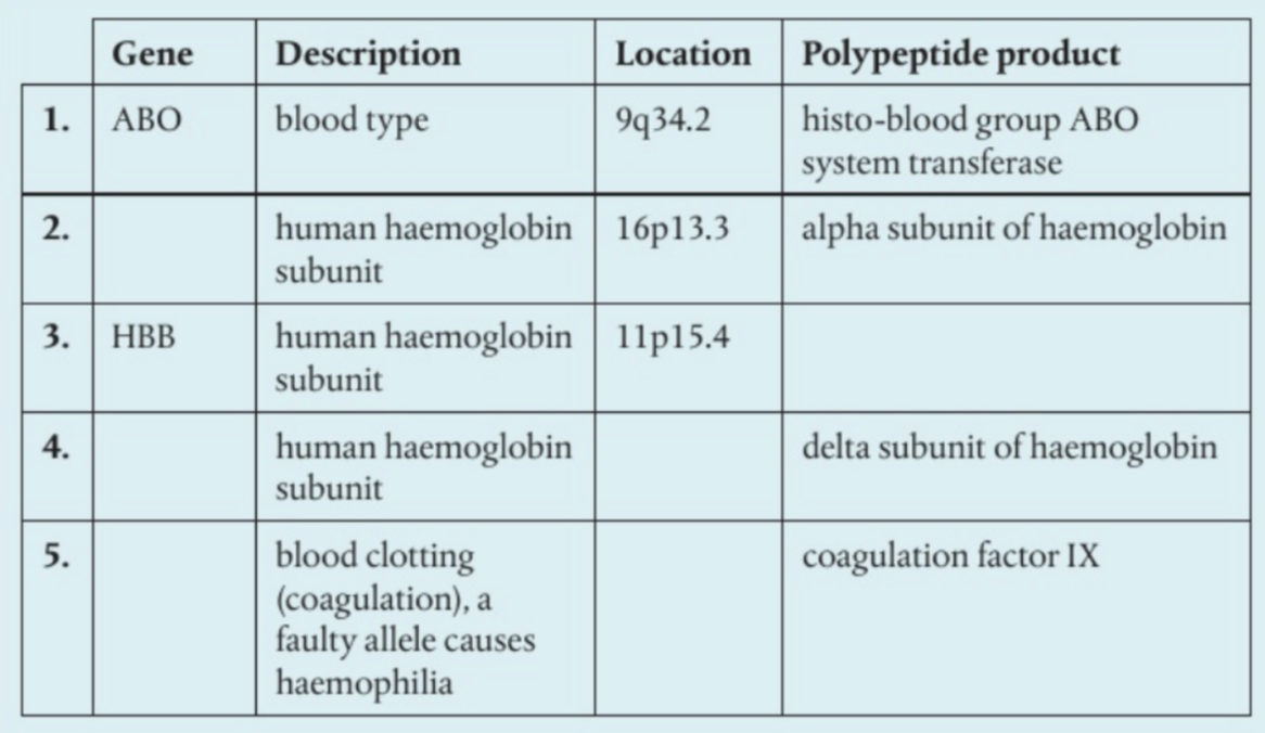
Exploring Gene Loci and Polypeptide Products
Segregation of Homologous Chromosomes in Meiosis
Segregation and independent assortment of unlinked genes in meiosis
Homologous chromosomes align and randomly separate into different daughter cells during meiosis
Two genes on separate, unlinked chromosomes will assort and distribute independently into gametes during meiosis → All possible combinations of two traits in offspring
Link between the movements of chromosomes in meiosis and the outcome of dihybrid crosses involving pairs of unlinked genes
Due to its ability to segregate and assort independently, the outcomes can be calculated using dihybrid crosses
Autosomal Gene Linkage + Notation
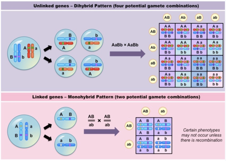
Linked = Genes located on same chromosome
Alleles of linked genes can fail to assort independently
Linked genes do not assort independently (Must travel together during meiosis) → More likely to be inherited together
Monohybrid pattern of gene inheritance
For example…
AB on one chr, ab on another chr → Only 2 possible gamete combinations
Gamete combinations are essentially the same across generations
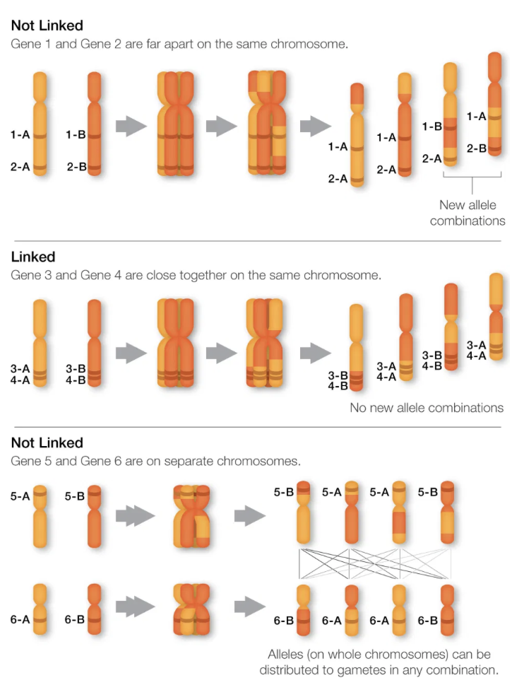
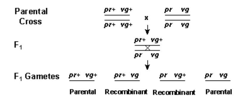
Homologous Recombination (Linked Genes w/ Differing Distances; Gametes, Genotype of Offspring, Phenotype of Offspring)
Recombination changes linked gene inheritance patterns
During recombination of linked genes, crossing over may occur, in which generating recombinant chromosomes → New genotypes
Where linked genes (AB || AB, ab || ab) are close to each other,
(Parental →) 48% AB, 48% ab, 2% Ab, 2% aB (← Recombinant)
Linked genes close together on the same chromosome has a lower probability of being separated by crossing over → Tend to be inherited together during meiosis
Gametes = AB, ab, Ab, aB
Offspring = AB || AB, ab || ab, AB || ab
Phenotype =
Dom. trait 1 + Dom. trait 2
Rec. trait 1 + Rec. trait 2
Where linked genes (AB || AB, ab || ab) are far apart on same chr,
25% AB, 25% ab, 25% Ab, 25% aB (← Recombinant)
Linked genes further apart on the same chromosome has a higher probability of being separated by recombination → All genotypes are equally likely to be created from meiosis
Same inheritance patterned as unlinked gene
Gametes = AB, ab, Ab, aB (1:1:1:1)
Offspring = AB || AB, ab || ab, AB || ab, Ab|| Ab, Ab || ab, aB || aB, aB || ab (New recombinant types: State of Trait1 ≠ State of Trait2)
Phenotype =
Dom. trait 1 + Dom. trait 2
Rec. trait 1 + Rec. trait 2
Dom. trait 1 + Rec. trait 2
Rec. trait 1 + Dom. trait 2
Different Types of Point Mutations
Gene Mutations = Structural changes to genes at the molecular level
Substitution
Insertion
Deletion
Inversion
Single-Nucleotide Polymorphisms (SNPs)
SNPs = Result of base substitution mutations; May (OR MAY NOT) cause different amino acid to be placed in polypeptide chain due to degeneracy of genetic code
Frameshift Mutations
Major insertions / deletions → Frameshift change; Affects every codon beyond the point of mutation → Likely cause polypeptides to cease to function
Cause of Gene Mutation
Mutagens ~ Errors in DNA replication or repair
Mutagenic forms of radiation, e.g. UV Radiation
High energy radiation knocks out base pair / break bonds → Modify DNA
Chemical mutagens, e.g. Cigarette smoke, Benzoyl peroxide, BBQ
May react / attack / replace DNA nucleotides
Infectious agents, e.g. H. pylori, HPV
Some bacteria produce reactive oxidative species → Induce inflammation + Damage DNA + Reduce efficiency of DNA repair in cell → Increase vulnerability to mutations
Randomness in Mutation
Mutation can occur anywhere in the base sequences of a genome, although some bases have a higher probability of mutating than others
Uncoiled DNA = More exposure → Increased susceptibility to mutations
Satellite DNA = Found at centromere and telomeres; Non-coding, structural function; High mutation rate (1 mutation per 1000 bp)
CpG sites = When methylation occurs, C can mutate into T
CpG islands = Repeated CpG sites
Mitochondrial DNA
RNA sequences
No natural mechanism is known for making a deliberate change to a particular base with the purpose of changing a trait
Consequences of Mutation in Germ Cells and Somatic Cells
Inheritance of mutated genes in germ cells and cancer in somatic cells
Somatic mutations = Occur in a single body cell; Cannot be inherited (Only tissues with mutation will be affected)
Germline mutations = Occurs in gametes; Can be passed onto offspring (ALL cells will be affected)
Mutation as a Source of Genetic Variation
Gene Mutation = Original source of all genetic variation
For an individual organism, most mutations are either harmful or neutral
In a species, they are essential for evolution by natural selection in long-term
For example…
Apolipoprotein A1 = Help transport cholesterol through bloodstream + Remove cholesterol from artery wall
Community in Italy has apolipoprotein A1-Milano mutant
More effective than normal type
Additional function as antioxidant → Prevent inflammation which occur in arteriosclerosis
Gene Knockout
Technique for investigating the function of a gene by changing it to make it inoperative
In a KO mouse model, the gene of interest is removed → Due to evolutionary similarity, allows investigation of function of gene in humans
Models often reproduce quickly and have few genes
CRISPR-Cas9
Use of CRISPR sequences and the enzyme Cas9 in gene editing
Cas9 enzyme is a protein that cleaves DNA at specific loci, guided by gRNA which functions by matching desired target sequence. Together, this allows the removal and replacement of genetic sequences.
An example of the successful use:
OB-mouse → Obesity (Leptin ~ Feelings of satiation)
Sickle cell anemia
Conservation of Genes
Hypotheses to account for conserved or highly conserved sequences in genes
Conserved sequences = Identical or similar across a species or a group of species
Highly conserved sequences = Identical or similar over long periods of evolution
Hypotheses include…
Functional requirements for the protein a gene produces
Essential gene where the lack of them or its mutation results in sever consequences which could prevent its inheritanceSlower rates of mutation
Transcription
Transcription = Synthesis of RNA using a DNA template
Only the antisense strand is transcribed and the mRNA strand synthesised is the sense strand
RNA is always shorter than DNA strand copied from as it is the complementary form of only one gene
Roles of RNA polymerase:
Binds to promoter region on template strand
Unzips DNA by breaking H bonds between two DNA strands
Transcribes mRNA molecule by complementary base pairing
Links RNA nucleotides
Hydrogen Bonding and Complementary Base Pairing in Transcription
Role of hydrogen bonding and complementary base pairing in transcription
The formation of the new RNA strand is allowed by complementary base pairing by arranging RNA nucleotides in the complementary sequence to the DNA template strand
Adenine → Uracil
H-bonds stabilise interaction between RNA polymerase and DNA, forming bonds between newly synthesised RNA strand
Stability of DNA Templates
Single DNA strands can be used as a template for transcribing a base sequence without the DNA base sequence changing. In somatic cells that do not divide, such sequences must be conserved throughout the life of a cell
In some somatic cells such as nerve cell which do not undergo mitosis but require proteins, sequences are conserved throughout its lifetime and do not mutate
Transcription ~ Gene Expression
Not all genes in cell are expressed at a given time.
As the first stage of gene expression, transcription is a key stage at which expression of a gene can be controlled (switched on and off).
Translation (tRNA and mRNA)
Translation = Synthesis of polypeptides from mRNA
Base sequence of mRNA is translated into amino acid sequence of polypeptide in units of 3 base pairs known as codons.
mRNA binds to the small unit of the ribosome
Two tRNAs bind simultaneously to the large subunit
The first tRNA carrying a specific amino acid attaches to the “A” site at the large subunit of the ribosome; tRNA forms hydrogen bonds with mRNA via complementary base pairing
A second tRNA enters the large subunit of the ribosome and repeats the process. These tRNA simultaneously bind to the large subunit of the ribosome
The enzyme catalyses the condensation reaction, resulting in a peptide bond between the two amino acids
The first tRNA detaches from its amino acid and the mRNA, leaving the subunit
The ribosome moves along the mRNA molecule in a 5’ to 3’ direction so that the second tRNA will now have been in the position of the first tRNA
A new tRNA molecule enters and pairs with the next codon sequence, repeating the process
RNA and Ribosomes
Role of mRNA, ribosomes, and tRNA in translation
mRNA: Carries the sequence of the sense strand from nucleus to ribosomes in cytoplasm
tRNA: Bring amino acids in the cytoplasm to ribosomes
rRNA: Combines with ribosomal proteins to construct cytoplasmic ribosomes
Complementary Base Pairing Between tRNA and mRNA
The antiocodon on a tRNA molecule is complementary t the codon of the mRNA. This complementary base pairing allows them to match up and produce proteins encoded by the mRNA.
Codon = A triplet in mRNA; Three bases in DNA that determines an amino acid
Anticodon = Three bases in middle loop of tRNA determining which amino acid is attached to it
Features of the Genetic Code
Degeneracy = Each amino acid can be identified by multiple codons
Universality = All organisms share the same genetic code with few minor exceptions
Reasons for a triplet code:
Provides many possible combinations (64) to encode all amino acids and the capability to cope with any expansion in number of amino acids during evolution
Degenerate → Allow for possibility of mutation (Even in the presence of a few substitution mutations, the same amino acid may be obtained)
Universal → Allows us to transfer genes between species via genetic engineering techniques → Treatment of diseases
Production of Growing Polypeptide Chain (Ribosome)
Stepwise movement of the ribosome along mRNA and linkage of amino acids by peptide bonding to the growing polypeptide chain
mRNA binds to the small subunit of the ribosome, and the two tRNAs to the large subunit.
The ribosome holds the tRNA and mRNA close together, allowing amino acids to be connected by peptide bonds
tRNA move sequentially through the binding sites of the ribosome during base paring between tRNA anticodons and mRNA codons along with the mRNA, one codon at a time (Remember, they are temporarily bonded together via hydrogen bonds)
Point Mutations
Point mutation = Mutations of one base in DNA code that change protein structure
Example of point mutation affecting protein structure is: Sickle-cell anaemia
Point mutation in the HBB gene sequence causes valine to be produced instead of glutamine → Abnormal haemoglobin forms strands that leads to sickle-shaped RBC → Decreased oxygen-carrying capacity
HBB gene = hemoglobin subunit beta
Normal HBB gene → (CCTGAGGAG) → Pro-Glu-Glu
Mutated HBBS gene → (CCTGTGGAG) → Pro-Val-Glu
Directionality of Transcription and Translation
5’ to 3’ Transcription (RNA makes sense strand; RNA synthesises a complementary and antiparallel strand to DNA template strand)
Read in 3’ to 5’, synthesise in 5’ to 3’ direction
5’ to 3’ Translation
Initiation of Transcription at Promoter
Promoters = Regions of DNA that RNA polymerase combines with to initiate transcription; Not transcribed
Transcription factors bind to the promoter and attracts RNA polymerase to attach to the promoter region and initiate transcription
Other transcription factors may also bind to other nearby areas
RNA polymerase functions like a helicase; The front unwinds and opens the DNA double helix / The back rewinds the DNA strands
RNA polymerase adds RNA nucleotides to enlarging mRNA
Non-Coding Sequences
Non-coding Sequences = DNA regions that do not code for polypeptides;
Regulators of gene expressions
Promoters, enhancers, silencers, insulators, etc. → Control transcriptionIntrons = Present in gene but removed before during RNA processing before it leaves nucleus, forming mature mRNA
Spliceosome excises introns into lariat loop shape, leaving a mature mRNA strand which goes to be translated into proteins
Telomeres = Ends of chromosomes made of repetitive non-coding sequences; Protects DNA
Genes for rRNAs and tRNAs in eukaryotes → Important for producing proteins
rRNA → Ribosomes // tRNA carries amino acids for translation
Post-Transcriptional Modifications in Eukaryotic Cells (Name 2 examples)
Removal of introns and splicing together of exons by spliceosome to form mature mRNA, leaving excised introns in lariat shape
Addition of 5’ caps and 3’ polyA tail to stabilise mRNA transcripts