Study Guide N ( test 3)
 Call Kai
 Learn
Learn Practice Test
 Spaced Repetition
 Match
Match Flashcards
 Knowt Play
1/142

There's no tags or description
Looks like no tags are added yet.
Name | Mastery | Learn | Test | Matching | Spaced | Call with Kai | Chat |
|---|
No analytics yet
Send a link to your students to track their progress
143 Terms
The functional filtration unit of the kidney is the
nephron
What is the correct branching sequence of arteries in the kidney?
renal artery → segmental arteries → interlobar arteries
The junction of the renal medulla and renal cortex is called the
corticomedullary junction
The major calyces merge to form a single, funnel-shaped
renal pelvis
Blood vessels, nerves, and the renal pelvis enter/exit the kidney at the
hilum
The main functions of the urinary system are
removal of waste and regulation of H2O and electrolyte balance
Blood vessels and the renal calyces are located in the
renal sinus
T or F
The renal cortex is more superficial than the renal medulla.
true
T or F
Urine is propelled along the ureters by peristaltic waves of muscular contractions.
true
What is the collective name for the layers of muscle in the wall of the urinary bladder?
the detrusor muscle
Reservoir for urine until micturition (urination) occurs ________________
urinary bladder
Transports urine from kidney to urinary bladder via peristalsis ___________________
ureter
Transports urine from urinary bladder to outside the body __________________
urethra
Filters blood and processes filtrate into tubular fluid, then urine __________________
kidney
T or F
One of the kidneys' functions is the regulation of the body's acid-base balance.
true
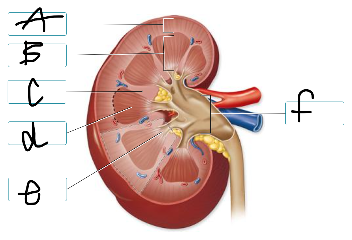
label the structures of the kidney
a) renal cortex
b) renal medulla
c) renal column
d) renal pyramid
e) renal papilla
f) renal pelvis
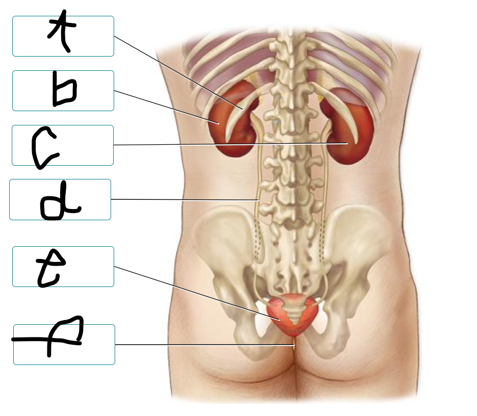
label urinary system
a) 12th rib
b) left kidney
c) right kidney
d) ureter
e) urinary bladder
f) urethra
An obstruction in the ________________ would prevent urine from leaving the kidney.
renal pelvis
Urine directly exits the body via the
urethra
The ____________ receives urine from the renal pyramid
minor calyx
The portion of the kidney in direct contact with the renal capsule is the _________________
renal cortex
The ureter and the renal artery and vein enter the kidney through the opening known as the ______
hilum
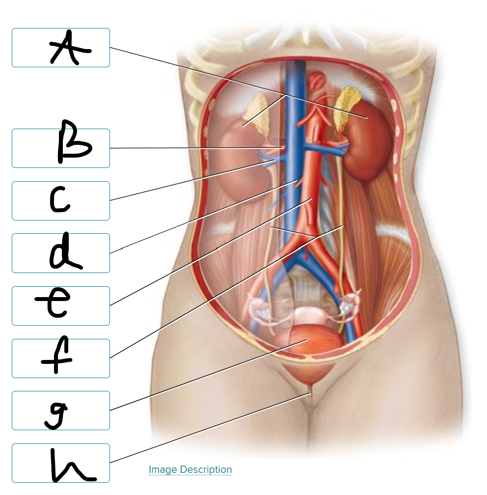
a) kidneys
b) renal artery
c) renal vein
d) inferior vena cava
e) descending abdominal aorta
f) ureters
g) urinary bladder
h) urethra
Which of the following is true concerning the location of the kidneys?
they are located behind the parietal peritoneum
What are the surround tissues of the kidney starting from the innermost to outtermost
1. fibrous capsule
2. perinephric fat
3. renal fascia
4. paranephric fat
T or F
Each kidney typically has eight to fifteen major calyces and two to three minor calyces.
false
The wide base of a renal pyramid lies next to
the renal cortex
Urine from the _________ is collected by the _________ and then drained into the _________.
renal pyramids → minor calyces → major calyces
The two components of a nephron are the
renal corpuscle and renal tubule
T or F
Filtration occurs in the renal corpuscle.
true
What is the correct sequence for parts of the renal tubule?
proximal convoluted tubule → nephron loop → distal convoluted tubule
Cortical nephrons can be distinguished from juxtamedullary nephrons by
their location within the renal cortex
The juxtaglomerular apparatus monitors?
blood Na concentration and adjusts glomerular filtration volume
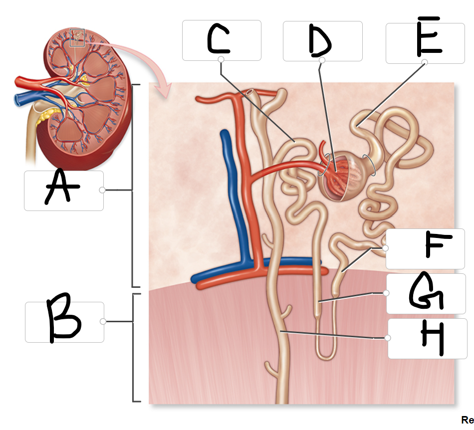
label the structures of a nephron
a) cortex
b) medulla
c) distal convoluted tubule
d) glomerulus
e) proximal convoluted tubule
f) descending limb
g) ascending limb
h) collecting duct
What part of the nephron:
Contains podocytes with pedicels and filtration slits?
glomerulus
What part of the nephron:
Last part of renal tubule; drains into collecting duct
distal convoluted tubule
What part of the nephron:
Contains a descending limb and an ascending limb
nephron loop
What part of the nephron:
First part of renal tubule; cell contain microvilli
proximal convoluted tubule
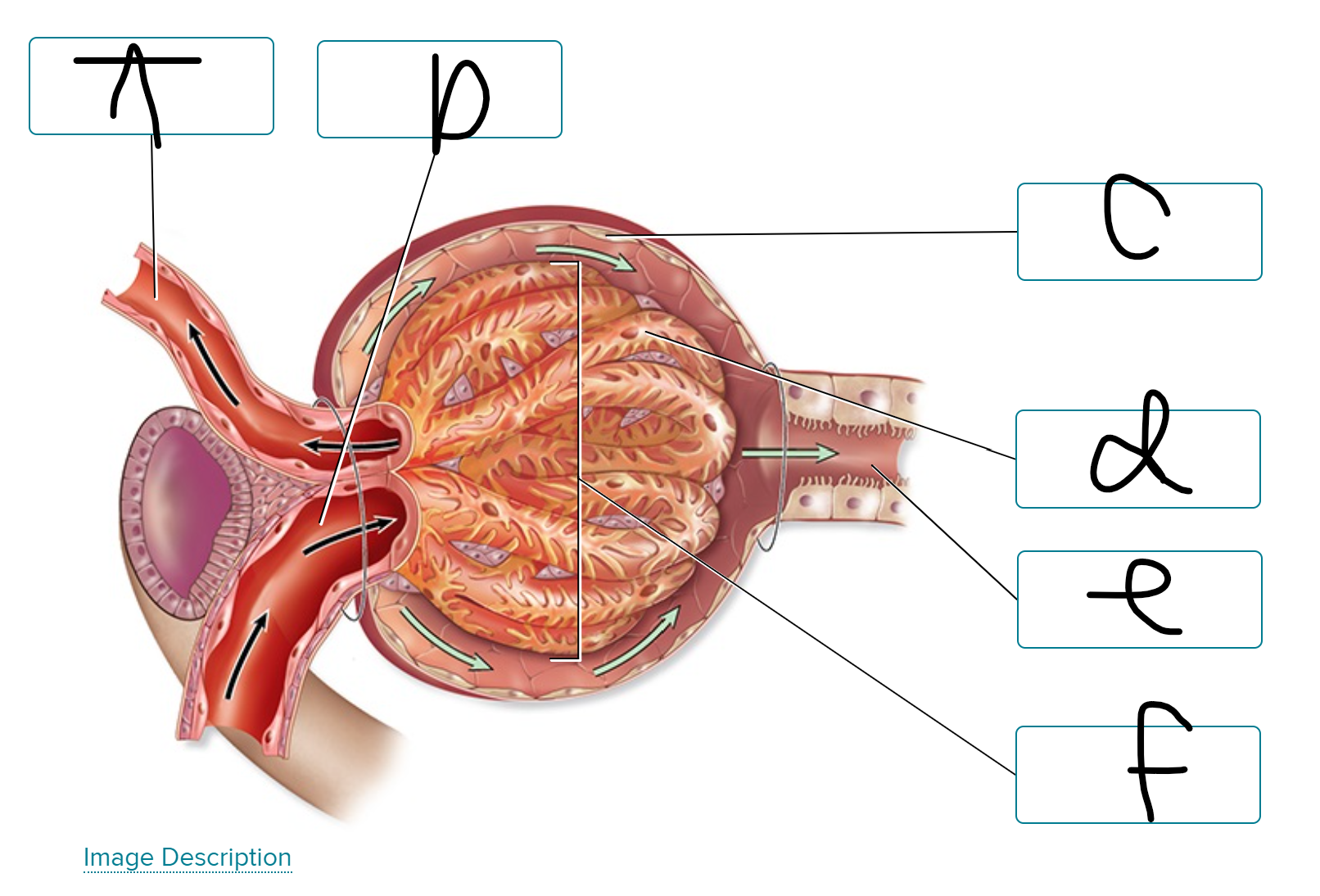
label renal corpuscle
a) efferent arteriole
b) afferent arteriole
c) parietal layer of glomerular capsule
d) visceral layer of glomerular capsule
e) PCT
f) glomerulus
A renal corpuscle includes the
glomerulus and glomerular capsule
What are characteristics associated with the 2 types of nephrons?
Cortical nephrons → make up about 85% of all nephrons
Juxtamedullary nephrons → make about 15% of nephrons
T or F
The juxtaglomerular apparatus is a structure of the nephron where the DCT contacts the afferent arteriole.
true
What are the primary components of the Juxtaglomerular apparatus?
macula densa
granular cells
T or F
Macula densa cells are modified smooth muscle cells of the afferent arteriole.
false
What are the actions of granular cells in the nephron?
synthesize, store, release renin
contraction when stimulated either by stretch or by sympathetic division
What are the functions of the macula densa?
detect changes in the sodium/chloride concentration within the DCT
signaling granular cells to release renin
Blood enter the glomerulus by an ______________ arteriole
afferent
What percentage of glomerular filtrate becomes urine?
1%
Most of the water in tubular fluid is reabsorbed in the
proximal convoluted tubule
Tubular secretion involves the movement of substances
from capillary blood to tubular fluid
The first capillary bed associated with the nephron
filters blood
Which of the following correctly lists the order of structures through which urine flows after its formation in the nephron?
calyx → renal pelvis → ureter → urinary bladder → urethra
In the kidneys, blood goes from the afferent arterioles to the _________, and then enters the _________, which then delivers the blood to the _________.
glomeruli
efferent arteriole
peritibular capillaries
What is the order of vein size that blood leaves the kidney from smallest → largest?
1. cortical radiate veins
2. arcuate veins
3. interlobar veins
4. renal veins
When blood flows through the glomerulus, both water and solutes are filtered from the blood plasma, moving across the wall of the glomerular capillaries and into the capsular space to form _________.
filtrate
Fluid found after leaving the collecting ducts
urine
Fluid found in the proximal convoluted tubule (PCT)
tubular fluid
Water and solutes filtered across the glomerular capillaries
filtrate
Tubular secretion occurs when
waste products from blood pass from the peritubular capillaries to the nephron tubules.
Filtration occurs when
filtered fluid leaves the glomerulus and enters the glomerular capsule.
Tubular reabsorption occurs when
filtered fluid passes from the nephron tubules to the peritubular capillaries.
What would “increase filtration”?
increase in BP
constriction of the efferent arteriole
What would “decrease filtration”?
obstruction in the proximal tubule
increase in plasma protein concentration
increase in capsular pressure
What “Will pass through” filtration of membrane?
glucose, Na, H2O, TH, Urea
What “Will NOT pass through” filtration of membrane?
platelets, TH bound to plasma protein, RBC, Albumin, Lymphocytes
What are the renal processes that form urine?
tubular secretion
tubular reabsorption
filtration
What are the pressures that oppose glomerular hydrostatic pressure to help determine net filtration pressure?
Blood colloid osmotic pressure
capsular hydrostatic pressure
An increased HPg causes what effects on the NFP, GFR, and reabsorption in the nephron?
NFP increases
filtrate reabsorption decreases
GFR increases
T or F
The myogenic mechanism of renal autoregulation involves the juxtaglomerular apparatus and regulation of NaCl.
false
Sympathetic stimulation results in what effects in the kidney?
decrease in GFR
vasoconstriction of the afferent arteriole
decrease in urine production
What are the organs of the urinary system?
kidneys, ureter, urinary bladder
What is the function of the kidney?
filter blood and convert the filtrate into urine
What is the function of the Ureter
transports liquid waste from the kidneys to the urinary bladder
What is the function of the Urinary bladder?
expandable, muscular sac that stores urine until it is eliminated from the body through the urethra
Describe the physiological processes that occur when the kidneys convert filtrate into urine.
Elimination of metabolic wastes: The kidneys remove waste that is present in the filtrate (e.g., urea, uric acids) to prevent these substances from reaching toxic levels within the blood.
- Regulation of ion levels: The kidneys help control the blood's ion balance, such as sodium ions (Na+) , potassium ions (K+ ) calcium ions (Ca2+ ) , and phosphate ions (PO43-) by eliminating varying amounts of these substances in the urine, depending upon dietary intake.
- Regulation of acid-base balance: The kidneys also help regulate blood pressure by excreting fluid in the urine. Regulating fluid lost in the urine helps to regulate blood volume. The kidneys also release the enzyme renin, which is required for production of angiotensin II, a hormone that increases blood pressure. Arguably, blood pressure regulation is one of the kidney's most important functions.
- Elimination of biologically active molecules: Loss of small molecules that are biologically active (e.g., hormones, drugs) also occurs as these substances are filtered, but are not reclaimed, and then become part of urine.
What are some other functions of the kidneys?
- Formation of calcitriol
- Production and release of erythropoietin
- Potential to engage in gluconeogenesis
What is the hilum?
A concave medial border where vessels, nerves, and the ureter connect to the kidney.
Describe the shape and location of the kidneys.
Kidneys are located along the posterior abdominal wall, lateral to the vertebral column. The left kidney is between the level of the T12 and L 3 vertebrae, and the right kidney is about 2 centimeters inferior to the left kidney to accommodate the large size of the liver. Both kidneys are only partially protected by the rib cage, making them vulnerable to forceful blows to the inferior region of the back.
Why are the kidneys said to be retroperitoneal?
Because they are posterior to the parietal peritoneum
Describe the fibrous capsule
is directly adhered to the external surface of the kidney. It is composed of dense irregular connective tissue and maintains the kidney's shape, protects it from trauma, and helps prevent infectious pathogens from penetrating the kidney.
Describe the adipose capsule
is external to the fibrous capsule and A contains adipose connective tissue. It provides cushioning and stabilization for the kidney.
Describe the renal fascia
is external to the perinephric fat and is composed of dense irregular connective tissue. It anchors the kidney to surrounding structures.
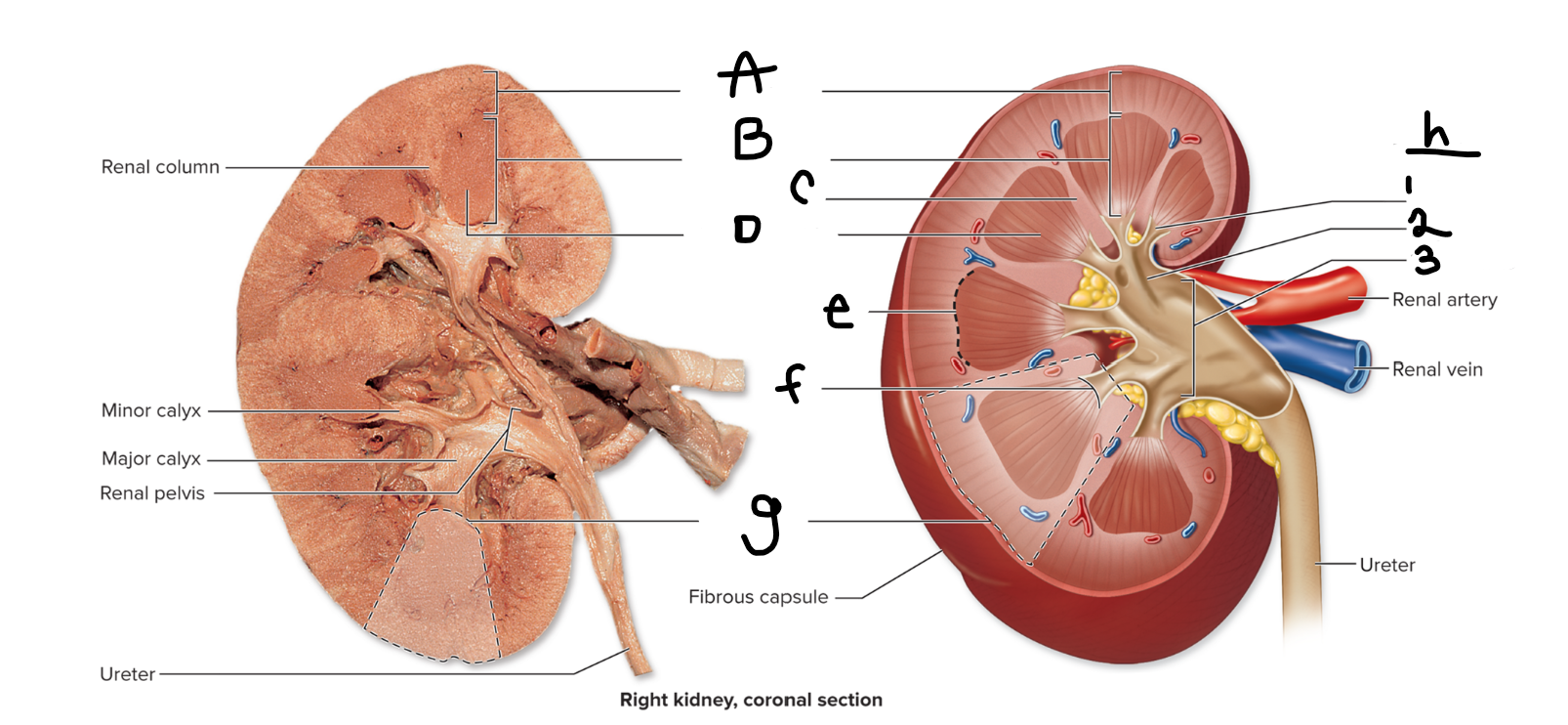
label the image
a) renal cortex
b) renal medulla
c) renal column
d) renal pyramid
e) corticomedullary junction
f) renal papilla
g) renal lobe
h) renal sinus
minor
major
pelvis
The kidney is innervated by which divisions of the autonomic nervous system?
Both divisions of the autonomic nervous system.
What is the functional unit of the kidney?
nephron
What are the two regions of the nephron?
renal corpuscle and renal tubule
What two structures compose the renal corpuscle?
The glomerulus and the glomerular capsule
Describe the glomerulus and the arterioles that serve and drain it.
Glomerulus: thick tangle of capillary loops called the glomerular capillaries
Blood enters the glomerulus by an afferent arteriole and exits by an efferent arteriole.
Describe the layers of the glomerular capsule.
Two layers:
Visceral layer: internal, permeable; directly overlies the glomerular capillaries
Parietal layer: external, impermeable; composed of simple squamous epithelium
Where is the capsular space located?
located between the visceral and parietal layer
What is the first region of the renal tubule?
What epithelium composes it?
What feature helps increase its reabsorption capacity?
1. Proximal convoluted tubule (PCT)
2. Composed of simple cuboidal epithelium
3. Apical microvilli that markedly increase its surface area and this its reabsorption capacity.
What are the two limbs of the nephron loop?
What composes the thick segments?
Thin segments?
Descending limb and ascending limb.
Thick: simple cuboidal epithelium
Thin: simple squamous epithelium
What is the last region of the renal tubule?
How does its lumen appear with a light microscope?
How does this compare to the PCT?
1. Distal convoluted tubule (DCT)
2. appears clear
3. compared to PCT DCT does not appear fuzzy
What are the two types of nephrons?
cortical nephrons and juxtamedullary nephrons
What are cortical nephrons?
are oriented with their renal corpuscles near the peripheral edge of the cortex and have a relatively short nephron loop that barely penetrates the medulla. Thus, the bulk of a cortical nephron resides within the cortex. Approximately 85% of nephrons are cortical nephrons.
What are juxtamedullary nephrons?
Their renal corpuscles lie adjacent to the corticomedullary junction, and they have relatively long nephron loops that extend deep into the medulla. Juxtamedullary nephrons are important in establishing a salt concentration gradient within the interstitial space that lies outside the nephron loop, the collecting tubules, and the collecting ducts thus allowing for the regulation of urine concentration by antidiuretic hormone (ADH).
What do nephrons drain into? (These in turn drain into larger structures.) See figure 2.9.
collecting tubules (these drain into collecting ducts)
The _____________ convoluted tubule contacts the _________ arteriole of the same nephron
(note that many of the nephron illustrations do not show this.
The __________________________ apparatus is a structure at this junction.
distal
afferent
juxtaglomerular (JG)
What are the components of the JG apparatus?
Granular cells and macula densa cells
What are granular cells?
Where are they located?
modified smooth muscle cells of the afferent arteriole
located near its entrance into the renal corpuscle.