HBS 1.2.1-1.2.2 Muscle anatomy & contraction cycle
 Call Kai
 Learn
Learn Practice Test
 Spaced Repetition
 Match
Match Flashcards
 Knowt Play
1/45

There's no tags or description
Looks like no tags are added yet.
Name | Mastery | Learn | Test | Matching | Spaced | Call with Kai | Chat |
|---|
No analytics yet
Send a link to your students to track their progress
46 Terms
Skeletal muscle
A muscle that is attached to the bones of the skeleton and provides the force that moves the bones. Striated and voluntary.

Cardiac muscle
Involuntary muscle tissue found only in the heart. Striated.

Smooth muscle
Involuntary muscle found inside hollow internal organs of the body, blood vessels, and ureters. Not striated.

ATP
(adenosine triphosphate) main energy source that cells use for most of their work made of carbon, oxygen, nitrogen, and phosphorous. Energy is harnessed when the third phosphate is removed.
Muscle contraction requires
ATP and ions like sodium (salt) & calcium
Binding of Calcium ion to troponin
Causes Troponin to release Tropomyosin so it can reveal the myosin binding sites

Myosin head flexes (pulls)- the power stroke
Occurs when Myosin releases ADP +P

ATP bonds to the myosin head
Causes Myosin Head to detach from Actin
ATP is converted into ADP +P
Causes Myosin heads to return to a high energy (ready to bond) position
actin, tropomyosin, troponin
What are the three protein molecules that make up the thin filaments?
Actin
What is part A?
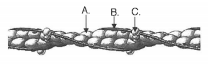
Length stays the same
Length of Thin Filaments during Muscle Contraction
Calcium ions being returned to Sarcoplasmic Reticulum
Causes troponin-tropomyosin to cover the actin binding sites
Length shortens
The length of a sacromere during muscle cell contraction
Tropomyosin
What is part B?
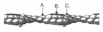
Troponin
Structure at Pink Arrow

Thick filament
Composed primarily of myosin
epimysium
a sheath of fibrous elastic tissue surrounding a muscle

perimysium
Connective tissue surrounding a fascicle
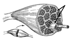
endomysium
Connective tissue surrounding individual muscle fibers
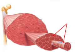
Muscle cramps and spasms
painful involuntary contraction due to fatigue/ATP loss, ion loss (sodium, magnesium), or dehydration.
antagonistic muscles
muscle pairs arranged to work against each other to move a joint in both directions - flexion and extension ex: biceps & triceps brachii

origin
attachment of a muscle that remains relatively fixed during muscular contraction
insertion
The attachment of a muscle tendon to a moveable bone or the end opposite the origin
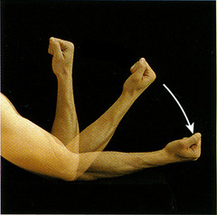
flexion
Decreases the angle of a joint, often ventral

extension
increases the angle of a joint
striations
alternate dark and light bands found on skeletal and cardiac muscle that run in the direction of/parallel to the direction of the action of the muscle from origin to insertion

biceps brachii
2 headed muscle on arm that flexes elbow

Percent Change Formula (the real one)
((final-initial)/initial) x 100
A leg in a cast for two months will
have decreases in size and function due to disuse of the muscle, which results in a loss of muscle fibers over time. "use it or lose it."
Prue diagnosis
Degenerative disc disease in back and neck
Ren diagnosis
Grade 2 Strain of distal leg muscles
Jackson diagnosis
Radius and Ulna fractures & Grade 1 Strain of distal arm muscles
Hazel diagnosis
Cerebral Palsy
Dillon diagnosis
Juvenile idiopathic arthritis of mainly the hands, knees, and elbows
Muscle rule 1
Must have 2+ attachments and cross at at least 1 joint
Muscle rule 2
Muscles only can "pull" and get shorter when they contract
Muscle rule 3
The attachment that moves is known as the insertion and the attachment that remains stationary is known as the origin, muscles need at least one of each type.
Muscle rule 4
Muscles that decrease the angle between bones at a joint are known as flexors. Muscles that increase the angle between bones at a joint are known as extensors
Muscle rule 5
Muscles usually work in opposing "antagonistic" pairs- at least one flexor and one extensor for each joint.
Muscle rule 6
Muscle striations point to the attachments and show the direction of pull/contraction
Muscle nomenclature
Muscles get their names for either their shape, size, location, orientation, action, attachments, or # of origins
Pectoralis major action
flexion, adduction, and medial rotation at shoulder

Intercostals origin and insertion
O: inferior border of rib above
I: superior border of rib below

brachialis action
flexes elbow (under & more distal to biceps brachii)
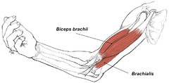
Brachialis origin and insertion
origin: distal portion of anterior humerus both medial and lateral
insertion: proximal posterior ulna