N - Motor Systems 1
1/20
Earn XP
Description and Tags
Descending control of spinal circuits and the motor cortex
Name | Mastery | Learn | Test | Matching | Spaced |
|---|
No study sessions yet.
21 Terms
the motor system
spinal cord:
motor neurons (upper/lower)
sensory input
local reflexes
descending motor pathways:
lateral = voluntary
ventromedial pathway = unconscious e.g. standing up (originates in brainstem)
cerebral cortex:
the motor cortex
sensory input
The motor system (flowchart)
Features:
hierarchical organisation
feedback loops
somatotopic representation
PUT IN IMAGE
Basic types of movement
reflex:
protective e.g. limb withdrawal
motor patterns generated in the spinal cord
‘closed loop’ (no input from brain)
rhythmic motor patters:
e.g. chewing, walking, breathing
a combo of reflex + voluntary
voluntary:
purposeful, goal-directed
command originates from brain
‘open loop’
Lower motor neurons
the alpha motor neuron (a-MN)
ONLY a-MNs directly control muscle contraction
only these can cause contraction at the NMJ
Spinal motor neuron activity - is governed by:
sensory input - local feedback (via dorsal roots)
spinal interneurons - circuitry generating motor programmes
1 + 2 are reflexes
upper motor neurons - initiation + control
Spinal cord circuits can generate movement in isolation
even when descending influences are severed - coordinated movements can occur
can see headless chickens running
Descending input from upper motoneurons
sophisticated, adaptable patterns of movement
voluntary + involuntary
involves input descending from the brain
The musculature: definitions + roles
Distal musculature:
hands, feet, digits
innervated by LATERAL motoneurons
fine motor
Proximal musculature:
elbow, knee
posture
innervated by MEDIAL motoneurons
Axial:
trunk muscles
posture
medial motoneurons
Summary of descending pathways
motor cortex - axon to spinal cord
motor cortex - to lateral pathways
= distal muscles, flexors, voluntary
motor cortex to ventromedial pathways
= proximal / axial muscles, extensors, posture
(include image)
Descending Tracts of the spinal cord
Corticospinal tract + Rubrospinal tract = lateral pathways
Medullary reticulospinal tract + pontine reticulospinal tract + tectospinal tract = ventromedial pathways
bc they run through ventral + middle of spinal cord
Lateral pathways: the corticospinal tract
pyramidal
a direct line contralateral projection from cortex → lateral spinal motor neurons
monosynaptic contact with a-motor neurons (aMNs)
majority of axons from neurons with cell bodies in the motor cortex
innervate aMNs controlled distal muscles
Lateral pathways: the rubrospinal tract
contralateral projections from red nucleus running down the lateral column of the spinal cord
similar role to corticospinal t.
much smaller component of the lateral pathway
Ventromedial motor pathways
‘extra pyramidal tracts’
all originate from the brain stem nuclei
both contralateral + ipsilateral descending projections
control of motor outputs to proximal + axial muscles
control of body position + posture
insert ss
Ventromedial motor pathways: pontine reticulo-spinal tract + medullary reticulo-spinal tract
Pontine reticulo-spinal tract:
enhances anti-gravity reflexes of spinal cord
facilitates leg extensors to maintain standing posture
Medullary reticulo-spinal tract:
has opposing effect
frees antigravity muscles from reflex control
allows voluntary override
Ventromedial pathways: Vestibulospinal tract + Tectospinal tracts
Vestibulospinal tract:
relays gravitational sensory info from:
vestibular labyrinth (inner ear) +
stretch receptors in axial muscles
maintains head + neck position + legs
Tectospinal tract:
relays visual sensory info from retina + visual cortex
orientates head + eyes to visual/auditory stimuli
Organisation of Cortical Motor Areas
control of voluntary movement involves neocortex
bc movement involves not just execution but also:
sensory input
planning
deciding action
holding plan in memory
the principle areas involved identified through electrical stimulation
Cortical motor + sensorimotor areas (image)
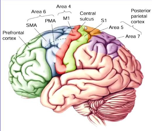
Cortical motor areas: terminology
The motor cortex includes:
area 6 inc: (more complex movement)
supplementary motor area
premotor area
area 4 inc: (lowest stimulus threshold)
primary motor cortex
Analogy:
Think of Area 4 as the trigger of a gun — very sensitive, and pressing it causes immediate action.
Area 6 is like the planning hand that positions the gun and decides how to aim — important, but not enough by itself to fire.
Roles of Cortical Motor Areas
Primary Motor Cortex (M1, Area 4):
control of distal musculature (fine motor control)
Premotor Cortex (Area 6, lateral):
control of proximal musculature (posture, balance)
control of movement sequencing
preparation for movement, initiation
Supplementary Motor Area (Area 6, fronto/medial)
planning + initiation
bi-manual co-ordination
Primary Motor Cortical Output Neurons (upper motor neurons)
‘upper motor neurons’
axons in the corticospinal tract
pyramidal type, cell body in cortical layer V (Betz cell)
somatotopically organised
activate small groups of muscles rather than single ones
individually encode the force OR direction of movement
Damage to Upper Motor Neurons
e.g. caused by stroke, tumour
initial muscle weakness
eventual spasticity
increased muscle tone (hypertonia)
increase reflex response (hyper-reflexia)
affects contralateral side to damage
recovery possible - PMC shows adaptive alterations