Path Exam 6 End of Chapter Questions and Practice Questions
 Call Kai
 Learn
Learn Practice Test
 Spaced Repetition
 Match
Match Flashcards
 Knowt Play
1/118

There's no tags or description
Looks like no tags are added yet.
Name | Mastery | Learn | Test | Matching | Spaced | Call with Kai | Chat |
|---|
No analytics yet
Send a link to your students to track their progress
119 Terms
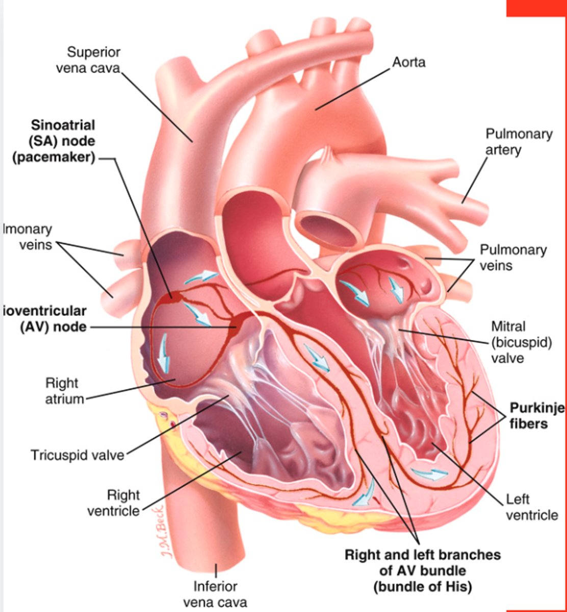
Study Image
1.Aorta
2.Pulmonary Artery
3.Superior Vena Cava
4.Inferior Vena Cava
5.Pulmonary Veins
6.Right Atrium
7.Left Atrium
8.Right Ventricle
9.Left Ventricle
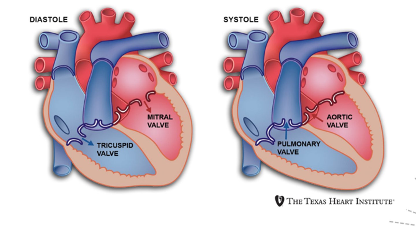
Mitral (bicuspid) valve
________________ delivers adequat amount of blood to all tissues in the body.
A. Cardiovacular system
B. Nervous System
C. Endocrine system
D. Exocrine system
A. Cardiovacular system
Which of the following controls rhythmic contractions of the heart?
A. Central nervous system
B. Autonomic nervous system
C. Automatic nervous system
D. Sympathetic nervous system
B. Autonomic nervous system
Acetylcholine does what to the heart?
A. Does nothing to the heart
B. Increases heart rate
C. Causes a heart attack
D. Slows the heart down
D. Slows the heart down
The ____________ nervous system stimulates the release of epinephrine, which accelerates the heart and increases the force of contractions.
A. central
B. autonomic
C. sympathetic
D. assympathic
C. sympathetic
The heart is lined with a delicate membrane called ________.
A. endocardium
B. myocardium
C. exocardium
D. mayocardium
A. endocardium
What separates the right and left side of the heart?
A. Septum
B. Ventricles
C. Atriums
D. Valves
A. Septum
________ are larger and thicker than atria, they have a heavier pumping load.
A. Cardial apex
B. Heart valves
C. Ventricles
D. Atriums
C. Ventricles
Match the word with definition:
1. Mitral (BiCuspid) Valve
2. TriCuspid Valve
3. Pulmonary Valve
4. Aortic Valve
A. is located between right ventricle and pulmonary artery
B. is located between the left atrium and left ventricle.
C. is located between the right atrium and right ventricle.
D. is located between the aorta and left ventricle.
1. B
2. C
3. A
4. D
Which of the following are preferred initial modalities for imaging the cardiovascular system? Select all that apply.
A. Ultrasound
B. Doppler
C. CT
D. X-rays
E. MRI
A, B, C, D.
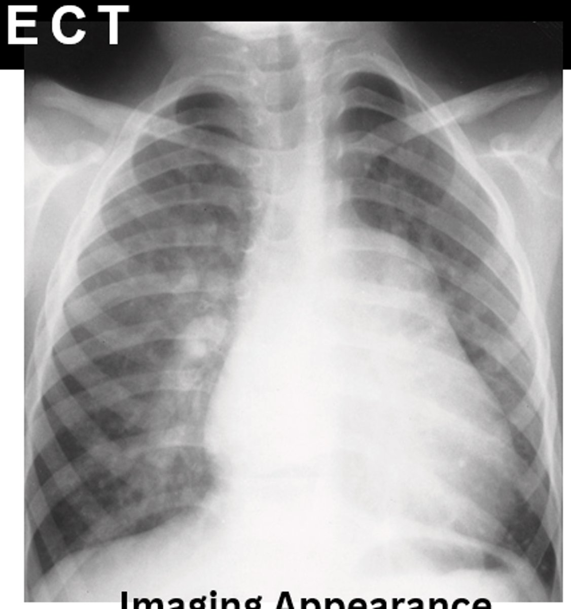
A chest radiograph shows the heart enlarged and somewhat triangular, and visualizes an increased pulmonary vascular volume. The pulmonary trunk is very large that it overshadows the normal-sized aorta. Which of the following would be the cause?
A. CHD patent ductus arteriousus
B. CHD ventricular septal defect
C. CHD atrial septal defect
D. Cyanotic congenital heart failure
B. CHD ventricular septal defect
Which of the following is the most common congenital cardiac leasion, permits free communication between the two atria due to foramen ovale not closing properly after birth or improperly close during gestation, and causes increased blood flow and overloading of the right ventricle?
A. CHD atrial septal defect
B. CHD ventricular septal defect
C. CHD patent ductus arteriousus
D. Cyanotic congenital heart failure
A. CHD atrial septal defect
________ is where a shunt, or hole, is between the two ventricles causing oxygen-rich blood to flow from the left ventricle to mix with the non-oxygenated blood from the right ventricle potentially causing the heart to work harder.
A. CHD patent ductus arteriousus
B. Cyanotic congenital heart failure
C. CHD atrial septal defect
D. CHD ventricular septal defect
D. CHD ventricular septal defect
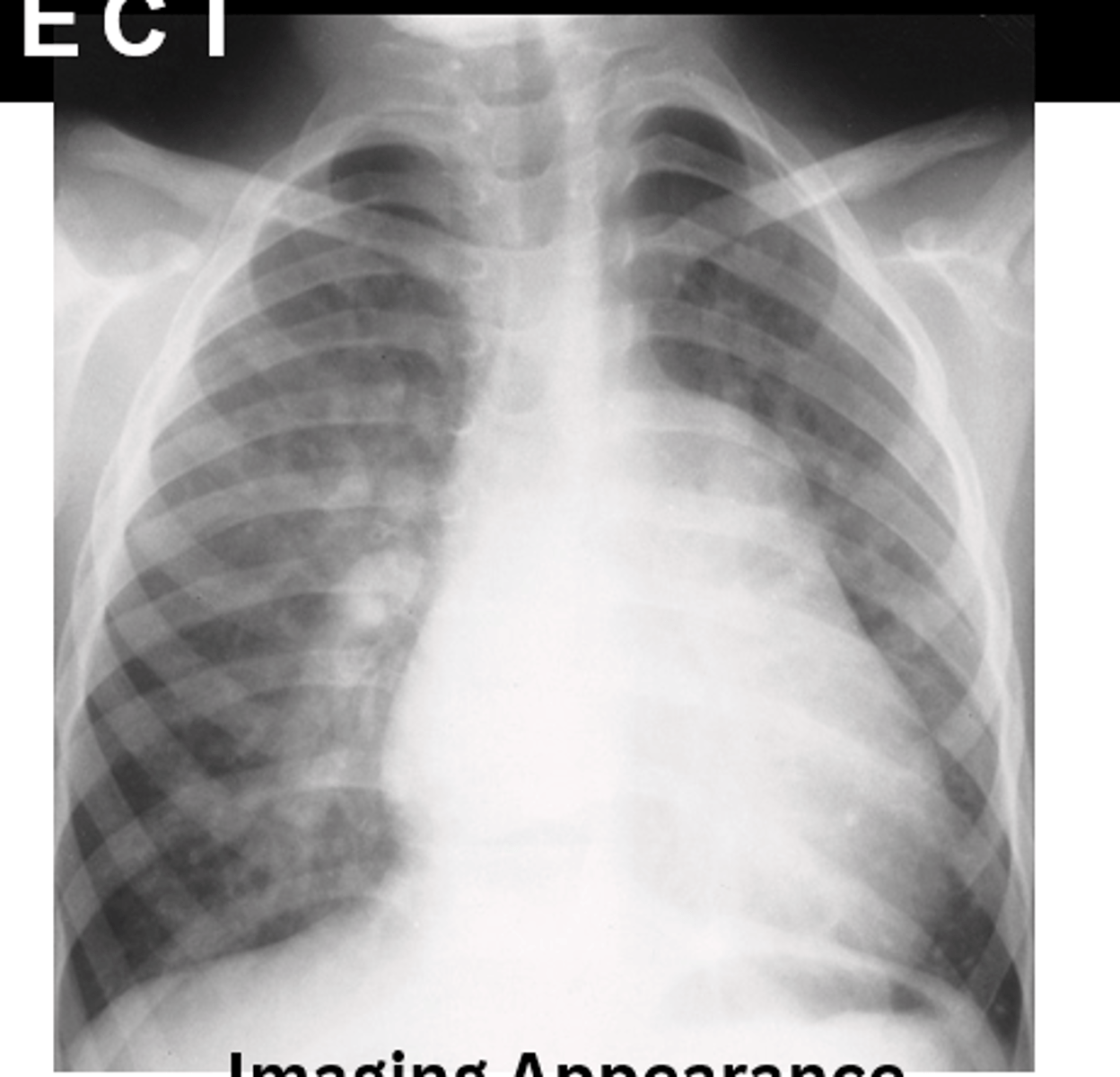
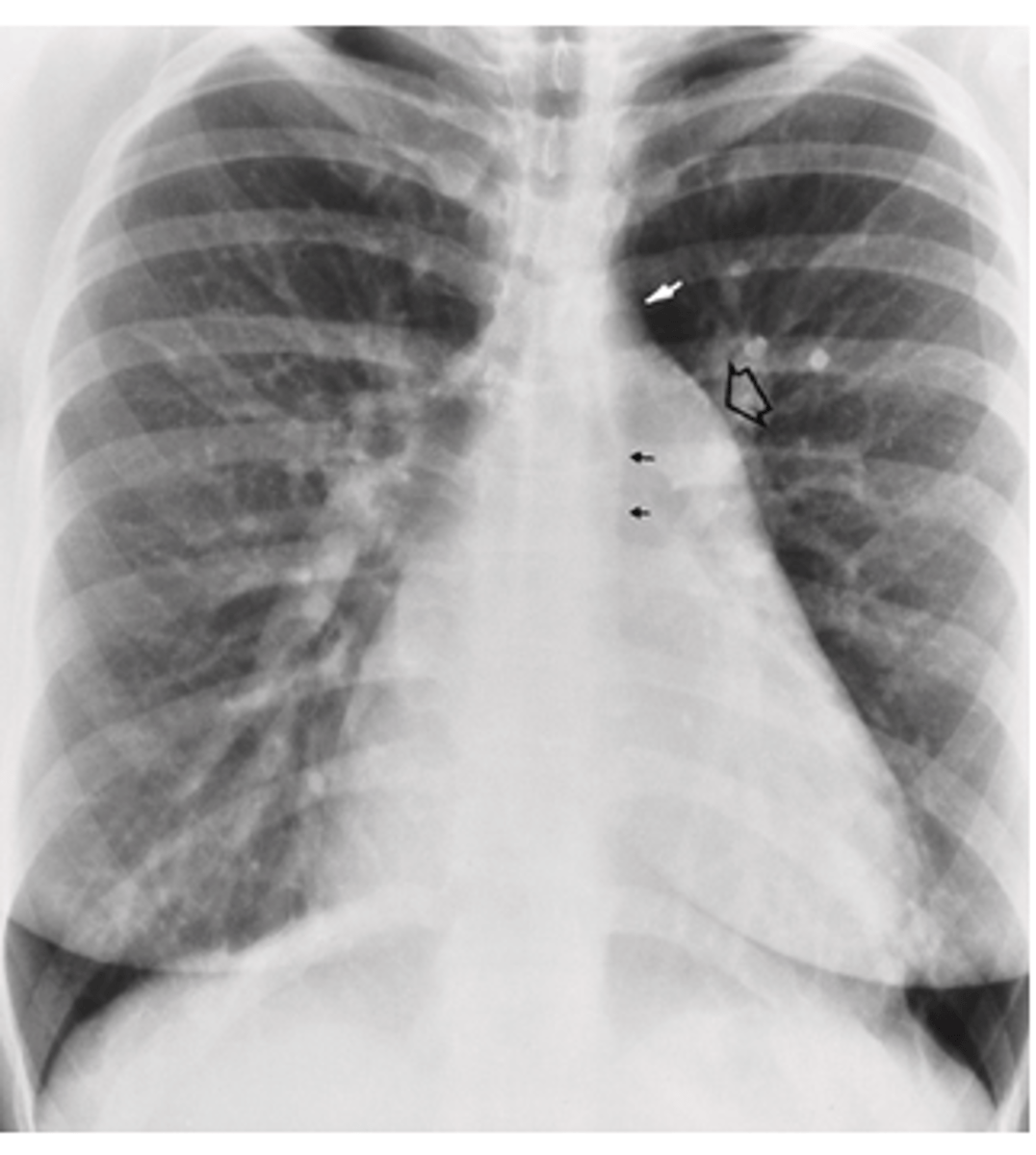
A chest radiograph shows an enlargement of the right ventricle, right atrium, and enlargment of the pulmonary outflow. Which of the following would be the cause?
A. Cyanotic congenital heart failure
B. CHD ventricular septal defect
C. CHD patent ductus arteriousus
D. CHD atrial septal defect
D. CHD atrial septal defect
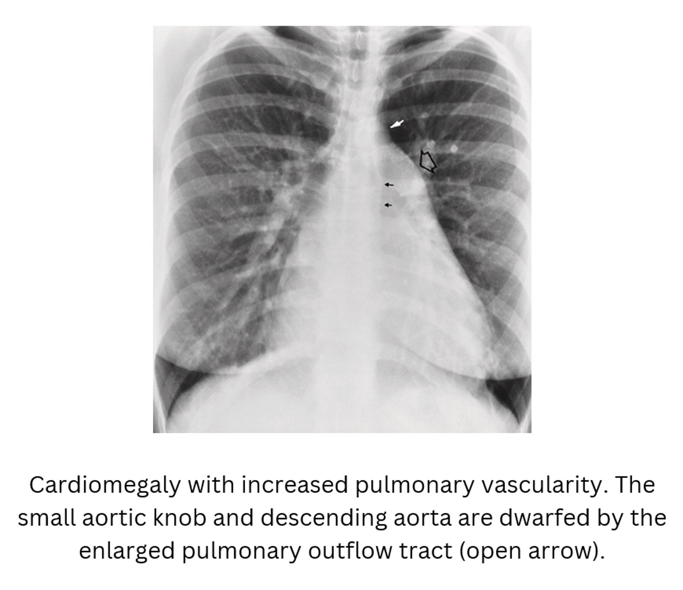
___________ is a vessel that extends from the bifurcation of the pulmonary artery to join the aorta and serves to shunt (to move) blood from the pulmonary artery into the systemic circulation during intrauterine life (in the womb)
A. Ductus arteriosus
B. Patent ductus arteriosus
C. Aortic notch
D. Expecto patroarteriosus
A. Ductus arteriosus
Which of the following is caused by the ductus artriosus not closing properly causing blood flow to flow from the high pressure aorta to the low pressure pulmonary valve causing an overflow of blood into the left atrium and ventricle?
A. Patient ducus arteriosus
B. Patent ductus arteriosus
C. Expecto patroarteriosus
D. Ductus aortic ateriosus
B. Patent ductus arteriosus
T/F: The leading cause of cyanotic congenital heart failure is CHD ventricular septal defect.
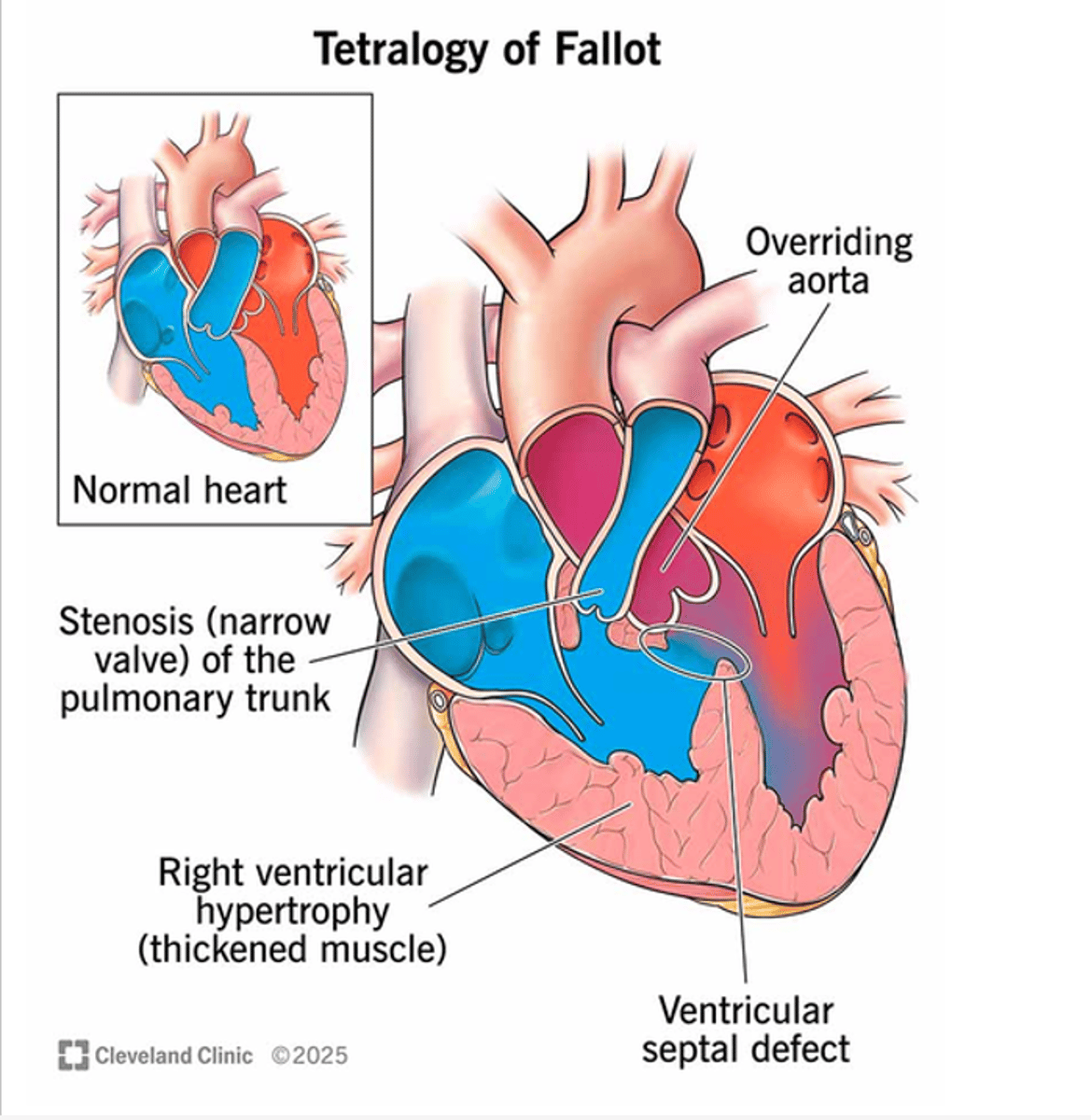
False (It's Tetrology of fallout: high ventricular septal defect, pulmonary stenosis, overriding aortic orifice above the ventricular defect, RT ventricular hypertrophy)
Which of the following is true about the Tetralogy of Fallot:
1. High ventricular septal defect
2. Left ventricular hypertrophy
3. Pulmonary stenosis
4. Overriding aortic orifice above the ventricular defect
5. Right ventricular hypertrophy
6. Without surgical repair, most patients will pass away before puberty.
A. Only 3, 4, 5, 6.
B. Only 1, 2, 3, 4.
C. Only 1, 3, 4, 5.
D. Only 1, 3, 4, 5, 6.
E. All answers are part of the Tetralogy of Fallot.
D. Only 1, 3, 4, 5, 6.
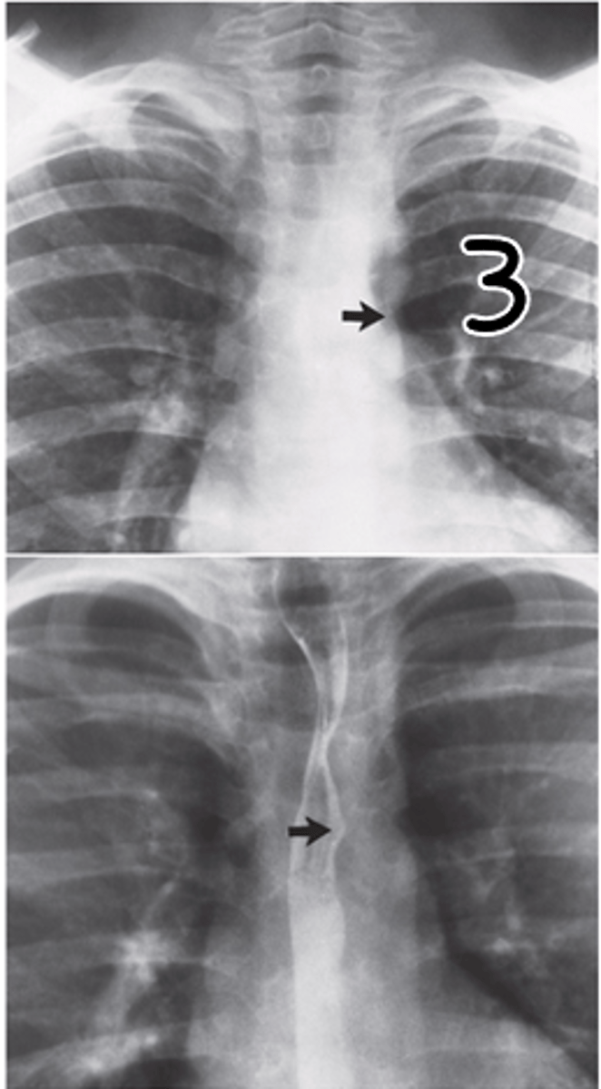
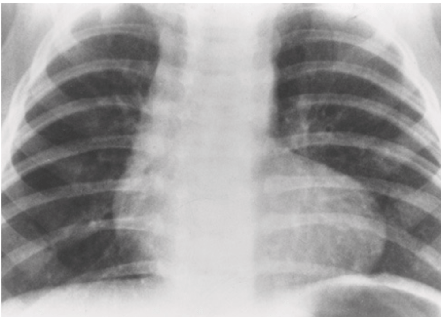
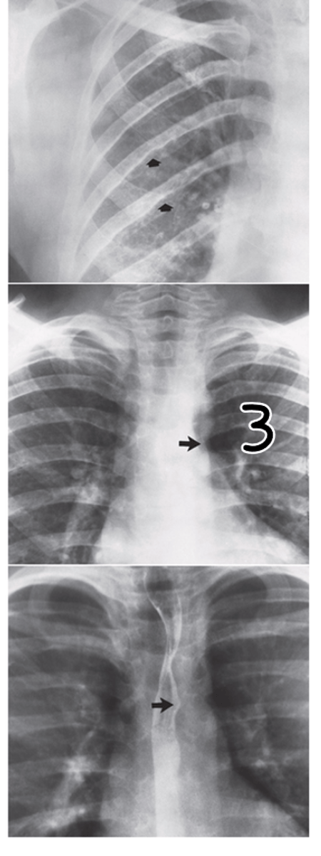
A pediatric radiograph has an image appearance of rib notching which produces a figure-3 sign. Which of the following would cause this on a radiograph?
A. Tetralogy of Fallot
B. CHD atrial defect
C. CHD ventricle defect
D. Coarctation
D. Coarctation
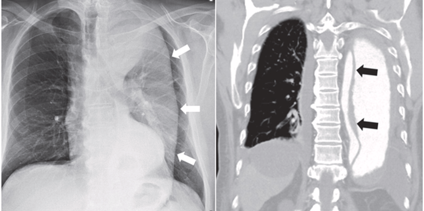
A chest radiograph is noted that there is prograssive widening of the aortic shadow which has an irregular or wavy outer border. Which of the following could cause this anomaly on the chest radiograph?
A. Aortic thrombosis
B. CHF
C. Dissection of the aorta
D. Thrombosis
C. Dissection of the aorta
T/F: In order to properly diagnose dissection of the aorta, CT aortography, MRI, or transesophagel echocardiography (TEE) must be done.
True
A lateral displacement and upward tilting of the prominent cardiac apex is found on a chest radiograph. This pathology decreases pulmonary vascularity and flattens the pulmonary outflow tract. Which of the following is the cause of this radiograph finding?
A. Tetralogy of Fallot
B. Triology of Fallot
C. CHD ventricular septal defect
D. CHD pulmonary arterial defect
A. Tetralogy of Fallot
T/F: Myocardial infarction is the blockage of a coronary artery depriving the myocardium of blood supply and can lead to death of muscle cells.
True
________ refers to a narrowing, or contriction, of the aorta.
A. Coarctation
B. Constrict
C. Stricture
D. Stenosis
A. Coarctation
Which of the following is true about coarctation of the aorta:
1. Blood pressure and pressure to the upper extremies are higher than normal.
2. Most commonly occurs just beyond the branching of the vessels to the head and arms
3. Most frequent cause of hypertension in children
4. Results in decreased blood flow through constricted area of abdomen and legs
5. Most frequent in adults with hypertension
6. Surgical repair or ballon dilation with stenting is requred for survival.
A. 2, 3, 4, 5
B. 2, 3, 4, 6
C. 1, 2, 3, 4, 6
D. 1, 2, 3, 4, 5
C. 1, 2, 3, 4, 6
Which of the following diseases is the narrowing of the arteries that causes oxyegen deprivation of the myocardium and ischemic heart disease?
A. CHD ventricular defect
B. Coarctation
C. Myocardial infarction
D. Coronary Artery disease
D. Coronary Artery disease
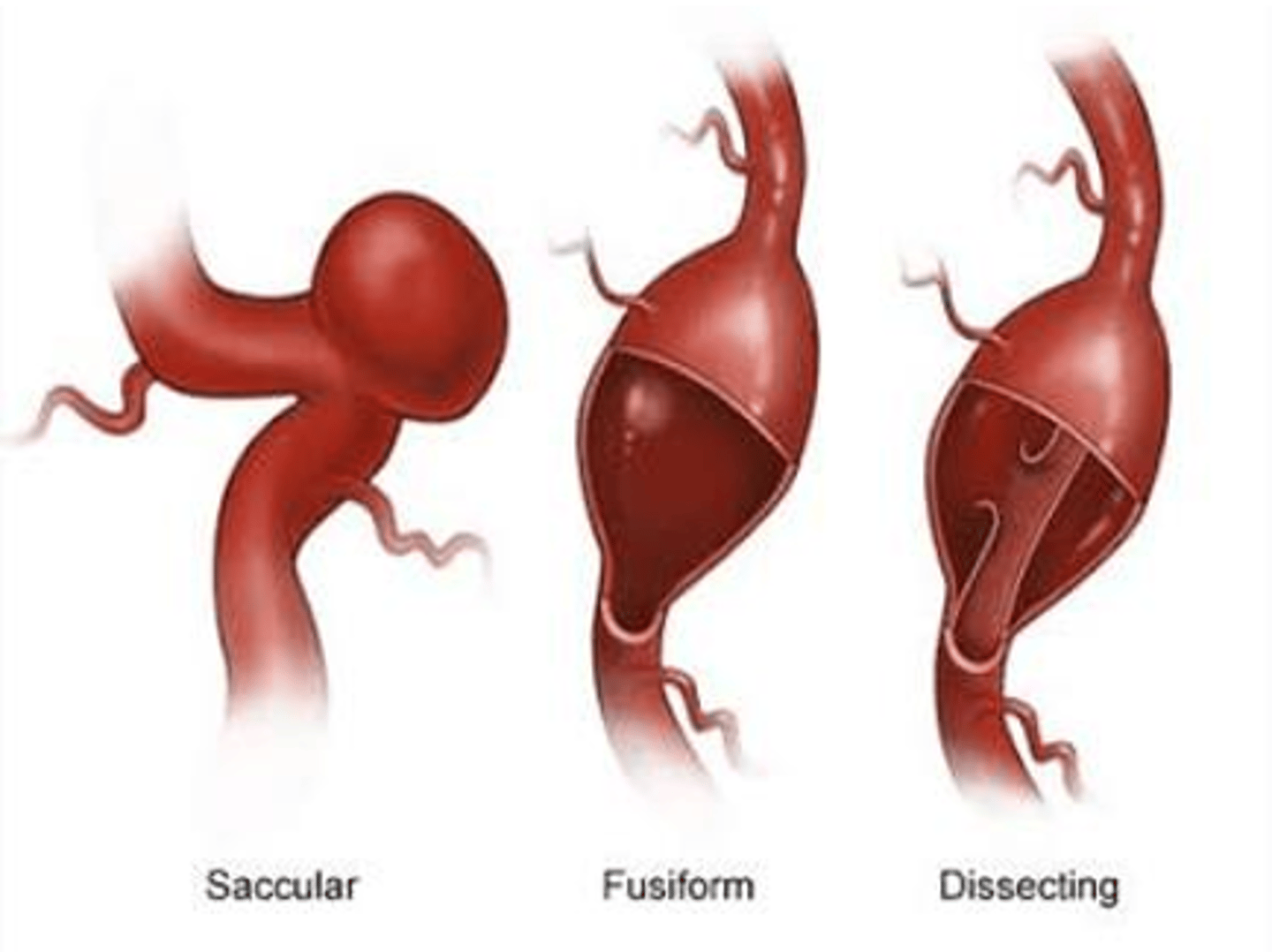
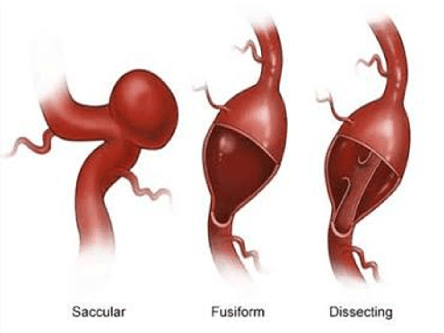
Bulging of the entire circumference of the vessel wall is termed a _________ aneurysm.
A. fusiform
B. saccular
C. pseudo
D. dissecting
A. fusiform
Which of the following is TRUE about coronary artery disease?
1. Narrowing of the arteries causing oxygen deprivation of the myocardium and ischemic heart disease
2. Most often caused by deposit of fatty material on the inner arterial wall (called artherosclerosis)
3. Only occurs in patients that are younger than 25 years old
4. Predisposed factors are hypertension, obesity, smoking, high-cholesteral diet, lack of exercise
5. The blockage of the coronary artery is called myocardial infarction
A. 1, 2, 5
B. 1, 2, 4, 5
C. 2, 3, 4, 5
D. All of the above are correct
B. 1, 2, 4, 5
T/F: CT is the perfered modality for abdominal aortic aneurysm.
False (US is the preferred imaging modality)
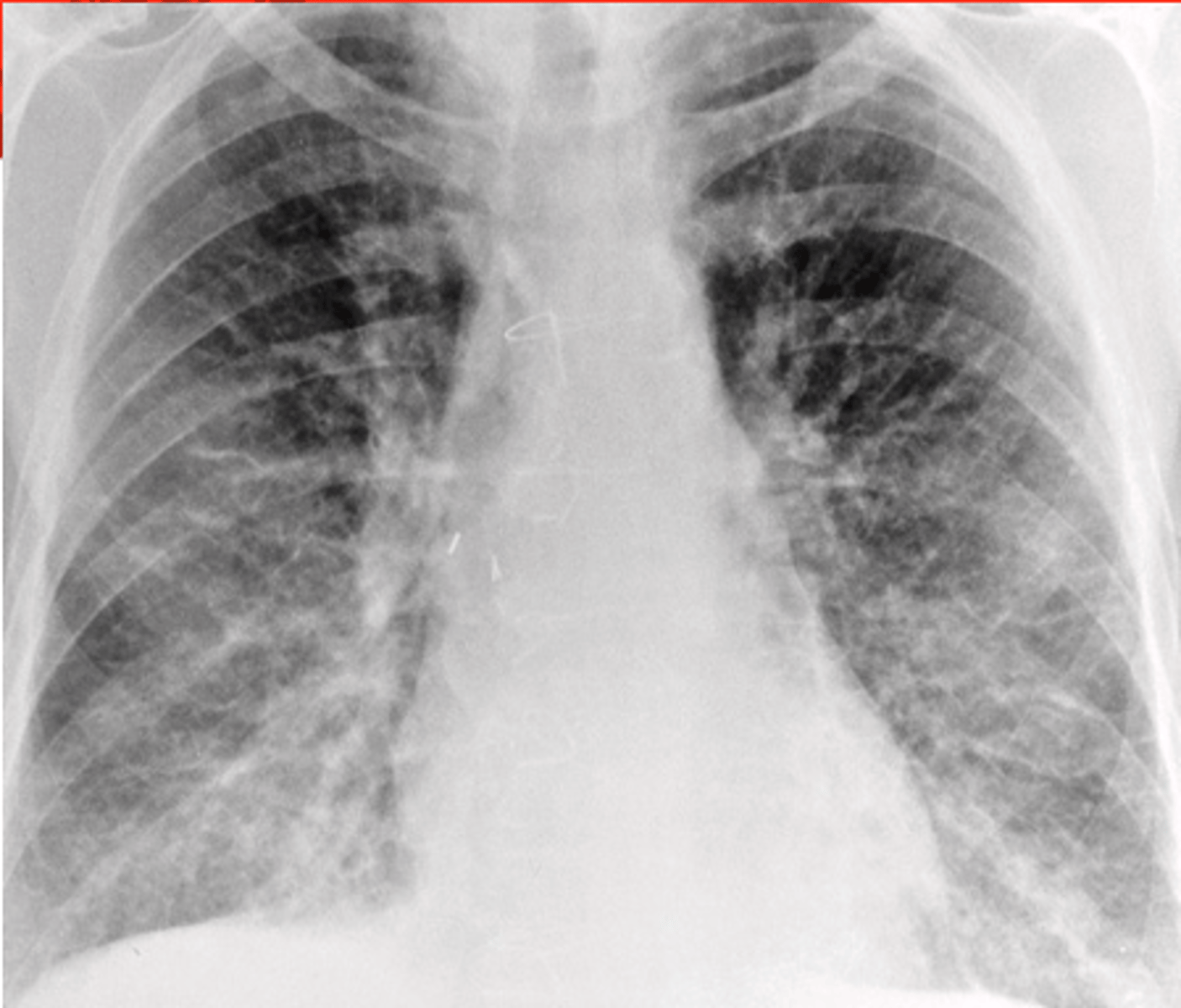
A radiograph shows fluid in the interstitial space of the lung causing loss of normal sharp definition of the pulmonary vascular markings and a perihilar haze. Which of the follow is causing the image to be less sharp?
A. Pulmonary edema
B. CHF
C. Primary broncial edema
D. Myocardial edema
A. Pulmonary edema
The vegitation of _____________ are filled with bacteria and tend to break apart easily to enter the blood stream and form septic emboli that travel to the brain, kidney and lung or other vital organs.
A. Varicose veins
B. CHF
C. Infective endocarditis
D. DVT (Deep vein thrombosis)
C. Infective endocarditis
The Thallium Stress Test is used to assess regional blood flow to the myocardium for which disease?
A. Coronary artery disease
B. CHD ventricular defect
C. CHF
D. Pericardial infarction
A. Coronary artery disease
A _____________ aneurysm involves only one side of the aterial wall.
A. fusiform
B. saccular
C. pseudo
D. vessel
B. saccular
___________ refers to an abnormal accumulation of fluid in the extravascular pulmonary tissues.
A. Bronchial edema
B. Periostinum edema
C. Pulmonary edema
D. Alveolar edema
C. Pulmonary edema
T/F: In CHF patients, if the cardiothoracic ratio is less than 50%, that means the patient has cardiomegaly.
False
__________refers to the Inability of the heart to pump at a rate and volume that would be sufficient to provide enough blood to the tissues.
A. CHD
B. CHF
C. CHF ventricular defect
D. None of the above
B. CHF
________ refers to the formation of nodules or vegetations on heart valves by deposits of bacteria or fungi which can be treated by antibiotics.
A. Infective endocarditis
B. DVT (Deep vein thrombosis)
C. Varicose veins
D. CHF
A. Infective endocarditis
T/F: US angiogram is the primary imaging to confirm a pulmonary embolism.
False (CT angiogram)
All of the following are areas in the lung that fluid buildup can occur except:
A. Lung lymphatics
B. Interstitium
C. Alveolar epithelial layer
D. Alveoli
E. Trachea
E. Trachea
All of the following are TRUE about pulmonary edema except:
A. Most common cause of pulmonary edema is an elevation of pulmonary venous pressure
B. Pulmonary venous pressure is most often attributed to left-sided heart failure
C. Pulmonary edema may be caused by pulmonary venous obstruction or lymphatic blockade.
D. Pulmonary edema may also be casued by narcotic overdose, exposure to noxious fumes, ARDS, etc.
E. All of the above are true
E. All of the above are true
An __________ is a localized dilation of an artery that most commonly involves the aorta, especially its abdominal portion.
A. aneurysm
B. saccular aneurysm
C. Pulyp
D. None of the above
A. aneurysm
__________ is the development of a blood clot in a vessel where blood slowly moves through it.
Thrombus
___________ is a potentially life-threatening condition in which disruption of the intima (inner layer) permits blood to enter the wall of the aorta and separate its layers into true and false lumen.
A. Aortic embolism
B. Tetralogy of Fallot
C. Thrombus and embolism
D. Disection of the Aorta
D. Disection of the Aorta
_________ occurs when a part or all of a thrombus unattaches from the vessel wall and enters the blood stream and potentially gets stuck in an artery.
Embolism
All of the following are TRUE about dissection of the aorta except:
A. Most dissections occur in patients with arterial hypertension
B. Patients present dull pain in the chest and abdomen areas.
C. Occurs from trauma or congenital defects, such as Marfan Syndrome
D. All of the above are true.
B. Patients present dull pain in the chest and abdomen areas. (Patients are in excruciating pain in the chest and abdomen area.)
Heart rate is controlled by the _________________ nervous system.
A. psychogenic
B. central
C. peripheral
D. autonomic
D. autonomic
The _________________ valve is located between the left atrium and the left ventricle, whereas the _________________ valve is located between the right atrium and the right ventricle.
1. tricuspid
2. mitral
3. pulmonary
4. aortic
A. 2, 1
B. 3, 1
C. 4, 1
D. 1, 2
A. 2, 1
The general circulation of the body is termed the _________________ circulation.
A. pulmonary
B. systemic
C. general
D. autonomic
B. systemic
The contraction phase of the heart is termed _________________, whereas the relaxation phase is termed _________________.
1. autonomic
2. diastole
3. systemic
4. systole
A. 1, 3
B. 2, 4
C. 2, 3
D. 4, 2
D. 4, 2
Contraction of which chamber of the heart forces oxygenated blood into the aorta?
A. right atrium
B. right ventricle
C. left atrium
D. left ventricle
D. left ventricle
Oxygenated blood reaches the heart muscle by way of the _________________ and _________________.
1. right coronary artery
2. left coronary artery
3. left coronary vein
4. right coronary vein
A. 1, 3
B. 4, 1
C. 2, 1
D. 2, 3
C. 2, 1
The heart has a specialized pacemaker called the _________________.
A. bundle of His
B. sinoatrial node
C. Purkinje fibers
D. atrioventricular node
B. sinoatrial node
Which of the following factors leads to coronary artery disease?
A. lack of exercise
B. obesity, hypertension
C. smoking, high-cholesterol diet
D. all of above
D. all of above
Temporary oxygen insufficiency to the heart muscle causes severe chest pain termed _________________.
A. angina pectoris
B. myocardial occlusion
C. myocardial pectoris
D. anginal occlusion
A. angina pectoris
Arterial disease caused by fatty deposits on the inner arterial wall is termed _________________.
A. arteriosclerosis
B. myocardial infarction
C. aneurysm
D. myocardial ischemia
A. arteriosclerosis
Which radiographic procedure is used to determine the presence of coronary artery disease?
A. angioplasty
B. chest imaging
C. coronary arteriography
D. CT
C. coronary arteriography
The procedure in which a balloon is used to dilate narrowed coronary arteries is termed _________________.
A. aortocoronary bypass
B. coronary arteriography
C. percutaneous transluminal angioplasty
D. fluoroscopy
C. percutaneous transluminal angioplasty
Which term refers to an inability of the heart to propel blood at a sufficient rate and volume?
A. congestive heart failure
B. pulmonary edema
C. valvular disease
D. valvular stenosis
A. congestive heart failure
An elevation of the pulmonary venous pressure is the most common cause of _________________.
A. congestive heart failure
B. pulmonary edema
C. valvular disease
D. valvular stenosis
B. pulmonary edema
The leading cause of strokes and congestive heart failure is _________________.
A. hypertension
B. low blood pressure
C. hypotension
D. cor pulmonale
A. hypertension
A localized bulging or dilation of an artery is termed _________________.
A. edema
B. effusion
C. aneurysm
D. fat emboli
C. aneurysm
What is the modality of choice for demonstrating an abdominal aortic aneurysm?
A. MRI
B. US
C. CT
D. plain image radiography
B. US
A congenital narrowing or constriction of the thoracic aorta is referred to as _________________.
A. an embolism
B. plaque
C. coarctation
D. tetralogy
C. coarctation
The _________________ of the heart is/are the major site of damage from rheumatic fever.
A. myocardium
B. valves
C. septum
D. endocardium
B. valves
The most sensitive and specific noninvasive method of diagnosing mitral stenosis is _________________.
A. US
B. echocardiography
C. cardiac arteriography
D. CT
B. echocardiography
The accumulation of fluid within the pericardial space surrounding the heart is termed _________________.
A. pericardial thrombosis
B. pericardial effusion
C. pulmonary edema
D. pulmonary effusion
B. pericardial effusion
The invasive procedure for determining deep vein thrombosis is _________________.
A. Doppler US
B. venography
C. CT
D. arteriography
B. venography
The invasive procedure for determining deep vein thrombosis is _________________.
A. Doppler US
B. venography
C. CT
D. arteriography
B. venography
The most accurate screening procedure for assessing renovascular lesions is _________________.
A. Doppler US
B. venography
C. CT
D. arteriography
D. arteriography
A potentially life-threatening condition that usually begins as a tear in the intima above the aortic valve is an _________________.
A. aortic stenosis
B. aortic coarctation
C. aortic thrombosis
D. aortic dissection
D. aortic dissection
______________ is the only noninvasive procedure that detect the valvular vegetatins that are hallmark of infective endocarditis.
A. US
B. CT
C. cardiac arteriography
D. echocardiography
D. echocardiography
All of the following are TRUE about DVT except:
A. Primerily involves the lower extremities.
B. Trauma, bacterial infection, prolonged bed rest, and oral contraceptives could be precipitating factors to develop DVT.
C. Major source of potentially fatal pulmonary embolism (PE).
D. Sometimes may be the earliest symptom of an unsuspected malignancy of the pancreas, lung, or GI system.
E. All of the above are true
E. All of the above are true
_______________ is characterized by an acute inflamation of the pia mater and arachnoid mater.
A. Meningitis
B. Gliomas
C. Bacterial Meningitis
D. Cerebrovascular disease
A. Meningitis
T/F: Pathogens can access the meninge through various routes such as middle ear, respiratory tract, or an infection in the frontal sinus causing meningitis.
True
What is the most common cause of bacterial meningitis in children?
A. Polio
B. Pneumococcl
C. Flu
D. Meningococcl
C. Flu
T/F: Viral meningitis is the most common form of meningitis.
False (Bacterial meningitis is the most common)
What is the most common cause of bacterial meningitis in adults?
A. Pneumococcl
B. Meningococcl
C. Flu
D. Both A and B
D. Both A and B
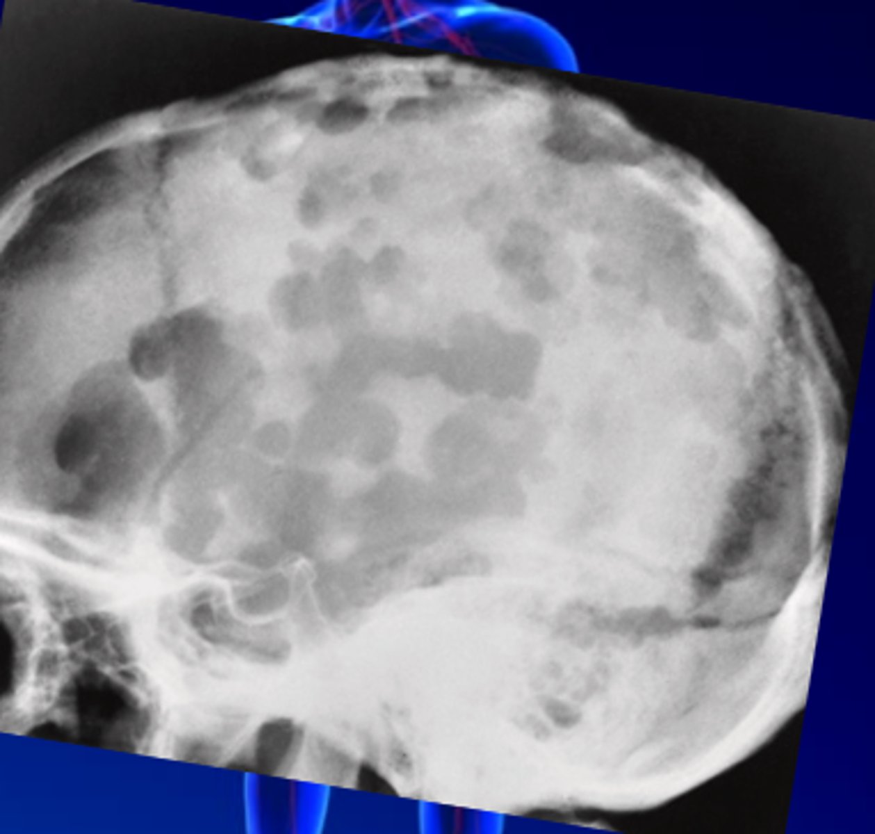
A skull radiograph shows multiple small, pooprly defined areas of lucency. Another radiograph was take a seveal weeks later, the lucencies enlarged and coalesced together to create larger lucencies. Which of the following disease would be the cause of the lucencies on the radiograph?
A. Osteomyelitis
B. Gliomas
C. Meningitis
D. Metastases
A. Osteomyelitis
What could cause viral meningitis?
A. Mumps
B. Polio
C. Herpes simplex
D. All of the above
D. All of the above
Which of the following imaging modalities best demonstrates Meningitis?
A. CT & MRI
B. US
C. Radiography
D. Doppler
A. CT & MRI
Which of the following is most commonly caused by direct extension of a suppurative process from the paranasal sinuses, mastoid air cells, or scalp in the skull?
A. Gliomas
B. Osteomyelitis
C. Metastases
D. Meningitis
B. Osteomyelitis
Which primary malignant brain tumors arise from glial cells, commonly occur in middle adulthood, and can spread between cerebral hemispheres through white matter tracts like the corpus callosum?
A. Osteomyelitis
B. Metastases
C. Astrocytomas
D. Gliomas
D. Gliomas
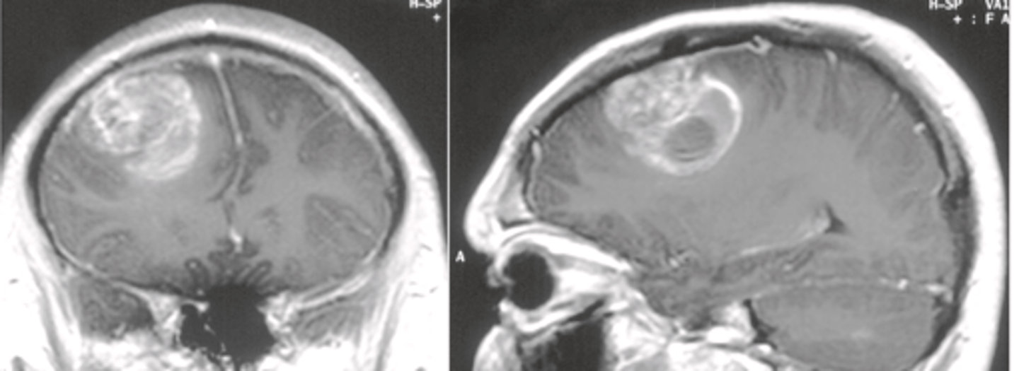
T/F: Astrocytomas make up 70% of Gliomas and are slow growing.
True
T/F: Grade 1 Astrocytomas are malignant.
False (Astrocytoma grade 1 is benign, Grade 2 - 4 are malignant)
T/F: Glioblastomas is the most common and aggressive type of malignant brain tumor in adults.
True
Which glioma is the most common and most aggressive malignant brain tumor in adults, characterized by rapid growth and infiltration that makes complete surgical removal difficult?
A. Glioblastomas
B. Astrocytoma
C. Gliosarcoma
D. Glioma
A. Glioblastomas
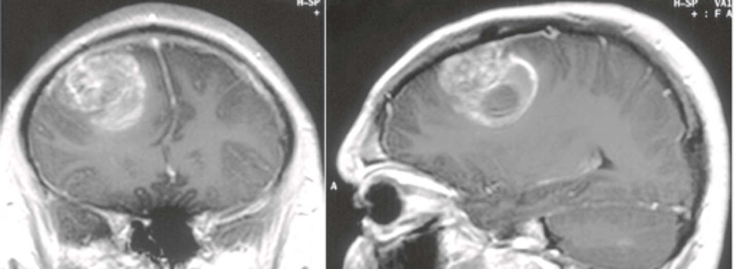
A _______________ skull fracture appears as a sharp lucent line that is often irregular or jagged and occasionally branches.
A. depressed
B. basilar
C. linear
D. open
C. linear
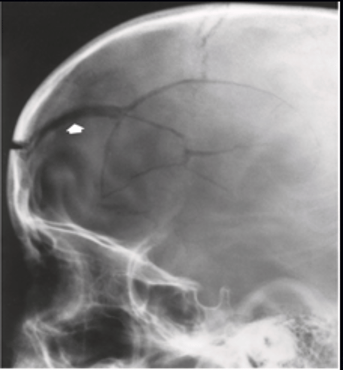
Which type of skull fracture appears stellate with radiating fracture lines, looks denser when viewed en face due to fragment overlap, and requires tangential views to evaluate the degree of depression?
A. Closed fracture
B. Basilar fracture
C. Linear fracture
D. Depressed fracture
D. Depressed fracture
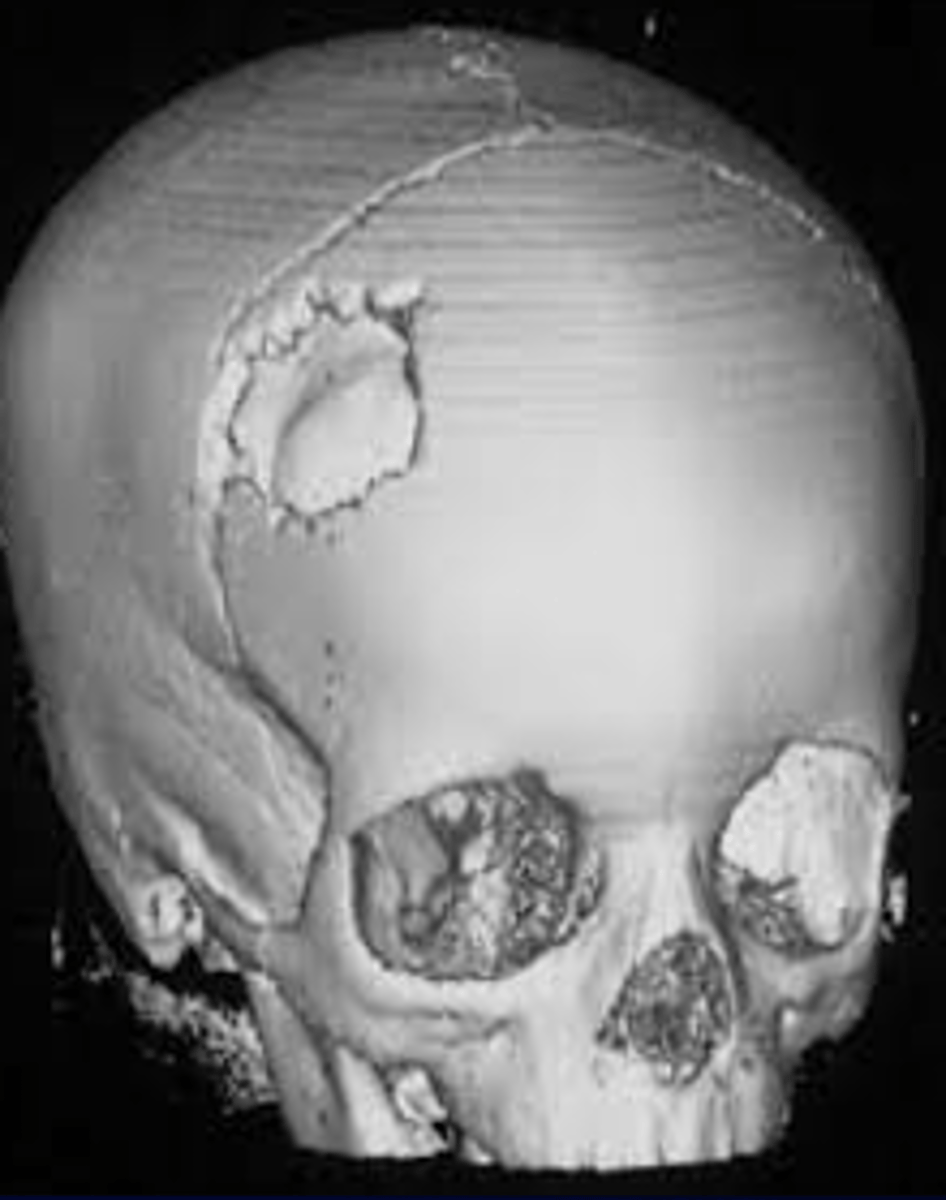
What condition involves a collection of blood between the dura mater and arachnoid mater that compresses brain tissue and can lead to severe damage or death if not treated?
A. Cranial hematoma
B. Subdural hematoma
C. Pia mater hematoma
D. Gliomas
B. Subdural hematoma
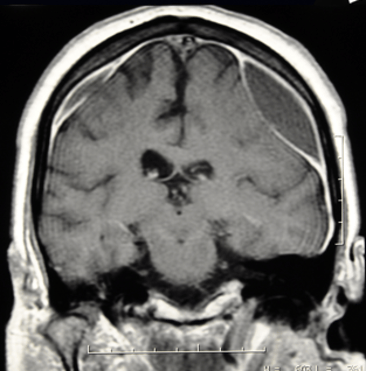
All of the following are causes of subdural hematoma except:
A. Head injury
B. Brain shrinkage
C. Blood thinning medication
D. Metastases of the blood
E. Hemophilia, cerebral aneurysm rupture, tumors, or cerebrospinal fluid leak.
D. Metastases of the blood
(Alcohol dependency is another cause)
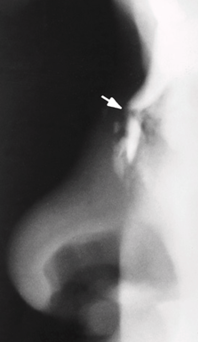
Which is the most common facial fracture, typically transverse with distal depression, and best demonstrated on right and left lateral soft-tissue nasal bone projections?
A. Nasal fracture
B. Zygomatic fracture
C. Blow-out fracture
D. Maxilary fracture
A. Nasal fracture
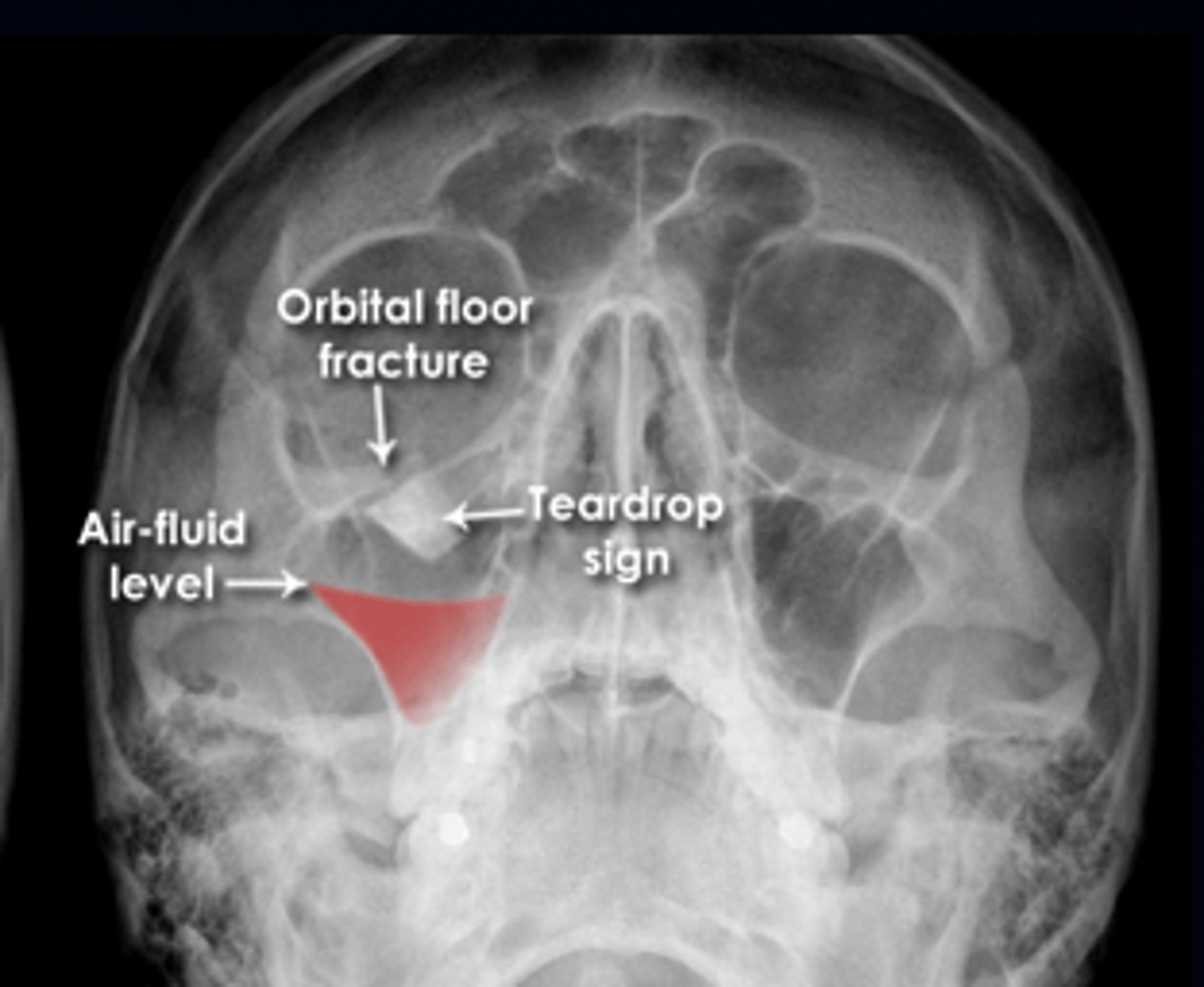
Which orbital fracture is caused by a direct blow that increases intraorbital pressure, breaks the thin orbital floor above the maxillary sinus while leaving the rim intact, and shows an orbital floor defect, air-fluid level in the maxillary sinus, and a teardrop sign on imaging?
A. Blow-out fracture
B. Tripod fracture
C. Le Fort fracture
D. Nasal Fracture
A. Blow-out fracture
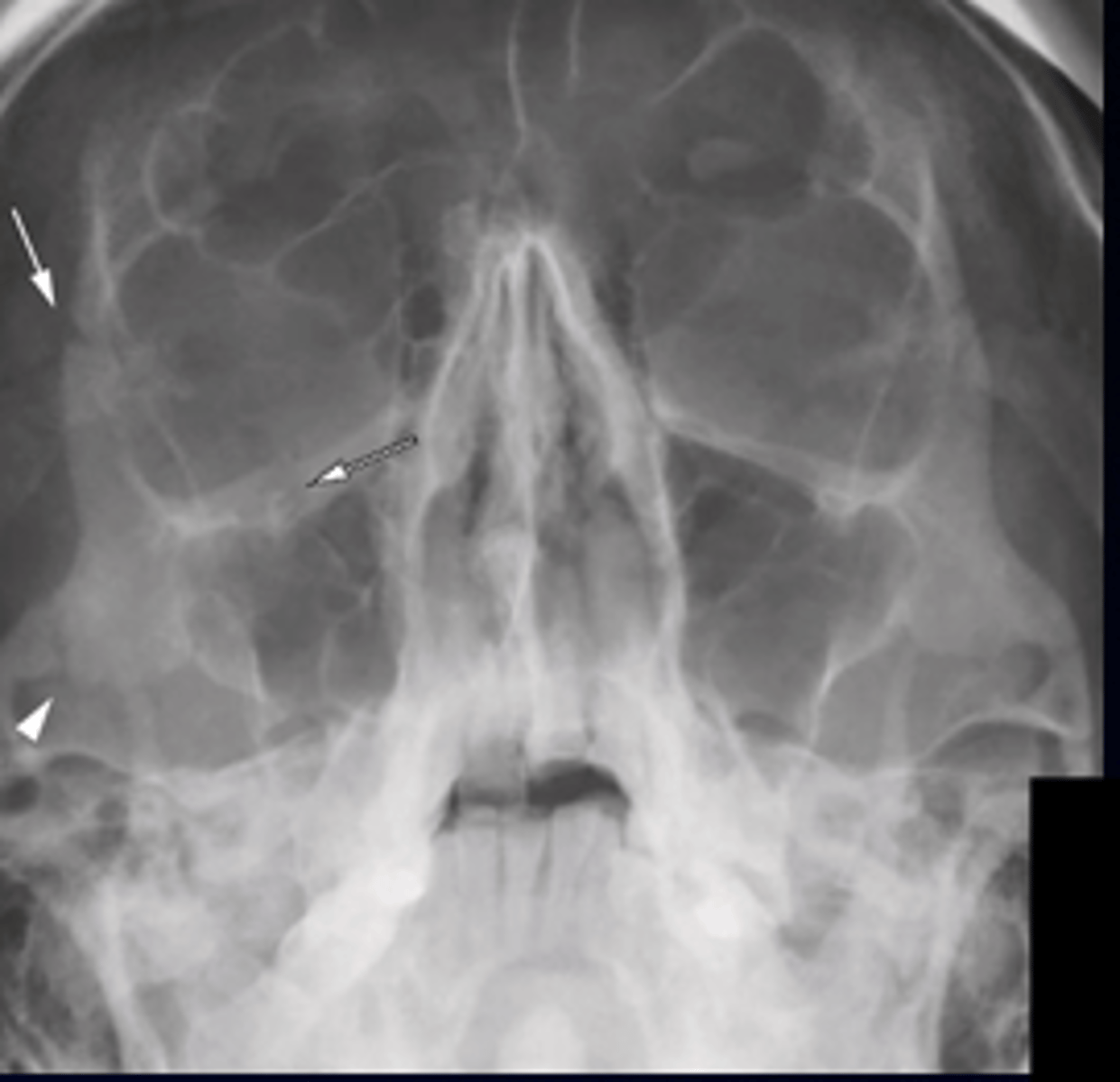
Which facial fracture involves separation of the zygoma from its three major attachments; fractures of the zygomatic arch, orbital floor or rim, and the zygomaticofrontal suture resulting in a free-floating cheekbone?
A. Nasal Fracture
B. Tripod fracture
C. Le Fort fracture
D. Blow-out fracture
B. Tripod fracture
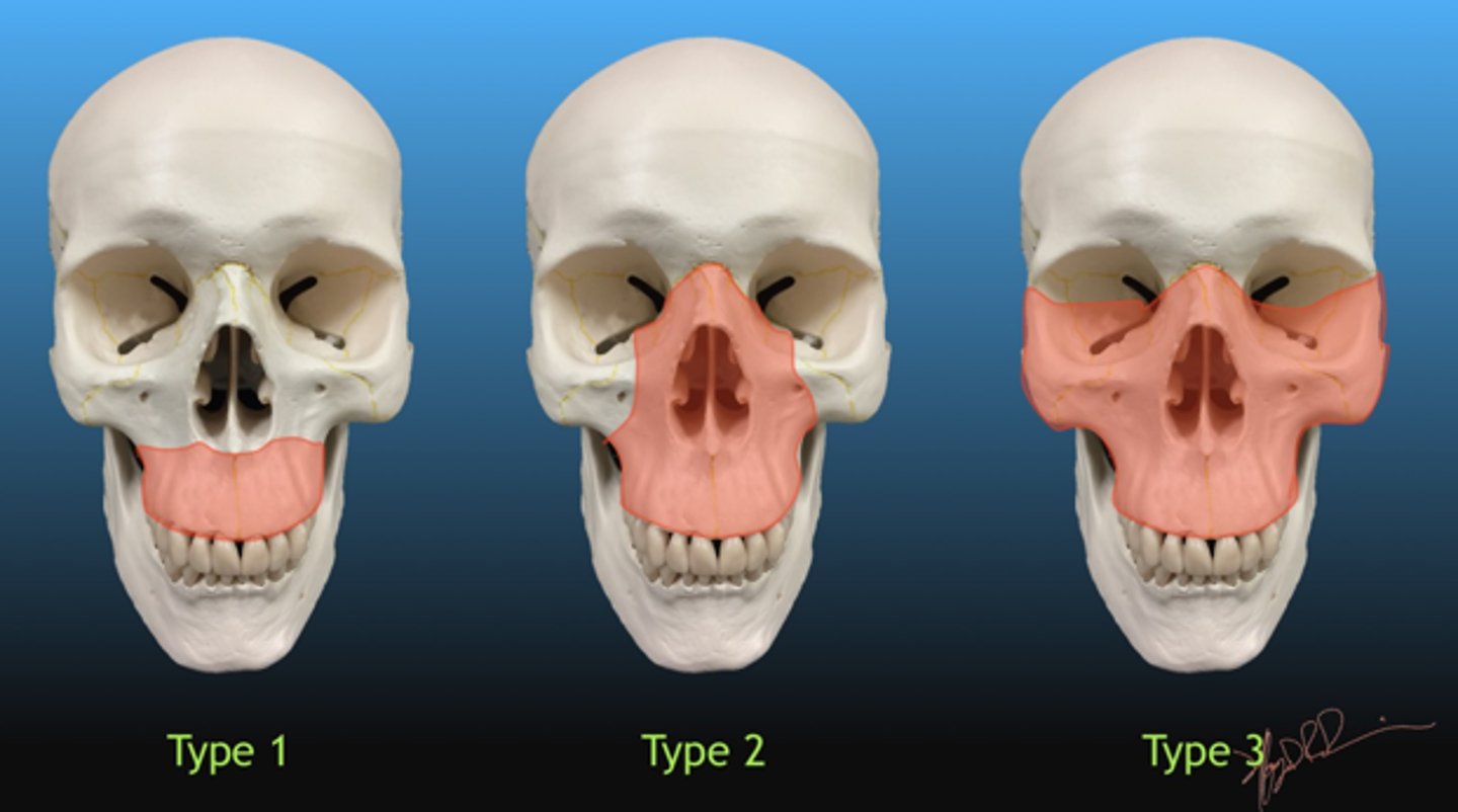
Which midface fracture involves separation of the facial bones from the skull base, is usually caused by blunt trauma, and is classified into three types (I, II, and III) based on the fracture pattern?
A. Nasal Fracture
B. Tripod fracture
C. Le Fort fracture
D. Blow-out fracture
C. Le Fort fracture
______________ refers to any process that is caused by an abnormality of blood vessels or blood supply to the brain.
A. Cerebrovascular disease
B. Stroke
C. Transient Ischemic attack
D. Intraparenchymal hemorage
A. Cerebrovascular disease
The imaging modality of choice to evaluate patients with suspected neurologic dysfunction caused by head trauma is _________________.
A. ultrasound
B. MRI
C. CT
D. skull radiographs
C. CT
Arterial bleeding sometimes associated with head trauma can cause _________________ hematomas.
A. intracranial
B. subdural
C. epidural
D. acute
C. epidural
Venous bleeding sometimes associated with head trauma can cause _________________ hematomas.
A. intracranial
B. subdural
C. epidural
D. acute
B. subdural
Movement of the brain within the calvaria following blunt trauma to the skull sometimes results in a cerebral _________________.
A. subdural hematoma
B. epidural hematoma
C. acute hematoma
D. contusion
D. contusion
Bleeding into the ventricular system caused by injury to surface veins, cerebral parenchyma, or cortical arteries can cause _________________ hemorrhage.
A. epidural
B. subdural
C. epiarachnoid
D. subarachnoid
D. subarachnoid