Chapter 4: Pulmonary, Bronchial, and Systemic Circulation and the blood
 Call Kai
 Learn
Learn Practice Test
 Spaced Repetition
 Match
Match Flashcards
 Knowt Play
1/69

There's no tags or description
Looks like no tags are added yet.
Name | Mastery | Learn | Test | Matching | Spaced | Call with Kai | Chat |
|---|
No analytics yet
Send a link to your students to track their progress
70 Terms
Pulmonary, Bronchial, and Systemic Circulation
• The movement of air into and out of the lungs provides oxygen to the lung tissue and a mechanism for the removal of waste gases such as carbon dioxide.
• The blood transports oxygen throughout the body and collects waste gases, returning them to the lungs for removal.
Pulmonary, Bronchial, and Systemic Circulation
What are the three circulatory systems that affect the lungs?
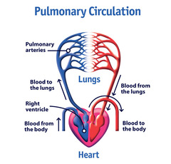
• Deoxygenated blood from the Right ventricle to Pulmonary Artery
• Branches into Capillaries that cover the alveoli and respiration occurs
• Oxygenated blood travels back to pulmonary vein and into the left atrium
Pulmonary Circulation
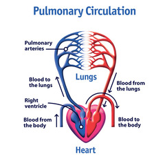
Begins in the right ventricle with deoxygenated blood >> pulmonary arteries >> pulmonary arterioles >> capillaries (respiration occurs here so the blood is now oxygenated) >> pulmonary venules >> pulmonary veins >> left atrium
Describe the pulmonary circulation (route of deoxygenated and oxygenated blood)
1. Supplies blood to the lung tissue itself
2. Originates from the aorta and carries oxygenated blood along the tracheobronchial tree to the terminal bronchioles. These arteries then merge with the pulmonary arteries and capillaries.
• After leaving the capillaries:
• 1/3 travels to right atrium via veins
• 2/3 drains into pulmonary veins
• Mixture of oxygenated and deoxygenated blood is called Venous Admixture
• Also supplies blood to esophagus, mediastinal lymph system, pulmonary nerves, and visceral pleura
Bronchial Circulation
1. Purpose?
2. Origin? Pathway?
venous admixture
describes the mixing of oxygenated blood with deoxygenated blood
Bronchial circulation
Which circulation does a venous admixture occur?
Systemic circulation carries the oxygenated blood throughout the body, collects the deoxygenated blood, and then returns the blood to the right atria of the heart where the process begins again
Describe the systemic circulation (function and pathway)
• Arteries• Arterioles• Capillaries• Venules• Veins
Pulmonary and Systemic vascular systems are both made up of which blood vessels?
Arteries
• Arteries divide into arterioles
Name the blood vessel type:
• Stiff vessels suited for transporting blood under high pressure
• Smooth muscle fibers integral to distribution and regulation of blood (called resistance vessels)
Arteries because of their ability to contract/relax in order to change the vessel diameter, and in turn change blood pressure and blood flow.
Which vessels are also known as resistance vessels? Why?
Capillaries
Name the blood vessel type:
• Where gas exchange occurs
• In Pulmonary System: external respiration; between blood and inspired air
• In Systemic System: internal respiration; between blood and tissues
External respiration: between the blood and inspired air
Internal respiration: between the blood and tissues
Where does external respiration occur vs. internal respiration?
capillaries
Which blood vessel type is responsible for gas exchange?
Veins because of their ability to collect large amounts of blood without changing the pressure too much (maintains).
Which vessels are also known as capacitance vessels? Why?
Veins
• Smallest Veins are called Venules - tiny veins continuous with pulmonary capillaries
Name the blood vessel type:
• Most carry deoxygenated blood back to the left atrium via pulmonary veins
• Less smooth muscle and elastic tissue than arteries
• Thinner walls, contain one-way valves
• Capable of collecting large amounts of blood with very little pressure change (called capacitance vessels)
• In pulmonary circulation, it is reversed.
Pulmonary arteries = deoxygenated
Pulmonary veins= oxygenated
Normally.....
• Arteries carry oxygenated blood away from the heart to the body
• Veins - carry deoxygenated blood to the heart.
In which circulation system is this untrue?
1. Echocardiography
2. Can be used to assess blood flow through the heart
1. What's the medical term for an ultrasound of the heart?
2. Why may this be used?
Intracardiac Echocardiography(ICE)
What is the medical term for when catheters are used to insert an ultrasound into the heart to view the internal structures?
Right Heart Catheterization
During this procedure, a specialized tube called a pulmonary artery (PA) catheter is inserted into a vein in the groin or in the neck and threaded through the right atrium and right ventricle of the heart into the pulmonary artery.
It allows practitioners to measure several parameters, including pressures and blood flow, that can be used for both diagnostic and treatment purpose.
1. The amount of blood the heart pumps through the circulatory system in 1 minute
2. Normal - 4-8 L/min
3. Can help determine the amount of oxygen being delivered to the cells of the body
1. What is cardiac output?
2. What is considered normal? (L/min)
3. Why would this be considered?
Venous Thromboembolism (VTE)
What's the medical term for a blood clot in the venous system?
Deep Vein Thrombosis (DVT)
What is the medical term for a blood clot in one or more of the deep veins of the body (usually in the legs)?
It's when a blood clot forms in the lungs or travels through the bloodstream to the lungs
*Life-threatening
What is a Pulmonary Embolism?
Pulmonary Hypertension (PH)
A group of progressive diseases characterized by pulmonary vascular resistance, remodeling, and occlusion of the pulmonary arterioles; right heart ventricular dysfunction; right heart failure; and often death
1. A PE can obstruct the blood supply to an area of the lungs (infarction), causing localized death of the tissue (necrosis).
• Because a PE can cause an infarction in the pulmonary parenchyma as well as prevent in adequate supply of oxygenated blood from flowing to the rest of the body, a PE is considered a life-threatening emergency.
Why are pulmonary embolisms so dangerous?
Infarction
Obstruction of the blood supply to an organ or body tissue can result in tissue damage and death due to a lack of oxygen. When this occurs, the obstruction is called a(n)....
necrosis
death of tissue is known as...
• Transport oxygen to tissue cells
• Transport carbon dioxide from tissue cells to lungs
• Transport nutrients and waste products
• Transport processed molecules throughout the body
• Transport regulatory hormones/enzymes
• Regulate pH and osmosis
• Maintain body temperature
• Protect against foreign substances
• Clot formation
Functions of the blood:
Makes up approximately 7% of total body weight
What percentage of body weight is blood?
Plasma -Liquid portion of whole blood (55%)
• Pale yellow
• 91% water
• 7% proteins
Formed Elements (45%)
• Erythrocytes (red blood cells/RBCs)
• Leukocytes (white blood cells/WBCs)
• Thrombocytes (platelets)
Plasma Proteins (7%)
• Clotting Factors
• Fluid balance
• Fighting Infection
Composition of blood
Plasma (55%)
Formed Elements (45%)
What are the 2 main components of blood? What percentage of blood is plasma vs formed elements?
Water (91%)
Proteins (7%)
Electrolytes - Na, Cl, K
Nutrients- amino acids, glucose, lipids, vitamins
Respiratory gases- O2, CO2, N2
Hormones
Waste Products- Urea (*BUN), uric acid, creatinine, bilirubin
What is plasma made up of?
1. Biconcave discs without a nucleus
2. Transport oxygen and carbon dioxide to and from body tissues.
3. Produced in the red bone marrow by hematopoietic stem cells
4. 120 day lifespan
5. Hemoglobin gives RBCs their red color
Erythrocytes (RBCs)
1. Structure?
2. Function?
3. Where are they produced? Origin?
4. What is their average lifespan?
5. What protein gives them their red color?
Increases surface area for gas exchange
What is the purpose of the biconcave shape for RBCs?
1. Hemoglobin is a protein component of RBCs; give RBCs their red color.
2. Hemoglobin is made up of four globulin chains (2 alpha-globulin chains and 2 beta-globulin chains), each of which contains an iron atom bound to a heme group.
3. Enables RBCs to carry oxygen from the lungs to the rest of the body and then return carbon dioxide to the lungs
Hemoglobin
1. What is hemoglobin?
2. Structure?
3. Function?
Males: 14-18 g/dL
Females: 12-16 g/dL
What are normal hemoglobin levels? (men and women)
Fetal hemoglobin is different structurally. Fetal hemoglobin (HbF) molecules are composed of two alpha chains and two gamma chains; they have a great affinity for oxygen.
-At birth and as the infant grows, the gamma chains are gradually replaced by beta chains, forming the adult hemoglobin structure of two alpha and two beta chains (about 1 year)
What is the difference between adult hemoglobin (HbA) and fetal hemoglobin (HbF)?
Mean corpuscular volume (MCV) - measures the size of the RBC
• Smaller than normal =anemia
• Larger than normal =Vitamin B12 deficiency, folic acid deficiency, liver disease, or hypothyroidism.
What is MCV? What does is measure?
Mean corpuscular hemoglobin (MCH) - amount of Hb
What is MCH? What does it measure?
1. Hematocrit (HCT) - # of RBCs compared to the total blood volume
2. Males: 40-54%
Females: 36-48%
1. What is hematocrit?
2. What is normal? (men and women)
• To protect the body against invading microorganisms (viruses, bacteria, parasites, and tumors)
• To remove dead cells and debris
What are the functions of WBCs?
Normal average number of WBCs: 5,000 to 9,000 cells/mm3
What is a normal WBC count?
abnormally high WBC count
What is leukocytosis?
abnormally low WBC count
What is Leukocytopenia?
The passage of blood cells through capillary walls into the tissues
What is diapedesis?
categorized according to cytoplasmic staining properties (granules)
How are leukocytes categorized?
1. Granulocytes: Neutrophils, Eosinophils, and Basophils
2. Agranulocytes: Lymphocytes and Monocytes
1. Which WBCs are granulocytes?
2. Which WBCs are agranulocytes?
Neutrophils (65% of WBCs)
Which WBC is the most common?
Fights infection by phagocytizing bacteria/pathogens
What is the function of Neutrophils?
Neutrophils (granulocyte)
Name the WBC described:
- Most numerous of all WBCs (65%)
-Large (2x the size of RBCs)
-Found at inflammation sites; kill bacteria through phagocytosis
-Numbers increase during bacterial infections
-Mobile and active phagocytic cells; contain lysosomes
Destroy protozoa and parasitic worms and release anti-inflammatory substances in antibody antigen reactions
What is the function of eosinophils?
Eosinophils (granulocyte)
Name the WBC described:
-2 to 5% of all circulating WBCs
-Numerous in tissues lining respiratory and digestive tracts
-Destroy protozoa and parasitic worms and release anti-inflammatory substances in antibody antigen reactions
-Elevated levels are commonly seen in asthma patients
1. Basophils
2. Histamine (inflammatory substance) causes vasodilation and attracts other WBCs to the inflamed site
3. Heparin is an anticoagulant
1. Which WBC releases histamine and heparin?
2. What does histamine do?
2. What does heparin do?
Release histamine and heparin to aid in inflammation and allergies
What is the function of basophils?
-T lymphocytes (T cells): act against tumors and virus-infected cells
-B lymphocytes (B cells): produce antibodies
What are the 2 types of lymphocytes and their functions?
1. Monocytes differentiate into Macrophages that are ready to phagocytize bacteria, dead cells, cell fragments and viral-infected cells
2. Increases seen in chronic infections (TB)
1. What is the function of monocytes/macrophages?
2. Clinical significance?
150,000 - 450,000 per mm3 of blood
What is the normal platelet range?
Minimizes bleeding by clumping together to form a platelet plug or blood clot
What is the function of platelets?
thrombocytopenia
medical term for low platelet count
1. CBC (Complete Blood Count) Test - counts and differentiates the number of RBCs, WBCs, and platelets in a blood sample.
2. Includes: WBC count and differential analysis, RBC count, Hemoglobin, Hematocrit, MCV, MCH, MCHC, RDW, platelet count, and mean platelet volume.
3. Important for understanding the comprehensive medical background of a patient; therefore, RTs often review a patient's CBC.
1. What is a CBC?
2. What values are included?
3. Why is this test important in respiratory care?
1. Lower than normal number of RBCs and/or low Hgb
2. Limits the body's ability to transport oxygen to the tissues
3. Symptoms include: weakness, shortness of breath, dizziness, headaches, irregular heartbeat
1. What is anemia?
2. What does this effect?
3. Symptoms?
• Nutritional Deficiency
• Hemorrhagic
• Folate deficiency- folic acid deficiency
• Pernicious- vitamin B12 deficiency
• Hemolytic- low # of RBCs
• Aplastic- bone marrow not producing enough blood cells
• Sickle-Cell- inherited disorder changing the structure of RBCs
What are the types of anemia?
Sickle Cell Anemia
• Inherited blood disorder
• Mostly seen in African American or Caribbean ancestry
• RBCs become stiff and sticky; curved, or sickle-shaped
• Results in plugging of small vessels
• Severe cases usually fatal before 30 yrs old
What is sickle-cell disease?
abnormally high numbers of RBCs
What is polycythemia?
lower than normal level of neutrophils
What is neutropenia?
Cancer of the blood that develops in the bone marrow, and produces abnormal leukocytes
What is leukemia? Origin?
Blood cancer that originates in the lymphatic system
What is lymphoma? Origin?
Thrombocytopenia
What term means below average platelet count?
Clinical-Style Application Questions
• Why would a patient with a PE have sudden shortness of breath and low oxygen saturation despite supplemental oxygen?
• If a patient has a low Hgb/Hct, what symptoms would you expect, and why?
• Why might an asthma patient show elevated eosinophil levels?
• How would sickle-shaped RBCs cause tissue hypoxia?
• If a trauma patient develops tachycardia, chest pain, and dyspnea, how could a pulmonary embolism be suspected?
• How would polycythemia increase the risk of clotting?
• Why would a CBC be one of the first tests ordered in a patient with suspected infection or anemia?