RAD 118 CH.2 General Anatomy
 Call Kai
 Learn
Learn Practice Test
 Spaced Repetition
 Match
Match Flashcards
 Knowt Play
1/51

Earn XP
Description and Tags
positioning
Name | Mastery | Learn | Test | Matching | Spaced | Call with Kai | Chat |
|---|
No analytics yet
Send a link to your students to track their progress
52 Terms
anatomy
structure of the body
physiology
function of body organs
osteology
study relating to the bones of the body
sagittal plane
divides body into right and left segments
midsagittal plane MSP
passes through midline and divides body into = right and left halves
Coronal plane
-Passes through body from side to side
-Divides body into anterior and posterior
midcoronal plane MCP
divides body into equal anterior/posterior halves
Horizontal plane (transverse) (axial) (cross sectional)
-Pass crosswise through body
-Right angle to sagittal and coronal planes
-Divides body into superior and inferior portions
Thoracic cavity
Subdivided into pericardial segment & two pleural portions
abdominal cavity
-Lower portion called the pelvic cavity
-Sometimes referred to as the abdominopelvic cavity
thoracic cavity contains..
-Pleural membranes
-Lungs
-Trachea
-Esophagus
-Pericardium
-Heart and great vessels
abdominal cavity contains..
-Stomach
-Liver
-Gallbladder
-Pancreas
-Spleen
-Intestines
-Kidneys
pelvic cavity contains..
-Rectum
-Urinary bladder
-Part of the reproductive system
right lower quadrant RUQ
liver, kidney, pancreas, gallbladder, colon
left upper quadrant LUQ
stomach, spleen, pancreas, kidney, colon,
right lower quadrant RLQ
colon, small intestine, ureter, appendix
left lower quadrant LLQ
colon, small intestine, ureter
sthenic
standard average body
hyposthenic
slightly thin/ tall body, organs lower/morevertical
asthenic
very thin/ tall, organs are lowest /most vertical
hypersthenic
short,broad, stocky organs higher/more horizontal
206
bones in the body
axial skeleton
80 bones
supports/protects head & trunk
appendicular skeleton
126 bones
provides means for movement
bone functions
Muscle attachment
Mechanical movement
Protection of internal organs
Support frame for body
Storage for calcium, phosphorus, and other salts
Production of red and white blood cells
long bones
Body and two enlarged articular ends
Examples: femur and humerus
short bones
Example: carpal bones (wrist) and tarsal bones (ankle)
flat bones
-bones consist of two plates of compact bones
-Examples: sternum and cranium
irregular bones
Examples: vertebrae and facial bones
sesamoid bones
Very small and oval
Develop inside and beside tendons
Protect the tendon from excessive wear
Largest is patella
arthrology
-Study of joints/articulations between bones
-Joints make it possible for bones to:
Support the body
Protect internal organs
Create movement
functional types of joints
-Synarthroses: Immovable
-Amphiarthroses: Slightly movable
-Diarthroses: Freely movable
fibrous
-structural
-strongest joints, immoveable
-ex: inferior tib/fib, sutures of skull, roots of teeth
cartilaginous
-structural
-no joint cavity, virtually immoveable
-ex: symphysis pubis (sm) & epiphyseal plates (nm)
synovial
-structural
-freely moveable with wide range of motion
-Most complex joints in the body
gliding, hinge, pivot, ellipsoid, saddle, ball & socket
open fracture
broken bone projected through skin
closed fracture
does not break through the skin
nondisplaced fracture
bone retains its normal alignment
displaced fracture
bones are not in anatomic alignment

Compression
a partial collapse or crack in a vertebra (the bone in the spine) caused by excessive force or weakened bone density.

simple “closed”
a broken bone that does not pierce or break through the skin
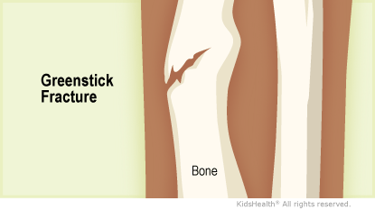
greenstick
an incomplete break where a bone bends and cracks on one side instead of snapping completely into separate pieces

transverse
break goes straight across the bone at a right angle

spiral/oblique
a complete break in a bone where the fracture line travels at an angle and twists around the bone shaft
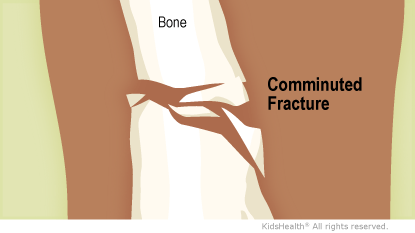
comminuted
a severe type of broken bone where the bone shatters into three or more pieces

impacted
when the broken ends of a bone are driven together by forceful impact.
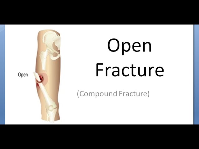
open compound
a severe broken bone injury where the sharp ends of the bone pierce through the skin and expose deep tissue to the outside environment
superficial
near the skin
projection
-Path of the CR as it exits the x-ray tube, passing through the patient to the IR
-Identified by entrance and exit points of the body
position
-Overall posture of the patient
-General body position
-Specific placement of the body or part in relation to table or IR
lordotic position
Upright position in which the patient is leaning backward
circumduction
circular movement of limb