Vascular 101 Ms Mac Exam 2 review
 Call Kai
 Learn
Learn Practice Test
 Spaced Repetition
 Match
Match Flashcards
 Knowt Play
1/94

There's no tags or description
Looks like no tags are added yet.
Name | Mastery | Learn | Test | Matching | Spaced | Call with Kai | Chat |
|---|
No analytics yet
Send a link to your students to track their progress
95 Terms
What are the 8 branches of the ECA?
1) superior thyroid
2) ascending pharyngeal artery
3) lingual artery
4) facial artery
5) Occipital artery
6) posterior auricular artery
7) maxillary artery
8) superficial temporal artery
Doppler
Evaluation of the presence, velocity (speed), direction and character of blood flow within the vessels
For the CCA and vertebral artery, box is steered to the
right
Beam steering
-steer the color doppler beam in the direction of the vessel
What happens if box is not properly steered?
Color will not fill the lumen of the vessel properly and it could be mistaken for plaque or an occlusion
Keep the box small if
A higher color frame rate is desired
Pulsed Repetition Frequency color scale (PRF)
- controls PRF scale of the color velocity limits
High velocity flow needs
higher PRF/Scale
*and other way around *
If PRF is set too high
Color will not fill the vessel
If PRF is set too low...
color will wrap around vessel (Alias)
What color should be on top of the color bar when the box is steered to the right?
red
What color should be on top of the color bar when the box is steered to the left?
blue
Sample volume gate (SVG)
Represents the area of flow assessed with Doppler
*placed mid stream
* set between 1.5 and 2mm
Steer Doppler angle cursor
controls the angle of the sample volume
what degree is the steer Doppler angle supposed to be set at?
45-60°
When dopplering in our lab class at what angle is the steer Doppler supposed to be at?
60°
Steer beam
Steer will control the direction of your Doppler cursor, Doppler beam line and color box
The steer beam should always be in the
Same direction as your color box and vessel
Doppler effect
Observed frequency change of reflected sound due to reflector movement relative to the sound source of the observer
Doppler shift
mode used to detect blood flow.
Do RBCs emit sound?
No, its considered a moving reflector
We rely on our transducers to
Send sound and recieve thd echo from moving reflectors
If the returning frequency is higher, the Doppler shift is positive. This is known as.
Antegrade flow
If the returning frequency is lower than the transmitted frequency, the Doppler shift is considered a negative shift. This is known as
Retrograde flow
On the Doppler display, the machine plots
Velocity (Y axis shown in CM) over time (X axis)
Puse Repetition Frequency (PRF)
Number of pulse echo cycles occurring in one second
(Hz)
Aliasing/Nyquist Limit
the wrapping around of the spectral waveform that can result from high velocity of blood flow-nyquist limit is exceeded
*try adjusting the baseline and then increase PRF scale to correct*
When does aliasing occur?
When the Doppler shift frequencies exceed 1/2 of the PRF (Nyquist limit)
Pulsed wave Doppler
uses a single element transducer that emits brief pulses of ultrasound energy
PW advantage
Ability to measure velocities at a specific location (range resolution)
PW disadvantage
Aliasing of velocities: inability to measure high velocities accurately.
CW Doppler
allows us to measure blood velocities along an entire line of interrogation
CW advantage
measures high velocities', no aliasing
CW disadvantage
not site specific, lacks range resolution (range ambiguity)
sweep speed
This switch changes the speed of the spectral display
dicrotic notch
represents the closure of the aortic valve and the onset of diastole
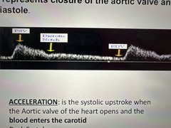
Acceleration
The systolic upstroke when the AOV opens and blood enters the carotid
CCA waveform characteristics
-Sharp upstroke and Peak
-Well defined dicrotic notch
-Diastolic flow
-Low Resistance
*color flow will reveal a lighter shade of red during systole and a darker shade of red during diastole*
Which vessels have a low resistance waveforms?
CCA, ICA, Vertebral, renal, splenic and hepatic arteries

High resistance waveform
This waveform is characterized by a sharp rise in flow velocity during systole, a rapid decline toward the baseline, and diminished diastolic flow
Which arteries have high resistance waveforms?
ECA, aorta, subclavian, extremity arteries

What's the normal peak systolic velocity of the CCA?
Less than 125cm/sec (
What's the normal vertebral artery peak systolic velocity?
20-60cm/sec in most pts
The vertebral artery
- Supplies blood to the posterior brain
- flow is antegrade
- low resistance vessel
What is the normal peak systolic velocity for ICA?
Less than 189cm/sec
What's the normal PSV in ECAs?
Less than 200c
Which vessels have a dicrotic notch?
ECA, CCA
What are some characteristics of the ECA?
- A lighter shade of red will be seen during systole. The normal color flow pattern will decrease during diastole and may disappear at the end diastole (no flow - flashes)
- more pulsatile Doppler signal
- flow pattern is a characteristic of flow patterns seen in high resistance distal vascular beds such as the face and scalp
ECA spectral waveform will oscillate with...
temporal tap
How to obtain the ECA and ICA?
Transducer is moved slightly anteriorly and posteriorly
Arteries and veins are both made up of what three layers?
Tunica intima: innermost layer
Tunica media: middle layer
Tunica adventita: Outer layer
The íntima of an artery is in contact with
Moving blood flow
The media of an artery is
The muscular layer that consists mostly of smooth muscle cells and allows rhythmic changes in the arterial size during cardiac cycle
Where is elastin and collagen found?
Media of an artery
What layer of the artery contains vasa vasorum?
adventitial layer
Large arteries
Elastic arteries with less smooth muscle, aorta and its largest branches
Small arteries
Approx 4mm in diameter, well developed smooth muscle layers. Include all arteries EXCEPT aorta and major branches
Arterioles
- smallest arteries
- Known as "stopcocks" of the vascular system
- circular smooth muscle layers control contraction and resistance
Capillaries
- Smallest blood vessels
- Place where nutrient exchange occurs
Venules
smallest veins
Small veins
1-10mm in diameter
- all veins EXCEPT portal vein, vena cava and their main branches
- walls are mainly connective tissue
large veins
- portal vein, vena cava and its branches
- thick adventitial layer w fibrous and elastic tissues
Veins
- thinner walls
- same laminate structures as artery but not as muscular
- have more elastic fibers and collagen than muscle fibers
Which vessels have valves?
veins
What's the purpose of venous valves?
Prevents retrograde movement of blood
systemic circulation pathway
Blood leaves the LV:
Aorta, arteries, arterioles, capillaries, venules, veins, and vena cava to enter the RA of the heart
Hemodynamics
the study of blood moving through the circulatory system
Laminar flow
normal blood flow
- consists of many laminae (layers)
- blood layers slide concentric
Disturbed flow
occurs when the parallel stream lines are disturbed (at the bifurcation)
Flow Separation
occurs when there is a sudden widening of a tube or vessel which causes the flow to reverse along the walls (at the bulb)
- known as the boundary layer

Turbulent flow
an irregular, mixing flow patter usually due to pathology in the artery
-occurs distal to a stenosis in a vessel
Plug flow
occurs when all of the layers and blood cells travel at the same velocity
- known as blunt flow
- occurs at branching origins and proximal to stenosis

Turbulent flow is characterized by
- chaotic flow patterns in many different directions and at many speeds
- often associated with pathologies and increased blood velocity, and beyond a stenosis
- spectral broadening on wave form
- May have a feathered appearance on wave form
Eddy currents or random vortices
appear as small hurricane like swirling rotational patterns

Proximal to a stenosis
velocity and pressure are baseline

Within the stenosis
velocity is high and pressure is low

Immediately distal to the stenosis
velocity will begin to decrease again and pressure will rise higher than in the stenosis
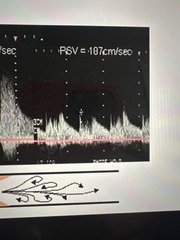
Tardus Parvus
a waveform of low amplitude and delayed rise to systole

When does turbulent flow occur?
When Reynold's number is greater than 2000
An increase in flow speed
Increases Reynolds number
Reynolds number (Re)
RE= (average flow x density x tube diameter) / viscosity
Bernoulli's Principle
describes the relationship between area, velocity, and pressure at a stenosis
When there is high velocity
There is low pressure
When there is low velocity
There is high pressure
Examples of Bernoulli's Principle
Areas of flow separation and stenotic (narrowed) vessels
Velocity and pressure share an
Inverse relationships
Pressure gradient
Pressure difference between two points
*there must be a pressure gradient present for flow to move*
What elements will flow from a higher pressure to a lower pressure?
Blood, air, and gas
Blood will move from an area of ___________ pressure to an area of ___________ pressure
higher, lower
Poiseuille's Law
describes the relationship between resistance, pressure, and volume flow
*gives insight into the complex task of regulation blood flow to different parts of the body*
Vasodilation by 19% will
double the blood flow
Vasoconstriction by 16% will
cut blood flow in half
The process of vasodilation and vasoconstriction offer
Powerful control mechanisms
Resistance to flow
-Due to changes in size of vessels (length & diameter).
-Influenced by length of tube, viscosity and radius (primary influence).
Collateral blood flow
heart makes new branches of blood vessels as we age so that damaged areas of the heart get good blood flow