15. lung cancer
 Call Kai
 Learn
Learn Practice Test
 Spaced Repetition
 Match
Match Flashcards
 Knowt Play
1/35

Earn XP
Description and Tags
review obstruction/restrictive lung patterns **
Name | Mastery | Learn | Test | Matching | Spaced | Call with Kai | Chat |
|---|
No analytics yet
Send a link to your students to track their progress
36 Terms

upper, middle, lower
lobes of the right lung
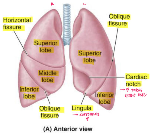
(2 lobes): upper, lower + lingula
lobes of the left lung
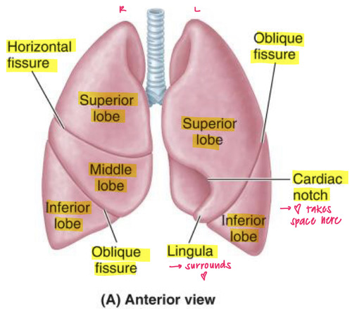
horizontal and oblique
fissures in the right lung
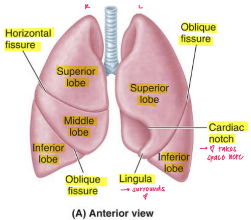
oblique only
fissures in the left lung

Right mainstem
= wider, shorter, more vertical
{RIGHT/LEFT} mainstem bronchus has a higher aspiration risk?
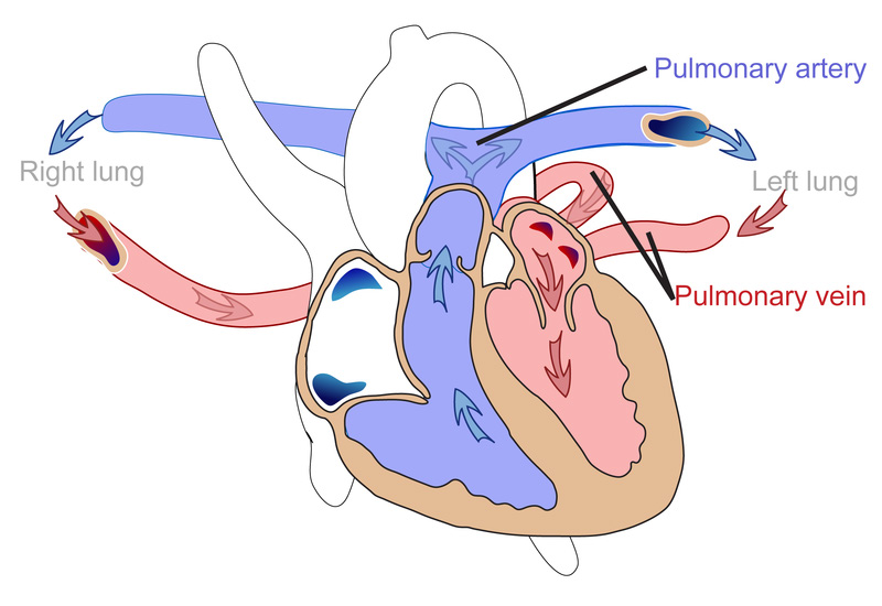
Pulmonary artery
Right: Anterior
Left: Superior
what is the RALS mnemonic?

type I pneumocytes
squamous, lines the aveoli
used for GAS EXCHANGE
pores of kohn = small openings between alveoli that allow air and macrophage movement. (& bac in pneumo)
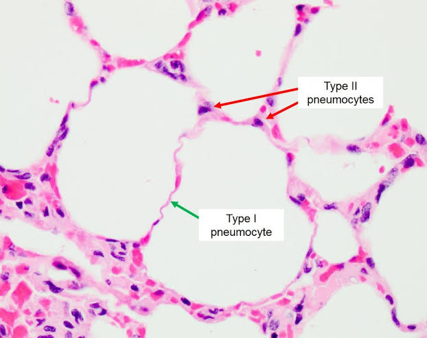
type II pneumocytes
cuboidal and clustered on aveoli
2 functions:
SURFACTANT (DDPC)
STEM CELL PRECURSOR (makes pneumocytes/repair)
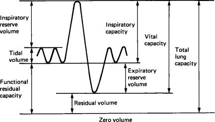
LUNG VOLUMES
know this
Ventilation → PaCO₂ / ETCO₂
Oxygenation → PaO₂ / SpO₂
Airflow/volumes → PFTs
Gas diffusion → DLCO
Test | What it evaluates | High-yield interpretation |
|---|---|---|
Spirometry ⭐ | Airflow | Measures FEV₁, FVC, FEV₁/FVC |
FEV₁ | Volume exhaled in first second | ↓ especially in obstructive disease |
FVC | Total amount forcibly exhaled | ↓ especially in restrictive disease |
FEV₁/FVC | Obstruction | ↓ = obstructive ⭐ |
TLC | Total lung capacity | ↓ = restrictive ⭐; ↑ may occur with hyperinflation |
RV | Air remaining after maximal expiration | ↑ = air trapping |
DLCO ⭐ | Alveolar-capillary gas diffusion | ↓ in emphysema, ILD, pulmonary vascular disease |
Pulse oximetry | Oxygenation | Measures SpO₂ |
ABG ⭐ | Oxygenation + ventilation + acid-base | PaO₂ = oxygenation; PaCO₂ = ventilation |
Capnography | Ventilation | Measures ETCO₂; especially important during anesthesia |
Exercise testing / 6-min walk | Functional cardiopulmonary reserve | Assesses exertional limitation/desaturation |
Obstructive | Restrictive | |
|---|---|---|
FEV₁ | ↓↓↓ | ↓ |
FVC | Normal/↓ | ↓↓ |
FEV₁/FVC | ↓ ⭐ | Normal/↑ |
TLC | Normal/↑ | ↓ ⭐ |
RV | ↑ | ↓/normal |
physiologic evaluation of the lung.
“Can the lungs move air, exchange gas, and oxygenate the patient?”
USPSTF: ALL 3 required ⭐⭐⭐
Criteria | Requirement |
|---|---|
Age | 50–80 years |
Smoking history | ≥20 pack-years |
Smoking status | Currently smokes OR quit within past 15 years |
criteria for annual low dose chest CT screening
Quit smoking ≥15 years ago,
OR
Health status substantially limits life expectancy or ability/willingness to undergo curative lung cancer treatment.
when to stop screening with low dose CT?
lung cancer
number one cause of cancer death worldwide
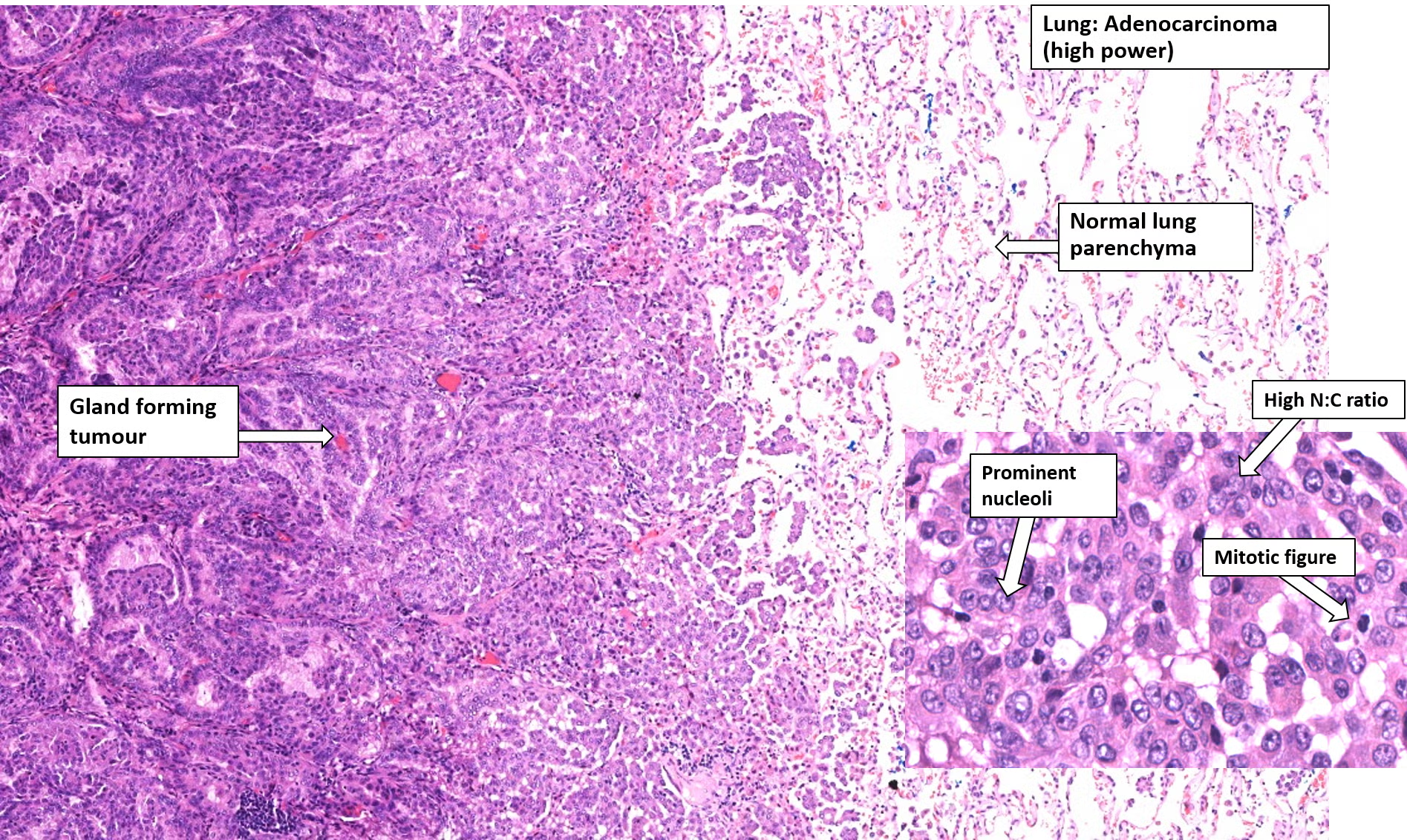
Adenocarcinoma
(non-small cell)
most common primary lung tumor: females / nonsmokers
associated with digital clubbing & loc’d peripheral
histo: glandular pattern & +mucin
KRAS / EGFR / ALK
Adenocarcinoma (non-small cell) — most common markers
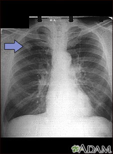
peripheral
(adeno = away)
loc of Adenocarcinoma (non-small cell)

Bronchioloalveolar subtype (Adenocarcinoma in situ)
CXR looks hazy similar to pneumonia;
aveolar “thickening” , tall columnar walls +mucus
*does NOT invade aveoli (more like a hug)
better prognosis 🙂

squamous cells carcinoma
Hilar mass arising from bronchus (centrally loc’d)
cavitation, cigs, hyperCa (due to PTHrP)
+keratin pearls, desmosomes (intracellular bridges)

Lab | Finding |
|---|---|
Ca²⁺ | ↑ |
PTHrP | ↑ |
PTH | ↓ |
Phosphate | ↓ |
seen in what cancer?
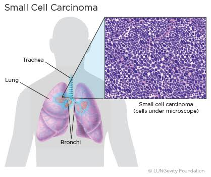
small cell (oat cell) carcinoma
central lung cancer
may cause neuro-paraneoplastic/endocrine syndromes: LEMS + SIADH + ectopic ACTH + Anti-Hu neurologic syndromes
MYC — very aggressive
small cell (oat cell) carcinoma is associated with what oncogene?
small cell (oat cell) carcinoma
kulchitchy’s cells —> small dark blue cells
+chromogranin A
neuro-specific: +enolase , +synpatophysin
History: Smoking history ⭐⭐⭐, age >50, secondhand smoke, radon, asbestos, occupational exposures, family history
PE: Persistent/new or changing cough ⭐, dyspnea, hemoptysis, chest pain, wheezing +/- constitunial symptoms
Older smoker + new/change in chronic cough ± hemoptysis + weight loss → suspect lung cancer.
Apical mass + shoulder pain + Horner syndrome → Pancoast tumor
Central lung mass + facial swelling/distended neck veins → SVC syndrome
Recurrent pneumonia in the SAME lobe → think obstructing lung tumor
P&E of lung cancer
sus CXR
—> CT chest
—> biopsy
Central mass → Bronchoscopy/EBUS
Peripheral mass → CT-guided transthoracic biopsy
Enlarged mediastinal nodes → EBUS-guided biopsy
determine hitso-path
—> PET/CT ± brain MRI for staging
→ molecular testing when indicated
→ treatment
diagnostic work up lung cancer?
PNEUMOTHORAX
Patient develops acute dyspnea + pleuritic chest pain + decreased breath sounds shortly after CT-guided lung biopsy → iatrogenic pneumothorax
Most common complication of transthoracic lung biopsy →
uncorrectable coagulopathy.
Major contraindication for transthoracic lung biopsy →
Pneumothorax — Upright CXR
Air in pleural space → partial/complete lung collapse
Visible visceral pleural line ⭐
• No lung markings peripheral to pleural line
• Hyperlucent affected side
• Collapsed lung toward hilum

Small + stable → observe ± O₂
Large/symptomatic → needle/catheter aspiration or chest tube
how to tx pneumothorax?
Pneumothorax — Supine CXR
Air travels anteriorly/basally when patient is supine
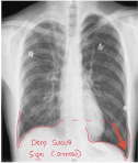
Deep sulcus sign ⭐⭐⭐
• Abnormally deep, hyperlucent costophrenic angle
• May NOT see classic apical pleural line
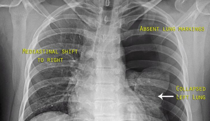
Tension Pneumothorax 🚨
Pneumothorax + mediastinal/tracheal shift AWAY from affected side
• Depressed ipsilateral diaphragm
• Widened intercostal spaces
obstructive shock
↑ intrathoracic pressure → compresses vena cava (+JVP)→ ↓ venous return → ↓ preload → ↓ CO → obstructive shock (hypotensive,tachycardia) → cardiac arrest
what type of shock is associated with tension pneumothorax
TENSION PNEUMOTHORAX
Clinical dx— DO NOT wait for CXR
⭐ Immediate decompression → chest tube
pt presents with:
Hypotension + tachycardia + severe respiratory distress + unilateral absent breath sounds + tracheal deviation AWAY
positive-pressure ventilation
Why?
PPV → pushes air into lungs → air can enter pleural space → ↑ pleural pressure → tension pneumothorax → ↓ venous return → obstructive shock.
⭐ Board pearl: Pneumothorax + mechanical ventilation = higher risk of tension pneumothorax.
If the patient is crashing and needs ventilation, don't withhold life-saving ventilation—simultaneously recognize and decompress a suspected tension pneumothorax.
Simple traumatic PTX can progress to tension pneumothorax by?
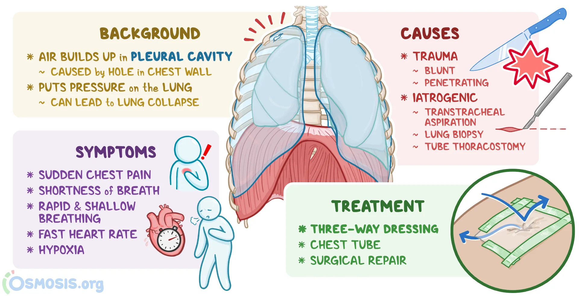
Open Pneumothorax
-Chest-wall defect communicating with pleural space
tx: Vented occlusive dressing + chest tube away from wound → definitive wound closure
“sucking chest wound”
TOWARD = volume LOSS → Atelectasis / pneumonectomy
AWAY = volume GAIN → Pleural effusion / large mass
NO SHIFT = volume preserved → Pneumonia
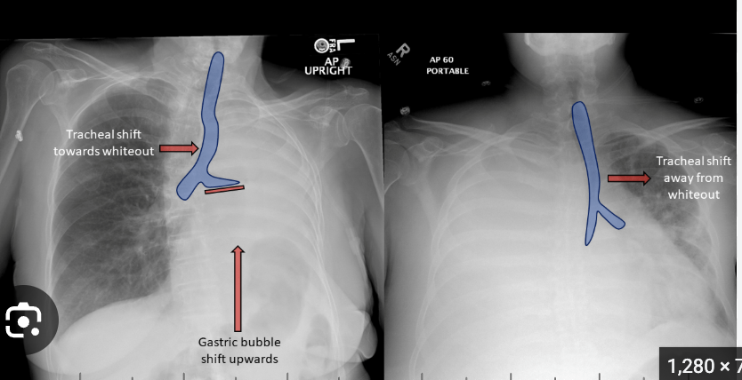
White hemithorax → look at the mediastinum
TOWARD = volume ____
AWAY = volume ____
NO SHIFT = volume ___