Joints
 Call Kai
 Learn
Learn Practice Test
 Spaced Repetition
 Match
Match Flashcards
 Knowt Play
1/53

There's no tags or description
Looks like no tags are added yet.
Name | Mastery | Learn | Test | Matching | Spaced | Call with Kai | Chat |
|---|
No analytics yet
Send a link to your students to track their progress
54 Terms
Joint
Structure within body which two parts of skeleton are fitted together
Allow articulation between bones, creating leverage to allow motion
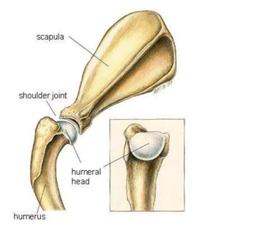
Simple joint
Joints comprise 2 articulating surfaces, connecting 2 bones that slide over each other,
E.g. shoulder, involving glenoid cup of scapula (shoulder blade) and humeral head
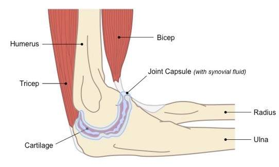
Compound joint
Joint comprise 3/more articulating surfaces, connecting multiple bones within joint
E.g. elbow, involving trochlear groove of distal humerus articulating with ulna and humeral condyles articulating with radius and ulna
Joint functions
Classified to function, location wide range of movement to enable locomotion, e.g. joint in limbs, need stability e.g. between two mandibles
Synarthrosis
No movement
Synostosis
Joints fused with age
Amphiarthrosis
Limited movement
Diarthrosis (synovial)
Wide movement
Fibrous (immoveable)
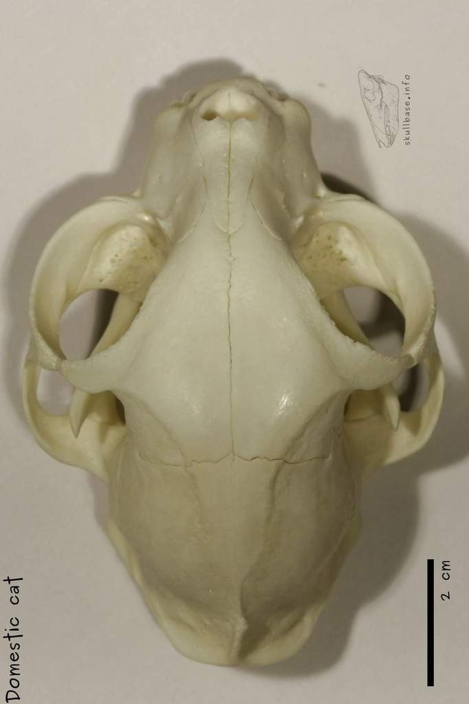
No discernible movement, rigid structure for protection
Bones joined by fibrous material, connecting bones along suture lines
Bones of skull fibrous joints, some degree movement during birth, fusing to provide solid cavity around brain
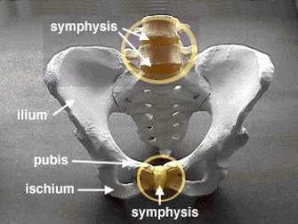
Cartilagenous joints
Cartilage between bones
Allows limited movement, fused once animal skeletally mature (pelvis symphysis)
Amphiarthrosis - Cartilagenous joints restricted movement. Vertebrae articulate through this joint
Synarthrosis - Cartilagenous joints no movement. Skeletal weakness, common fracture sites. e.g. mandibular and pelvic symphyses
Synovial joint
Most frequent joint within skeleton
Throughout body, highly mobile, movement and locomotion
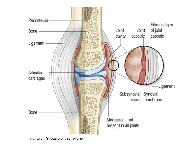
Joints involve number of specialist structures to ensure smooth articulation of bones, suppress development of degenerative changes (arthritis)
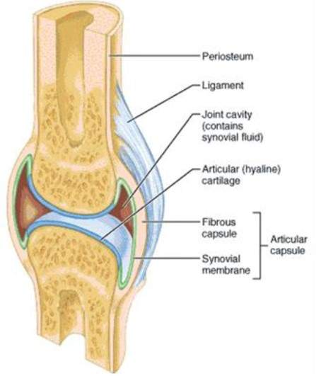
Surrounding synovial joint
Protective fibrous joint capsule surrounds joint and synovial membrane (secrete synovial fluid)
Cavity within capsule (joint space) filled with thick synovial fluid to lubricate, nourish, protect joint
Condylar joints
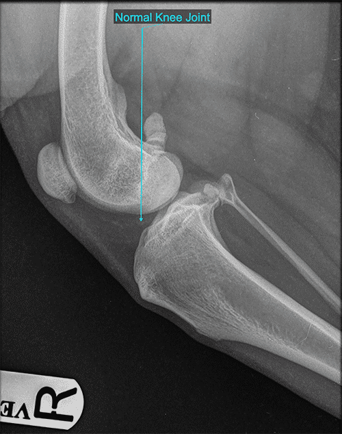
Curvature of bones to create joint. Bony projections (condyles) rock against adjoining bone to allow movement (flexion and extension)
e.g. stifle
Hinge joints
“hinge” open/closes door.
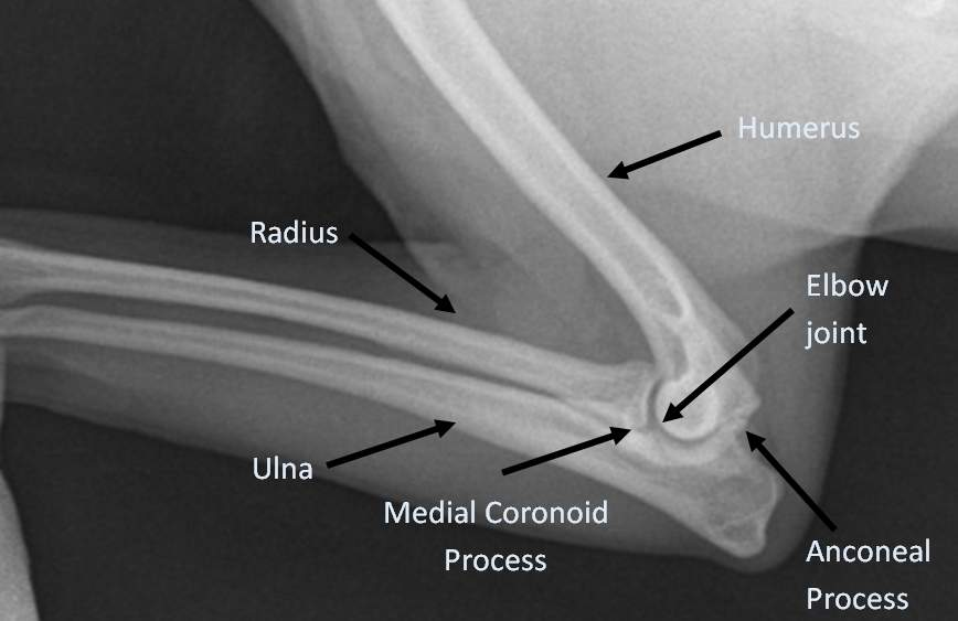
Contours of bones within joint allow movement (flexion/extension) in defined way, often providing stable joint
e.g. elbow and hock (calcaneus)
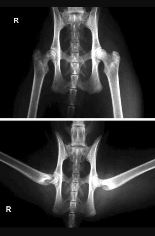
Spheroidal Joints
ball-and-socket arrangement, allows range of movements (flexion, extension, rotation, abduction (movement away from body) and adduction (movement towards body).
e.g. hip
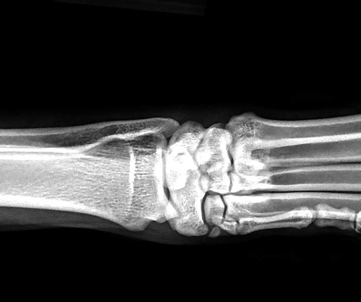
Plane joints
Simple joint, allowing movement one place/direction i.e. backwards and forwards. Architecture between articulating joint mainly involves flattened surfaces
e.g. within carpus and tarsus
Pivot joints
Allow twisting movement. May involve rocking movement as two articulating bones slide over each other, e.g. radius ulna to twist wrist/carpus
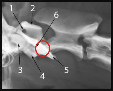
Odontoid peg
More defined anatomical feature within pivot joint is odontoid peg (odontoid processl odontoid dens) of second cervical vertebra (axis) that articulates with first cervical vertebra (atlas) - hidden under wings of axis
Stubby finger like projection allows sideways rotation of vertebrae, giving shaking head ‘no’ action
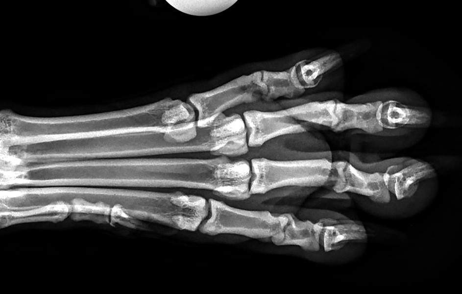
Saddle joints
Movement in one direction, involve concave architecture so articulating bone rocks within cup (saddle) rather than glide over straight surface
e.g. connections between phalangeal bones (digits, toes)
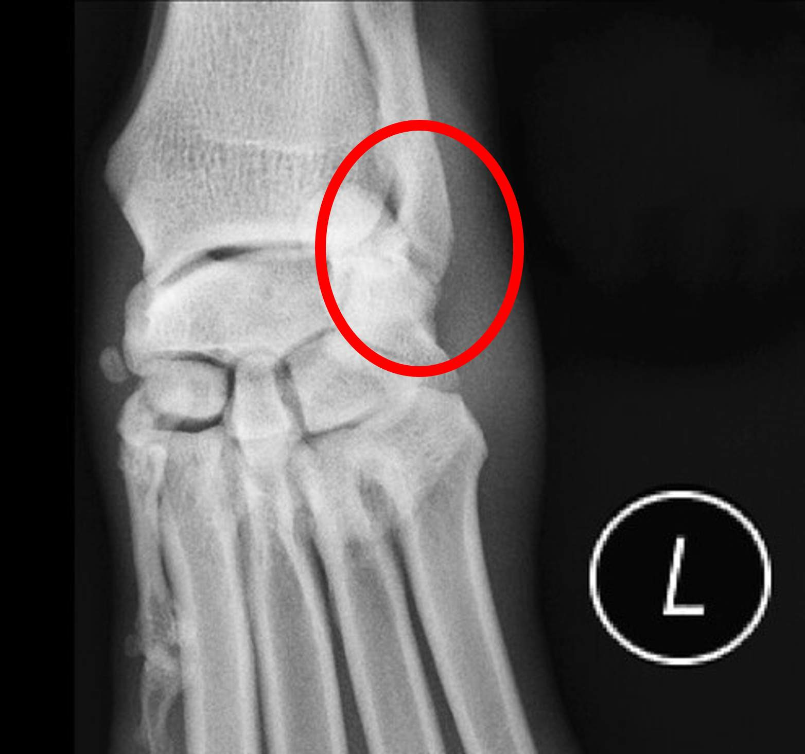
Ellipsoidal Joints
Sliding movement between articulating bones. Ellipsoidal action with joints (egg-shaped) allow flexibility in range of movement along horizontal plane
e.g. radiocarpal joint, connecting rotating radius with carpal bones
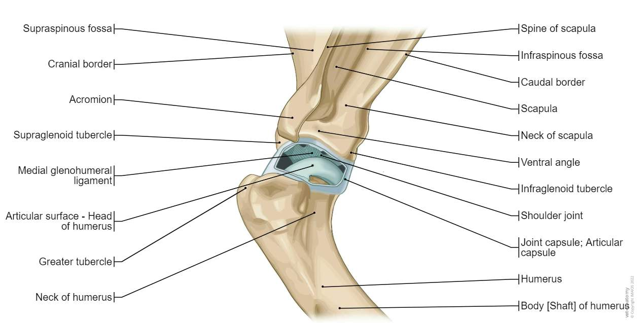
Shoulder
Largest joint in thoracic limb, main weight-bearing joint
ball-and-socket joint, movement circular and flexion/extension
Articulation occurs between glenoid cavity in scapula (socket) and humeral head (ball), with acromion at point of scapula spine limited lateral movement (abduction)
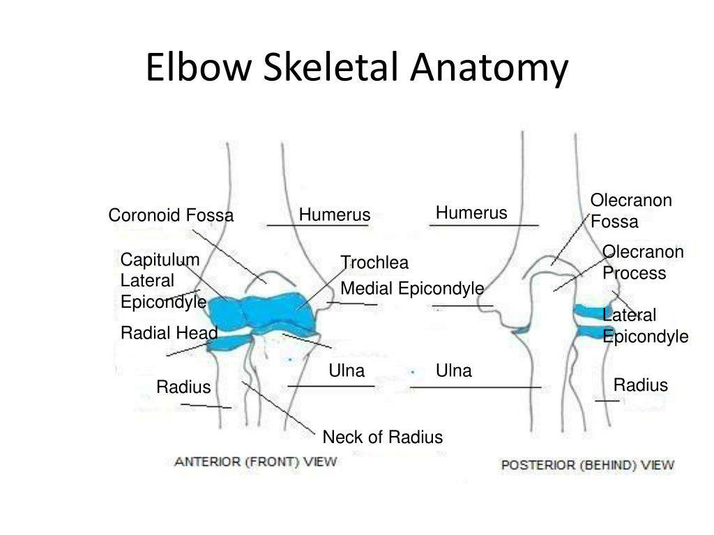
Elbow
Skeletally complex joint, with number of bones and projections
Distal humerus had number of condyles that lock into contours of proximal radius ulna to increase stability within joint
Within joint, trochlear groove provides rocking surface of hinge joint, with olecranon fossa (hole), allow anconeal process on ulna to slide into space on extension of joint
Radius and ulna highly sculptured to increase stability within joint, ulna taking more active role in stabilisation, radius primarily bearing animal weight
Proximal ulna essentially cup, anconeal process (proximal to joint) and coronoid processes (distal to joint) securing distal humerus
Arrangement of projections produces semi-circular cavity (trochlear notch), Which articulates with humeral trochlear groove to produce movement
Finally, olecranon (elbow point) prevents over-extenion of joint when weight-bearing
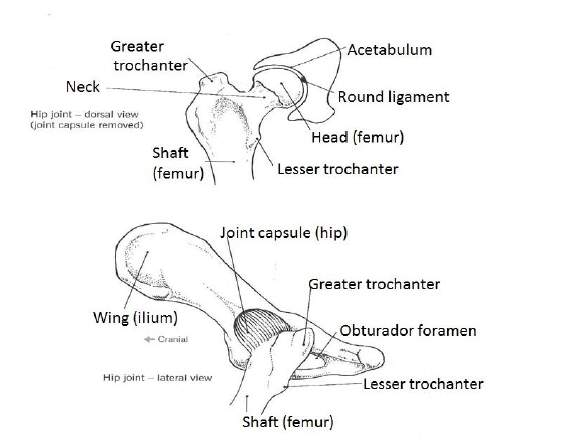
Hip
Main joint in pelvic (hind) limb, responsible for power behind running/jumping
ball-and-socket joint simple anatomical structure, development can be affected by conditions e.g. hip dysplasia, severly affect animal’s ability to move
Socket is a curvature of pelvis (acetabulum) that articulates with femoral head (ball), which rotates wide range movement - more than shoulder
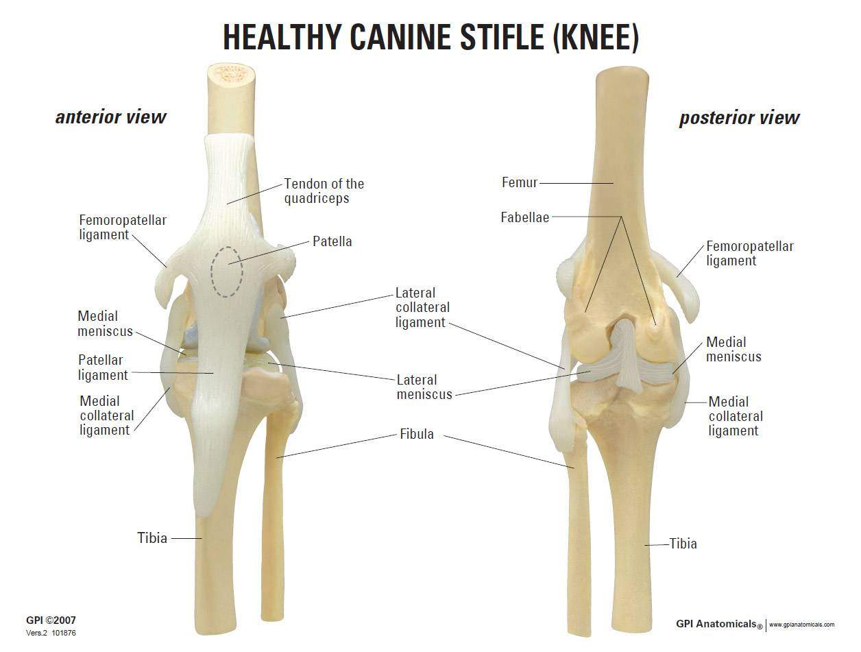
Stifle
Simple joint with condyles of distal femur articulating with flat proximal surface of tibia (tibial plateau)
Lack of skeletal contouring makes joint unstable -
To compensate, stifle has ligaments to hold joint together
Small bones (sesamoid bones) placed within tendons to provide additional support
Patella - patella tendon, cranially over stifle joint
Fabellae - pair of bones within gastrocnemius tendon, caudally to joint
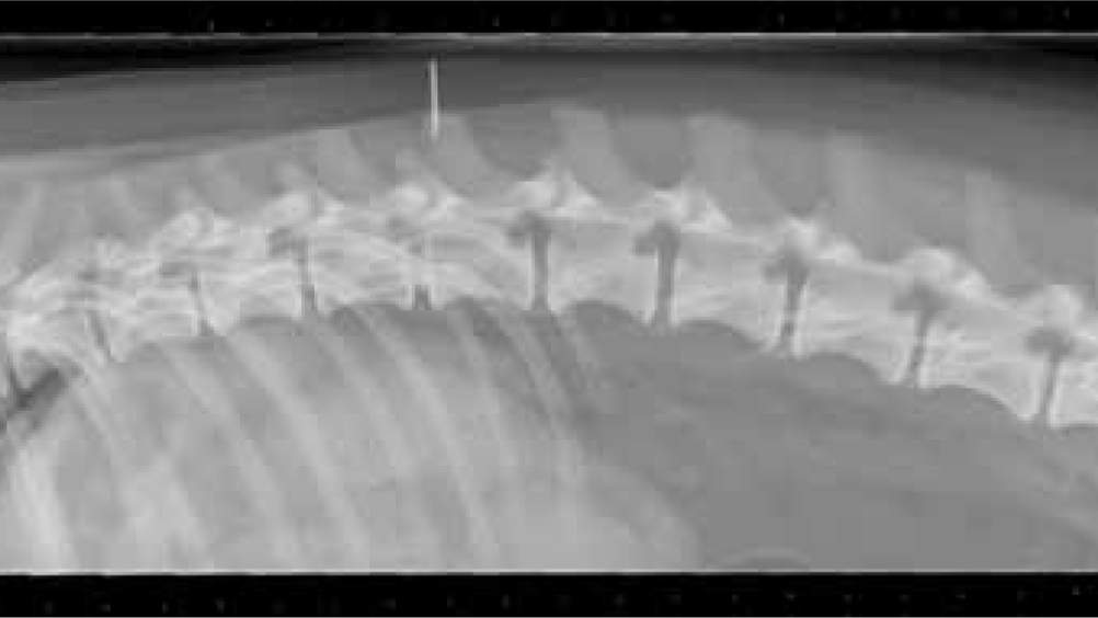
Thoracolumbar joint
Fibrous joint of vertebrae
Architecture of thoracic vertebrae different from lumbar, resulting in forces of walking/jumping/pulling different directions
Particularly seen long-backed short-legged breeds, e.g. dachshund, consequent development of intervertebral disc disease (IVDD)
Tendon and ligaments
Intermediaries between bones and muscles, leverage and stability within joint
Tendon
Closely packed, dense connective tissue used to connect muscle to bone
Ligament
Elastic fibrous tissue used to connect bone to bone
Tendons
Densely packed connective tissue, high tensile strength and good shock absorption. Muscle to bone distal to joint, provide leverage when muscle contracts, producing movement
Movement can cause friction against bones, potential damage tendon
Bursa
Found where tendon located close to bone, synovial fluid ‘cushion’ providing protection against pressure/friction.
e.g. bicipital bursa that cushions biceps tendon from point of shoulder
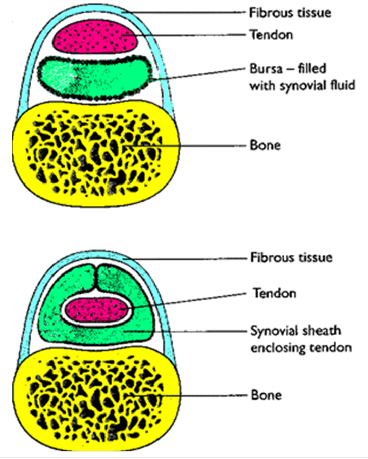
Tendon sheath
Extends fluid-filled structure to completely enclose tendon, provide friction-free movement against both bone and overlying tissues.
e.g. tendon sheath seen in carpal flexor tendon, sheath overlying protruding accessory carpal bone (covered by carpal pad)
Achilles tendon
Most clinically significant tendon seen
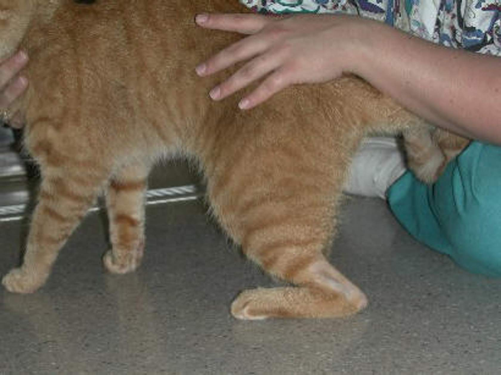
Tendon attaches gastrocnemius (calf) muscle to calcaneus (hock point). Muscle contraction extends joint, maintains hind limbs in digitigrade (toes) stance.
Damage to tendon results in characteristic plantigrade (dropped hocks) stance, seen in image.
Can occur due to mechanical injury e.g. acute injury during high impact sports e.g. agility or flyball
May occur due to physiological/neurological changes, e.g. characterisitc posture diabetic cat
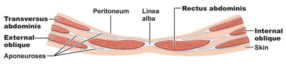
Aponeurosis
A special form of tendon found at linea alba - seen performing lap spays, ‘white line’
Tissue structure similar to joint tendons but aponeurosis joins muscles of longitudinal rectus abdominis muscles (muscle to muscle) rather than attaching to bone
Ligaments
Consist thick bands of fibrous connective tissue to provide strength & stability to joint, elastic to allow joint movement when contract/relax.
Connect bones within joint to ensure articulating surface remains close contact during standing/moving
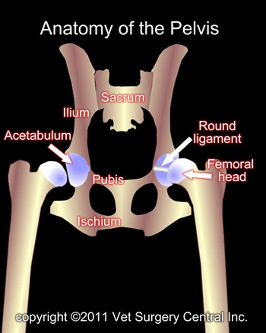
Teres ligament
Sits within hip joint, attach acetabulum of pelvis to femoral head.
Round ligament keep ball of femoral head within socket of acetabulum
Ligament strength
Elastic, ineffective at retaining strength if stretched beyond normal limits.
Hip dysplasia - increased movement within joint can ‘stretch’ ligament, causing increased laxity of ligament, resulting in further degeneration of joint
Prolonged ligament stretching reduces ability to stabilise joint, displacement of joint occurs
Stifle
Minimal skeletal architecture to stabilise joint. Uses ligaments to ensure close connection between femur, tibia, fibula
slight curve in stifle when standing increases pressure on ligaments to sustain correct alignment of bones
Ligament rupture
Common in dogs, incidence increasing with exercise (dynamic injury) or conformation (straightness of leg/angle of tibial plateau)
Muscles within stifle
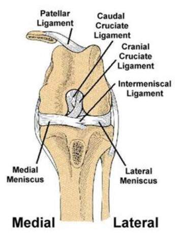
Medial Collateral ligament
Lateral Collateral ligament
Cranial Cruciate ligament
Caudal Cruciate ligament
Medial collateral ligament
Lies along inner surface of stifle to prevent inwards collapse of joint
Lateral Collateral ligament
Lies along outer surface of stifle to prevent outwards collapse of joint
Cranial Cruciate ligament
Runs from front of stifle to back to prevent excessive forward movement of joint
Caudal Cruciate ligament
Runs from back of stifle to front to prevent excessive backward movement of joint
Range of movement
Primary purpose of arrangement of bones, tendons and ligaments - create movement
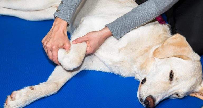
Flexion
Closes/decreases angle of joint by bending limb
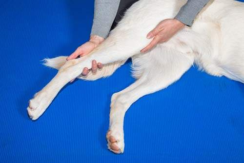
Extension
Opens/increases angle of joint by straightening limb
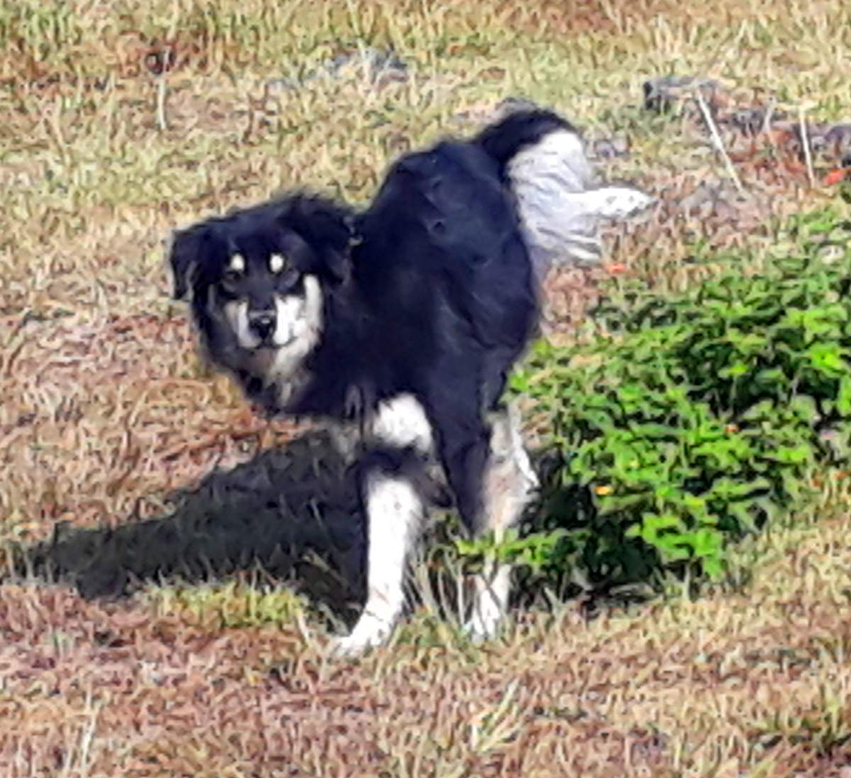
Abduction
Moves whole limb away from body, moving laterally at hip/shoulder
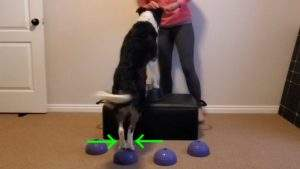
Adduction
Moves whole limb closer to body, moving medially to centre from hip/shoulder
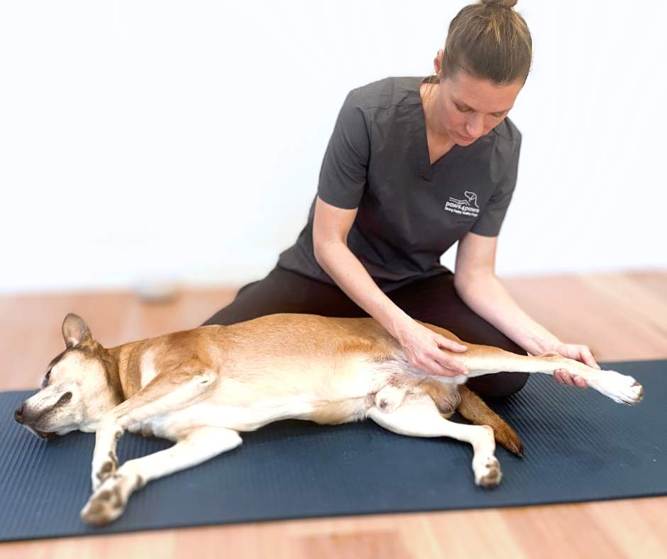
Retraction
Moves whole limb backwards, moving caudally from hip/shoulder
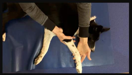
Protraction
Moves whole limb forwards, moving cranially from hip/shoulder
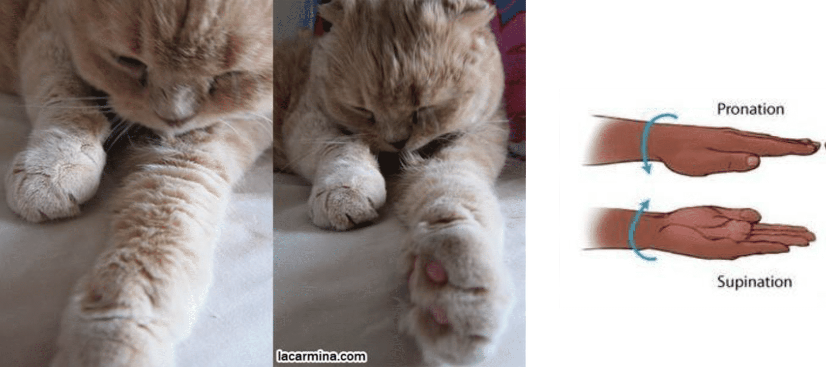
Pronation
Turning forepaw downwards, pads ventral
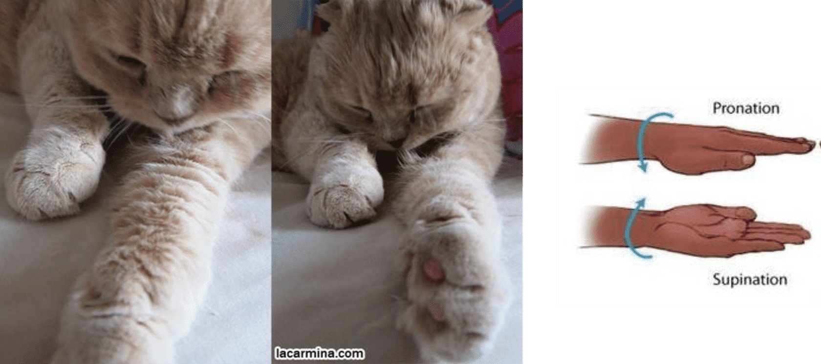
Supination
Forepaw turned upwards, pads dorsal
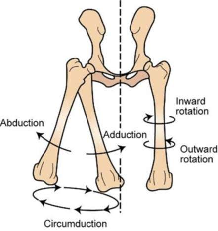
Rotation
Twisting bone along length e.g. rotation of radius/ulna when paw pronated/supinated
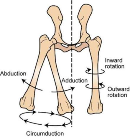
Circumduction
Whole limb moving circular from shoulder/hip