Immunology Exam 1
 Call Kai
 Learn
Learn Practice Test
 Spaced Repetition
 Match
Match Flashcards
 Knowt Play
1/110

There's no tags or description
Looks like no tags are added yet.
Name | Mastery | Learn | Test | Matching | Spaced | Call with Kai | Chat |
|---|
No analytics yet
Send a link to your students to track their progress
111 Terms
The first vaccination was created by Edward Jenner to fight
smallpox
The first vaccination came from
the vaccinia virus (cowpox)
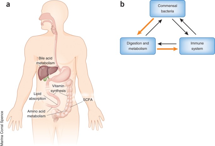
Commensal species
Typically do not cause harm to the host.
Likely become killed off from a round of antibiotics.
Can be present in many different parts of the body.
Gram-positive bacteria example
Staphylococcus
Gram-positive bacteria
Thick cell wall containing teichioc acids
Innate Immune system
Sets up the adaptive immune system (after breach)
Fast response
Activates the adaptive with dendritic cells and FDC’s
Activates inflammatory response
Fixed (does not change or adapt to recognize new pathogens
Uses different type of receptors
Operates in both first and second immune response
Adaptive Immunity
Slow response
Variable (adaptive)
remembers microbes
Uses one type of receptor
Operates during second immune response
Inflamation
occurs when a pathogen gains entry through the surface of a wound
causes redness, heat, pain, and swelling
can be triggered by non-infectious factors
results in increased vasodilation (to bring more blood and immune cells to the area) and increased vascular permeability (to let fluid and white blood cells exit the bloodstream into the tissue).
Vaccines:
Provide protective immunity against a specific pathogen, by stimulating the adaptive immune response for the body to gain memory of it.
gold standard: neutralization (surrounding the pathogen with antibodies to prevent it from causing harm to us)
Clonal selection and expansion
the process the immune system uses to find and multiply the exact B cells and T cells needed to fight a specific infection (adaptive immune response)
Lymphocytes
include 20-50% of WBC: T-cells, B-cells, and NK cells
all are lukocytes
most have never been activated
include both innate and adaptive response
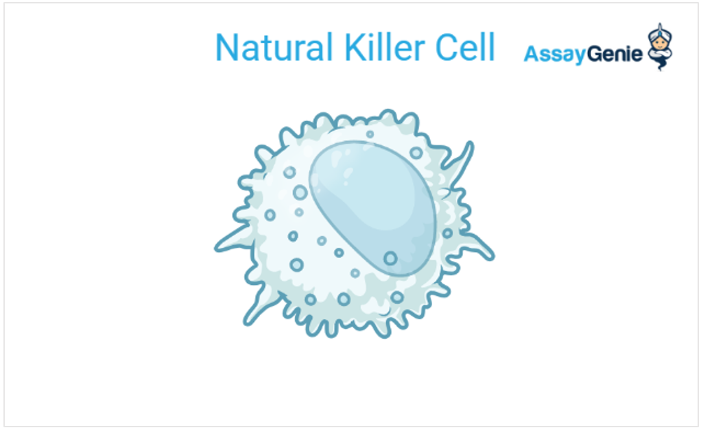
Natural Killer Cells (NK cells)
Innate Viral Fighters
large granular lymphocytes
full nucleus
secrete cytokines
help prevent viral replication + activate t-cell mediated immunity
Kills infected host cells by apoptosis ( tells them they must die)
Neutrophil
Best innate killing machine; first out in mass
granulocyte
neutral staining (lightest)
most abundant
stored until needed, released in mass numbers when needed
Phagocytic (capture and kill microorganisms)
short-lived
Oxygen dependent mechanism of destruction: O2 turn into radicals (weapon killing pathogen)
Oxyget independent mechanism of destruction: Defensins (poke holes into pathogen)
accumulation of dead neutrophils →pus

Monocyte (mononuclear Phagacytes)
precursor of macrophages, dendritic cells, or mast cell
located in blood (steal two o’s from monocyte)
phagocytic

Macrophage
located in the tissue
phagocytosis and killing of organisms
activation of T-cells and initiation of immune responses

Dendritic cells
recognize and present antigens (using MHC II) to T-cells in lymphnodes
“dendritic” comes from the similar appearance (folds) of a dendrite of the nervous system
folds allow for maximum interaction with other cells of the immune system
Most have MHC class II and class I molecules
Can be found in secondary lymphoid tissues
use many PRRs (pattern recognizing receptors)
Follicular Dendritic cells
only found in follicle of lymphnode
B-cells will be directed to FDC in lymphnodes
Harbor antigen for B-cell to check
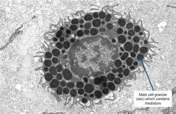
Mast Cell
tissue bound
bind immunoglobulin E (IgE) antibodies on a surface
innate
get rid of parasites by the release of granules containing histamine and other active agents
mediator→ releases histamine result of allergies
Eosinophils
granulocyte
redish staining (middle)
kill antibody coated parasites through the release of granule content
innate but work w adaptive cells
red granules
Basophil
granulocyte
Darkest staining
control immune response to parasites
Histamine related
innate but work w adaptive cells
least abundant type of leukocyte
blue granules
megakaryocyte
make platelets
come off erithro pre cursor
Erythrocytes
carry lose antigen + antibody to the liver to be cleared by kupffer cells
rumba of immune system
Naiive lymphocytes
(B+T cells) never been activated
Effector cells
plasma cell: terminally differentiated affectpr B-cell
cytokine producing T-cell: helper T-cell/ CD4 cell
Cytotoxin T-cell (TC) = CD8 cell
Hematopoiesis
the generating of cellular elements of blood
occurs in the bone marrow
B-lymphocytes arise and mature in bone marros
T-lymphocytes arise in bone marrow but mature in thymus gland
common myeloid cell precursor
made from Hematopoietic cells
precursor of megakaryocyte-erythroid precursor cell, granuloocyte precursor cell, Monocyte, dendritic cell, and mast cell
will eventually generate Monocytes/macrophages, dendritic cells, mast cells, neutrophils, eosinophils, basophils
Megakaryocyte-erythroid precursor cell
Precursor of Erythroblasts (make erythrocyte) and megakaryocyte (make platelets)
Granuloocyte precursor cell
precursor of basophil, eosinophil, and neutrophil
Common lymphoid cell precursor
will eventually generate B cells, T cells, NK cells, innate lymphoid cells (ILCs)
Common ILC precursor
Precursor of NK cell, ILC1, ILC2, ILC3, LTi
Common CD4 T-cell precursor
precursor of T-reg, TH17, TH2, TH1
Phagocytosis
The process where phagocytic immune cells are specialized to capture, engulf, and kill microorganisms
receptor recognition → pseudopodia wrap around target → target enters a phagosome → lysosomes fuse → phagolysosome → destruction
T-cell receptors
are highly specific
made of 2 chains
membrane bound
Antigen
foreign substance
BCRs
direct/ native antigen recognition
membrane bound form of immunoglobulin
secreted by activated B-cells
differentiated from antibody by C-term
Ig’s receptors (immunoglobulin’s receptors)
Helper T-cell/ CD4
release cytokines (signals) to activate other immune cells
Plasma cell
specialized, fully differentiated B cells that factory-produce and secrete large amounts of antibodies
T-cell activation
When a T cell is activated, it undergoes clonal expansion (cell division) and differentiates into effector and memory T cells
Secondary Lymphoid tissues
Lymph nodes: serve as the main sites where mature T and B lymphocytes encounter antigens and become activated, can swell during infection
Spleen: filters blood, traps blood-borne pathogens, and initiates immune responses against them
Peyer’s patches: located in the mucosa of the small intestine, trigger immune responses and stimulate the production of immunoglobulin A (IgA), which neutralizes pathogens along the mucosal lining
Pathogen associated molecular patterns (PAMPs)
molecules on microorganisms that the innate immune system recognizes as foreign and triggers a rapid defense response
Lipopolysaccharides (LPS/gram-negative), Peptidoglycans and Lipoteichoic Acids (gram-positive), Flagellin, Carbohydrates and Glycans (fungal)
Gram-positive
thick peptidoglycan and taichoic acids
memory trick: look for T
Include:
Bacillus
Staphylococcus aureus
Streptococcus
Mycobacterium (kinda)
Gram-negative
thin outer membrane and lipopolysacharides (LPS)
Include:
E. coli
Pseudomonas
Salmonella
Shigella
Process of phagocytosis
1) Bacterium becomes attached to membrane envaginations called pseudopodia
2) Bacteria is ingested, forming phagosome
3) Phagosome fuses with lysosome
4) Lysosomal enzymes digest captured material
5) Digestion products are released from cell
Primary Lymphoid tissue
Bone marrow (where B-cells mature)
Thymus (where T-cells mature)
Intracellular pathogen in the nucleus/cytosol vs in the vesicle
Pathogens in the nucleus/cytosol: attacked by killing infected cell
Pathogens located in the vesicles: are attacked by the infected cell by increasing antimicrobial activity
Complement
It helps tag pathogens and extracellular molecules for destruction
Can destroy pathogens directly by poking holes in the outer membrane or cell wall
ubiquitous in blood and lymph
Involves soluble proteases called zymogens
a molecular defense that can be used immediately
Anaphalatoxins
C3a, C5a (recruit Neutrophils to site of infection)
recruit phagocytes
lead to inflammation
chemoattractant (recruits cells to site of infection)
C5a is more potent
Complement receptors
CR3 and CR4 recognize iC3b
CR2: a B-cell co-receptor, recognizes C3d
CR1 recognizes C3b and C3bBb
type of bond exposed upon cleavage of C3
Thyoester bond
iC3
the product of C3 hydrolysis
iC3Bb
iC3Bb is the soluble C3 convertase of the alternative pathway
Pattern recognition receptor (PRR)
spot signs of infection or cell damage to trigger immune response
Variola virus
causes small pox
opportunistic pathogen
cause disease if the body’s defenses are weakened or is found in an unusual place in the body (ex.aids)
Bacteria (ex)
mycobacteria tuberculosis
causes tuberculosis
Viruses (ex)
HIV → AIDS
Influenza → Flu
Variola → small pox
Fungi
Candida albicans → Thrush, systemic candidiasis
Parasites
Trypanosoma brucei → sleeping sickness
Defensis =
antimicrobial peptide
ILC3
secrete IL-17 (cytokine)
innate vertion of T helper 17
recruits neutrophils → promotes phagocytosis
secretes antimicrobial peptides
responds to extracellular infections, bacteria and Fungi
ILC2
secretes IL4 and IL5
innate version if T helper 2
responsible for non inflammatory macrophage activation
responds to intestinal parasites, infections
ILC1
secrete interfering gamma
innate version of T helper 1
inflammatory macrophage activation
respond to extracellular infections and bacteria
NK
secrete interfering gamma
Innate version of CD8
use cell mediated cytotoxisity
respond to intercellular infections by viruses and some bacteria
alpha-2-macroglobulin
A protease inhibitor
cuts its “bait” by enclosing the protease and creating an “eat me signal” for phagocytes
where are acute phase proteins made
Liver
where are M Cells located
in the lining of Peyer's patches
Steps of inflammation
Inflammatory cytokines activate endothelium → vasodilation (widening of blood cells) + increased vascular permeability (rushing blood to injured area) + new adhesion molecules → leukocytes can leave blood and enter tissue.
Endema
swelling from fluid/cells/molecules entering tissue
Barrier breach→
Macrophage recognize PAMP with PRRv→ Macrophage realease cytokines/chemokines → endothelial activation (activation of inflammation) → vasolation/permeability/ adhesion (inflammation) → neutrophil recruitment → destruction of pathogen
Opsonin
a host molecule that tags/coats a target to make it easier for a phagocite to recognize and ingest
C3b and C4b act as a opsonin (recognized by CR1)
C3b and C4b attach covalently to microbial surfaces by thioester bond after nuophilic attack
Certain antibodies can act as opsins
Erythroid precursor
will generate Erythrocytes and megakaryocytes → platelets
Naivve B/T cells
not yet activated
little cytoplasm (not yet producing large quantities of effector molecules)
Kupffer cells
Macrophage in liver
Microglia
Macrophage in brain
Tfh (follicular helper cells)
innate version of Tfh (follicular helper cells)
Activate B cells
suppress immune system
MHC (major histocompatibility complex)
TCR recognizes the combination of MHC amino acids + presented peptide
amino acids
MHC I
Used by essentially all nucleated cells
Presents intracellular peptides
Virally infected cells can present to effector CD8 T cells
MHC II
Restricted to professional antigen-presenting cells: dendritic cells, macrophages, and B cells.
Extravasation
movement of cells of of the blood across endothelium to tissue or lymphoid tissue
sequence: rolling → adhesion/tight binding → diapedesis → chemotactic migration
Afferent
Arrive
Efferent
Exit
Spleen → red puld
red cell handling
Spleen → white pulp
organized secondary lymphoid tissue for blood born antigen / pathogen
MALT
mucosal associated lymphoid tissue (mucosal surfaces)
GALT = gut associated lymphoid tissue ( contains IgA = immunoglobuline A
BALT = bronchus associated lymphoid tissue
Peyer’s Patches = organized GALT with B/ T cell regions
continuously samples external body and supports antibody mediated neutralization
Neutralization
Antibody binds / covers pathogen so it cannot cause harm
Opsonization
Antibody binds to pathogen and is recognized by an Fc receptor leading to phagocytosis
Complement activation
certain antibodies initiate complement and lead to opsonization
Primary vs secondary adaptive immune response
Primary = creates memory
Secondary = acts faster + stronger
“student surpasses the master”
TCR
Need MHC + peptide;
Draining lymphnode
lymph node receiving material from an infected site
a node where the adaptive response / B- and T-cell activation is occurring.
Secondary Lymphoid
Lymph nodes, spleen white pulp, organized MALT such as Peyer's patches
antigen meet B/T cells
Defensin Production
epithelial cells + neutrophils
poke holes
Acute Phase Response
Macrophage recognizes PAMP with PRR and produces cytokines → IL-6 will travel to liver
“IL-6 binding to hepatocytes leads to the production of Acute Phase Proteins (CRP + MBL) → more complement
Hepatocytes = Liver cells
CRP → classical complement pathway (complement receptor protein)
MBL → Lectin complement pathway ( Manose binding lectin)
DAMP’s
damage associated molecular patterns
signals from damaged/dead host tissue that promote clearance and wound healing even without infection.
Recognition of a PAMP by a signaling PRR drives
six inflammatory cytokines: TNF-alpha, IL-1, IL-6, CXCL8, CCL2, IL-12
TFN-alpha
Endothelial activation
cting on nearby endothelium = paracrine
IL-1-beta
Endothelial activation(Identical to TNF)
induces fever
acting on nearby endothelium = paracrine
IL-6
acts on Liver/ hepatocytes to make acute phase proteins
“I Love 6 pack abs” (abs=core=liver)
traveling to liver = endocrine
TNF-alpha, IL-1, IL-6 are
pyrogens: act on hypothalamus/ body systems to increase temp and promote bone marrow output
CXCL8
chemokines that recruite neutrophils
Neutrophils 8 bacteria