Anatomy and Physiology 2 Brain Structure and Function lab quiz
1/102
There's no tags or description
Looks like no tags are added yet.
Name | Mastery | Learn | Test | Matching | Spaced | Call with Kai | Chat |
|---|
No analytics yet
Send a link to your students to track their progress
103 Terms
Identification of Major Brain Regions
There are four major brain regions
Cerebrum
It is large, is the dominant brain structure, with many folds and crevices enveloping the diencephalon.
Cerebellum
Posterior to the brain stem.
Diencephalon
Superior to the brain stem in the center of the brain.
Brain stem
Connected to the superior part of the spinal cord.
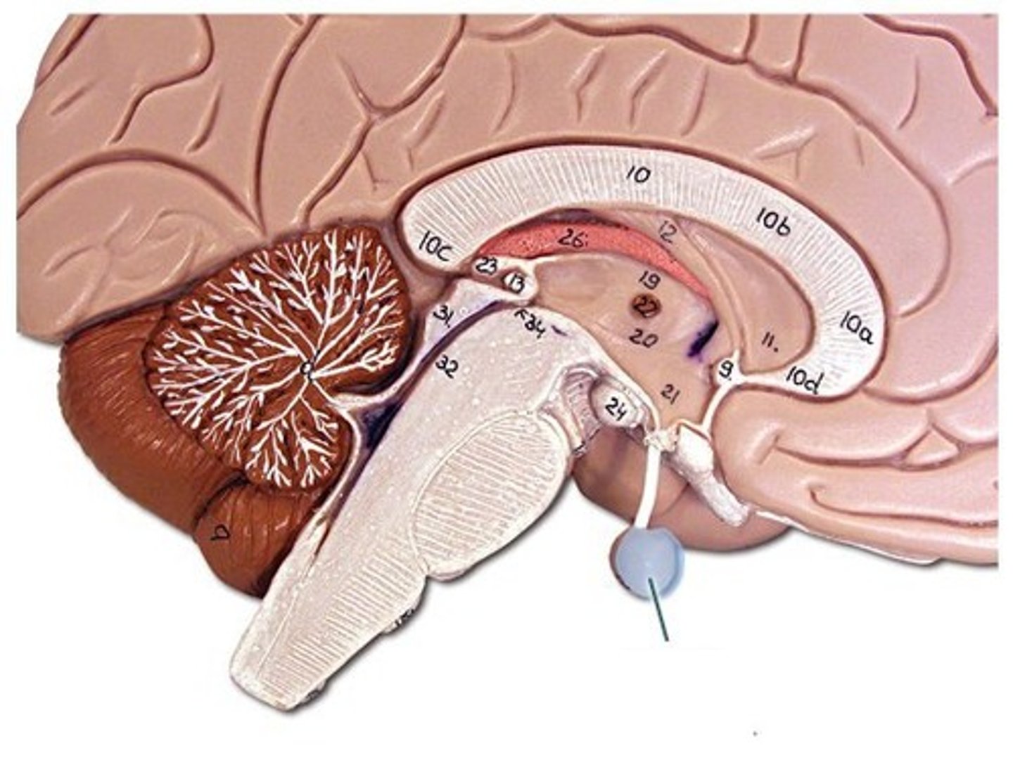
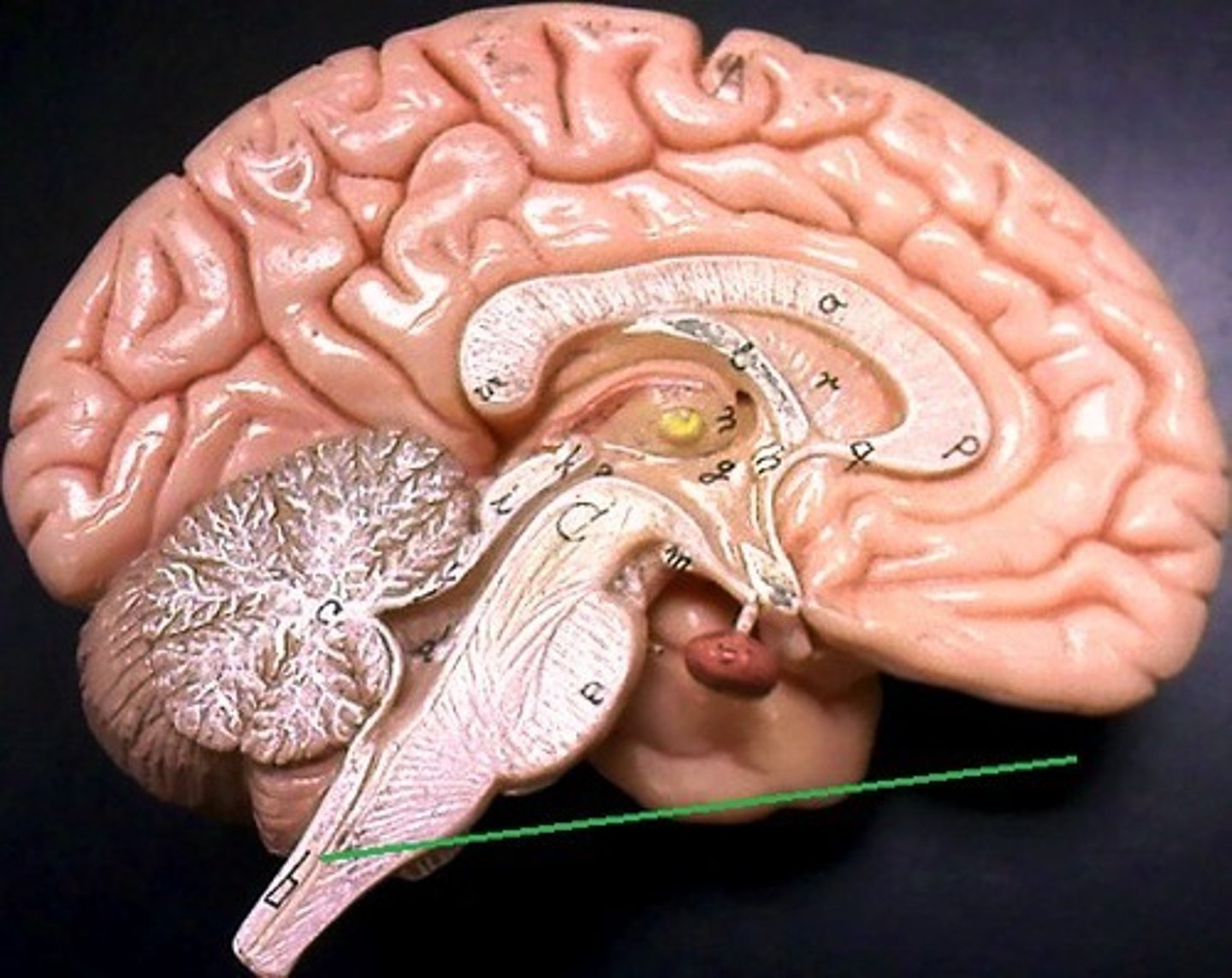
The Brain Stem (Sagittal section, medial view)
It is composed of 3 structures.
Corpora quadrigemina
Composed of 2 superior colliculi and 2 inferior colliculi.

Midbrain
A smaller area superior to the pons and inferior to the diencephalon, consisting of cerebral peduncles and the corpora quadrigemina.
Pons (Bridge)
An expanded structure located superior to the medulla oblongata and anterior to the cerebellum, and has respiratory centers that assist the medulla oblongata in controlling breathing.
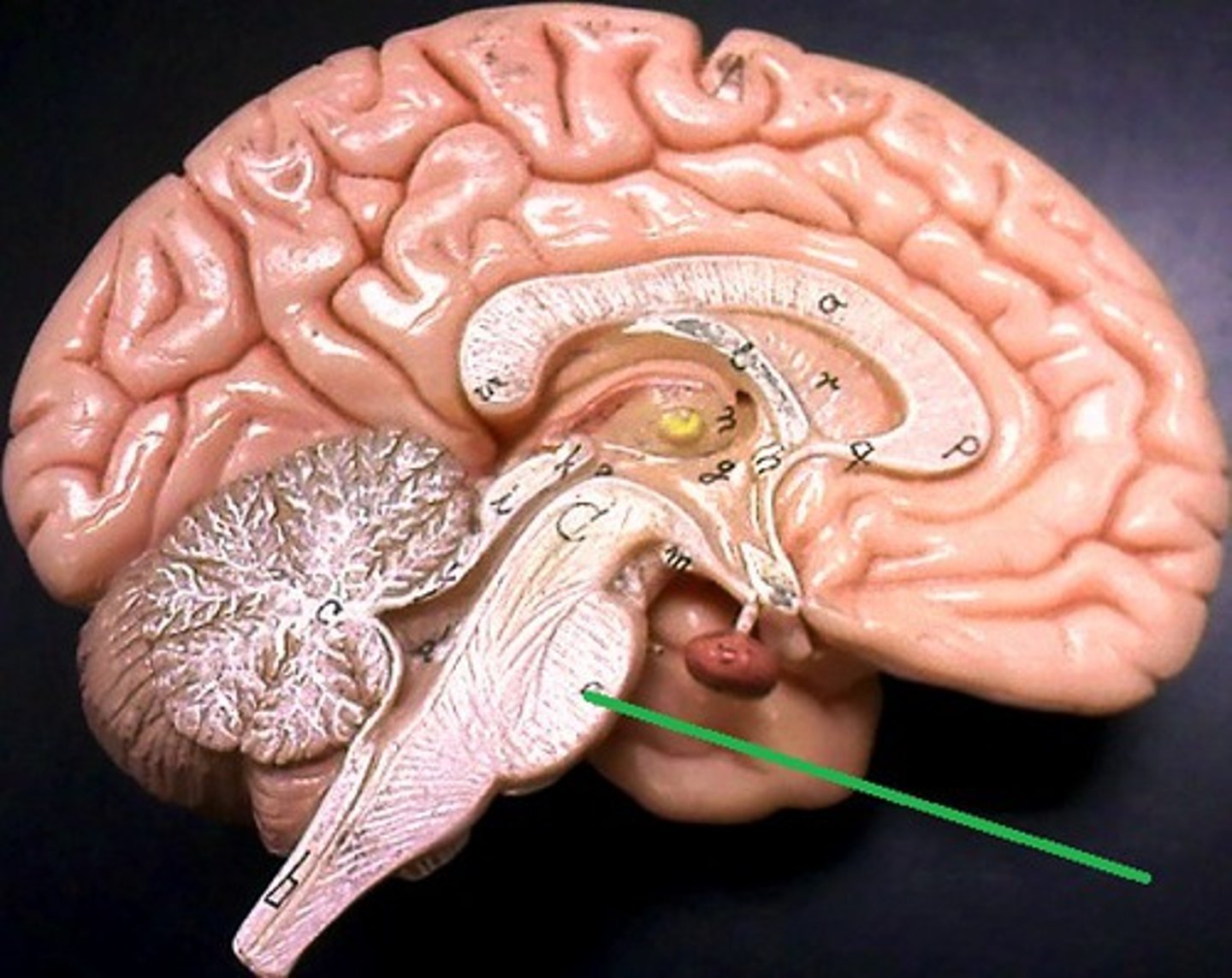
Medulla oblongata
The first brain structure, is immediately superior to the spinal cord and is the most vital part of the brain because it houses the respiratory and cardiovascular control centers.
Cerebral peduncle
White fibers that connect the upper and lower brain areas.
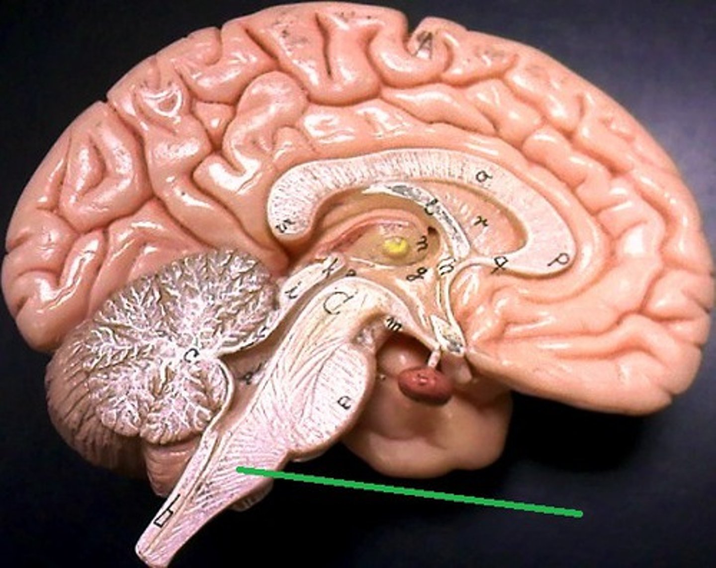
The Brain Stem (Inferior aspect of brain)
Posterior view continued
Midbrain
A smaller area superior to the pons and inferior to the diencephalon, consisting of cerebral peduncles and the corpora quadrigemina.
Pons (Bridge)
An expanded structure located superior to the medulla oblongata and anterior to the cerebellum, and has respiratory centers that assist the medulla oblongata in controlling breathing.
Medulla oblongata
The first brain structure, is immediately superior to the spinal cord and is the most vital part of the brain because it houses the respiratory and cardiovascular control centers.
Spinal cord
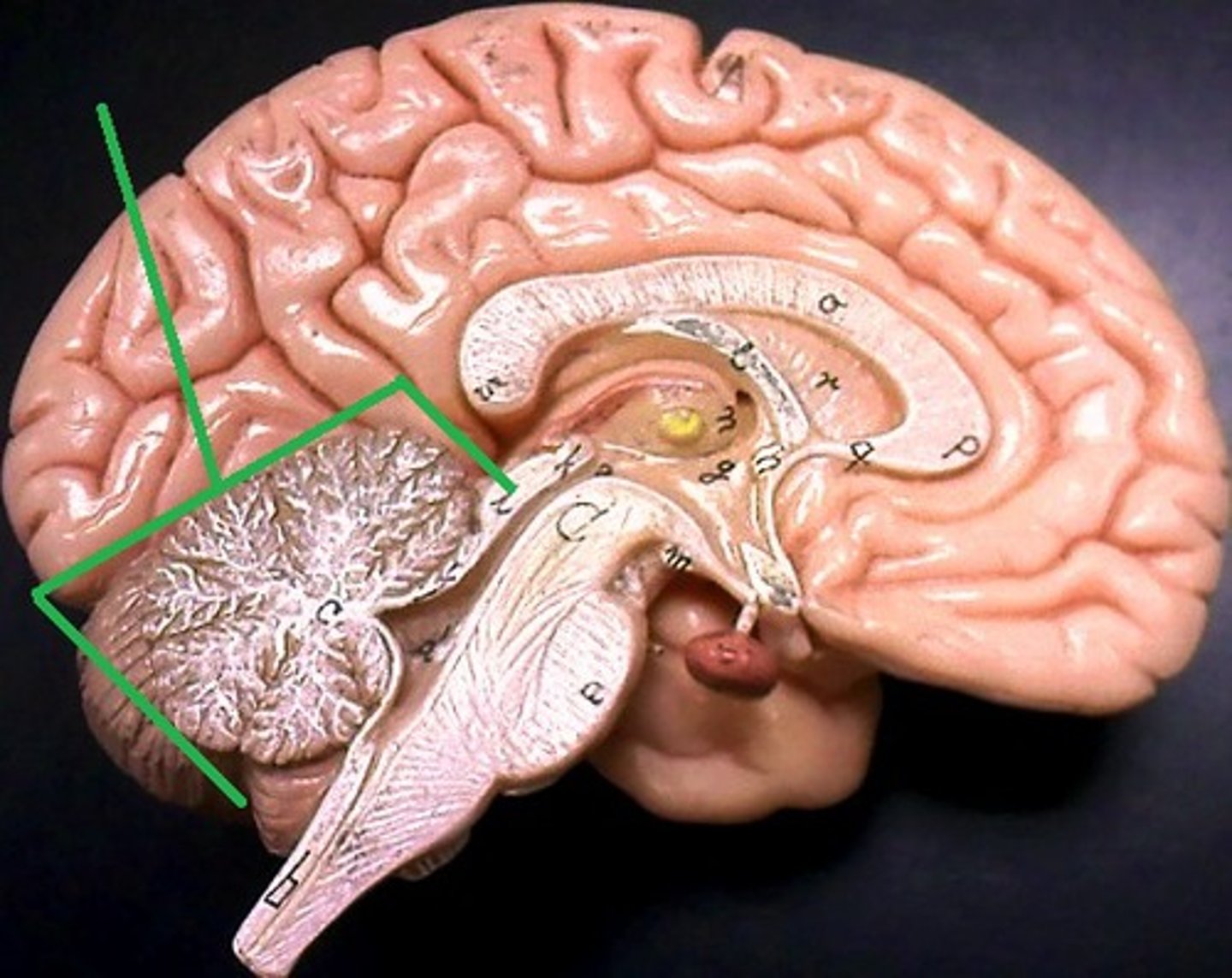
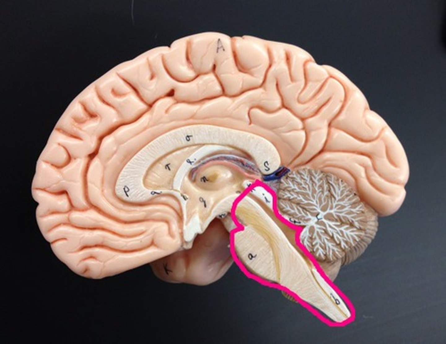
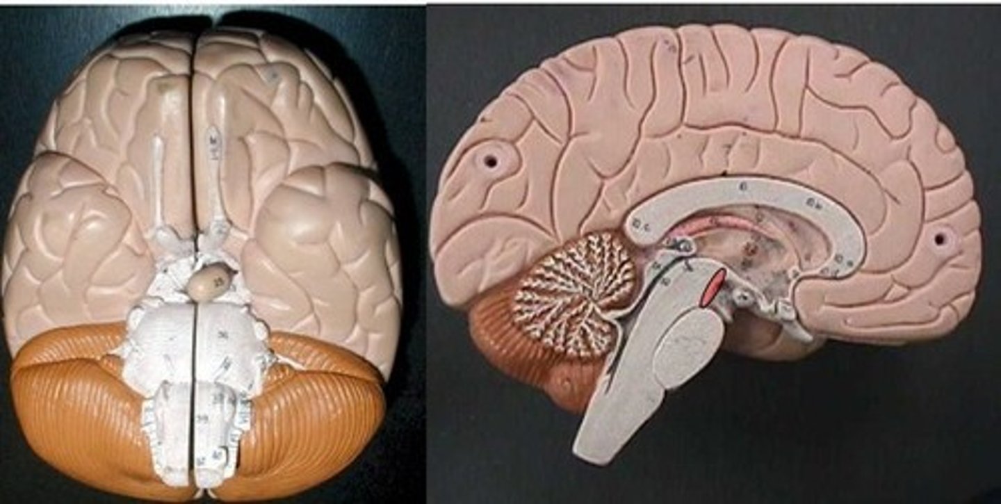
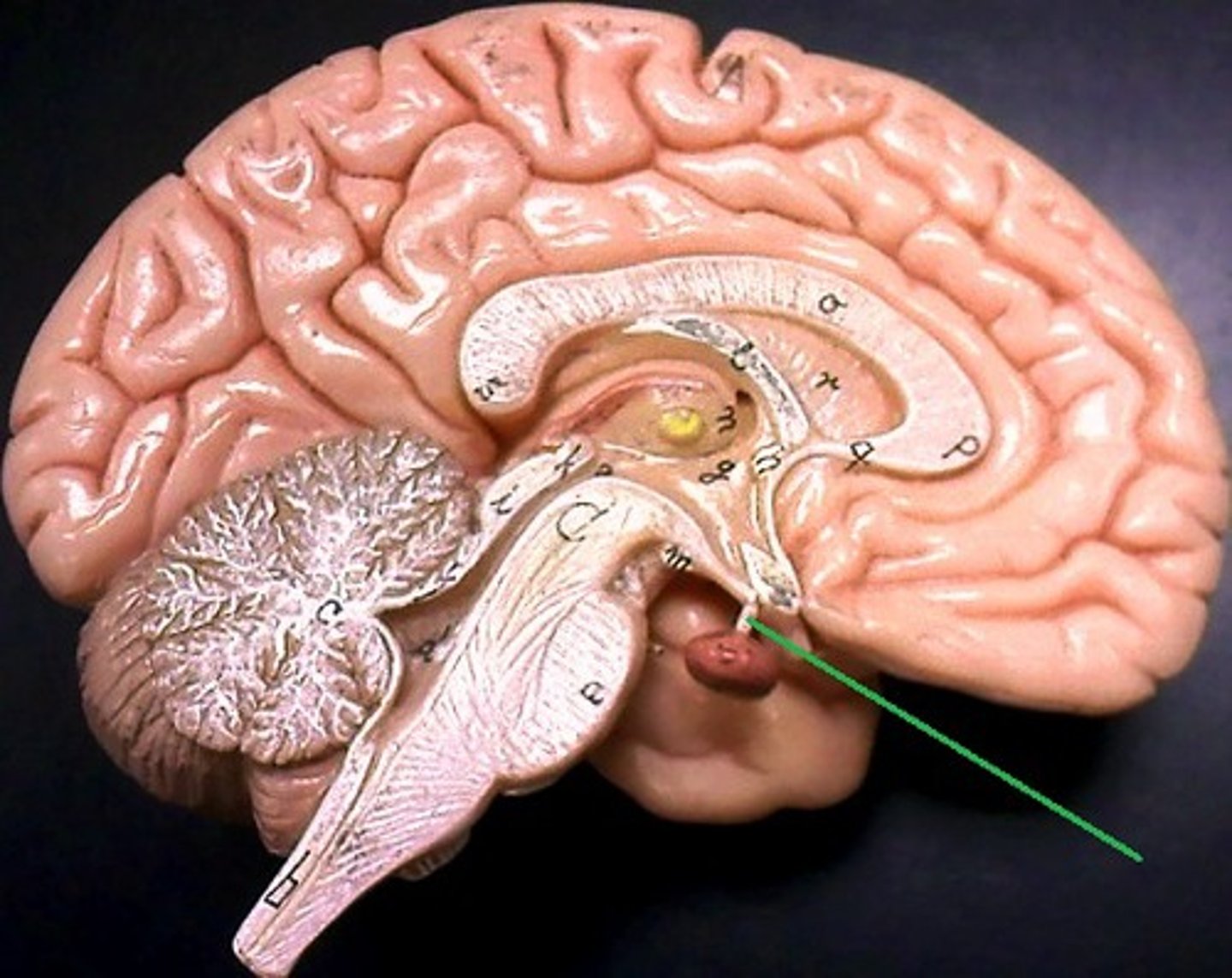
Cerebellum (Little brain) superior view
Second in size to the cerebrum and is located inferior to it and posterior to the medulla and pons.
Cerebellar hemispheres
There are 2 of them with a central area.
Vermis
It connects the 2 cerebellar hemispheres.
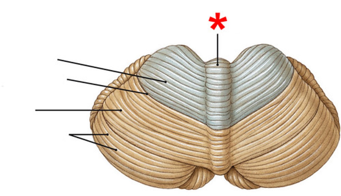
Folia (Leaves)
The cerebellar folds are slender, plated gyri that look similar to pages in a book.
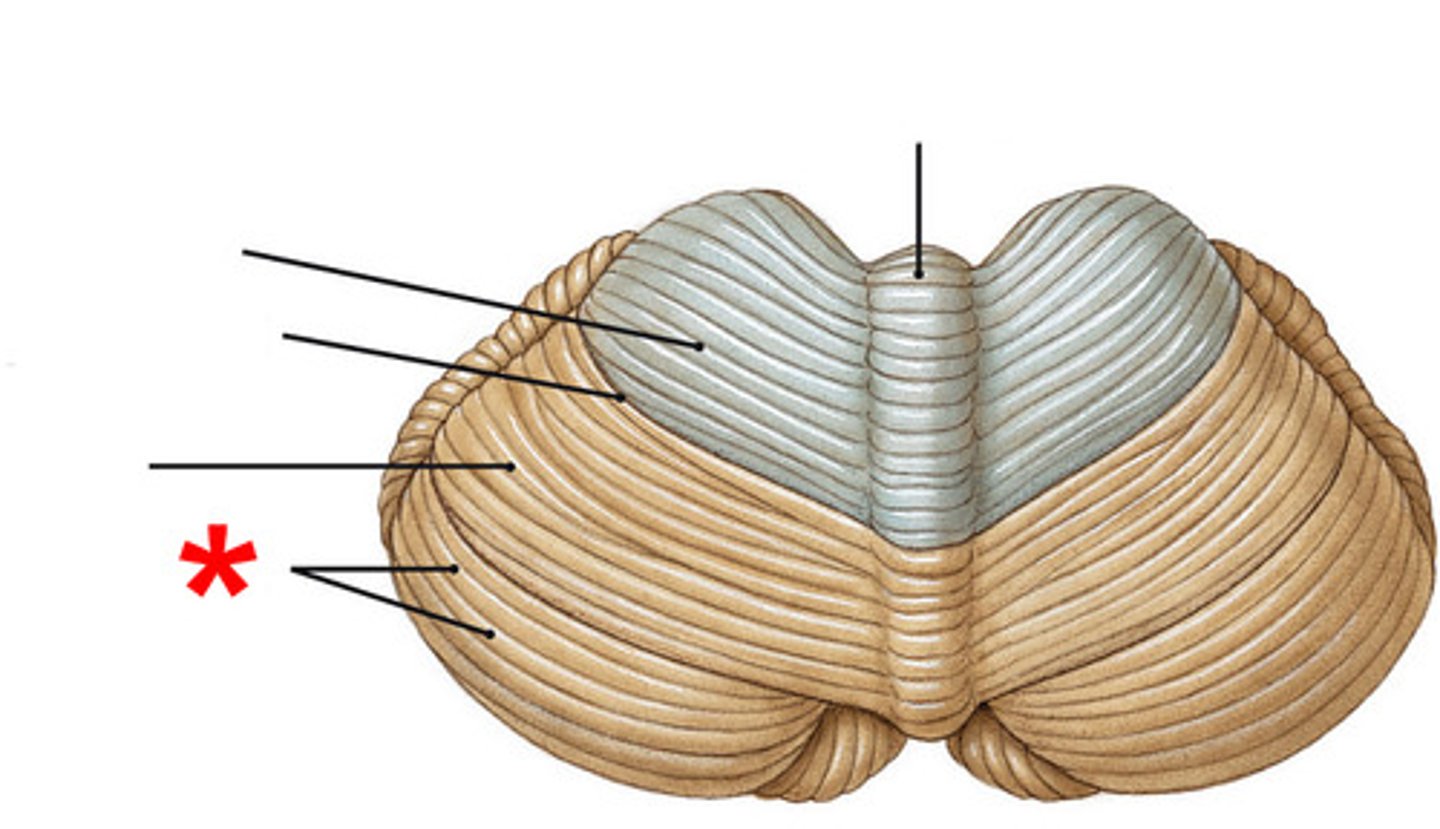
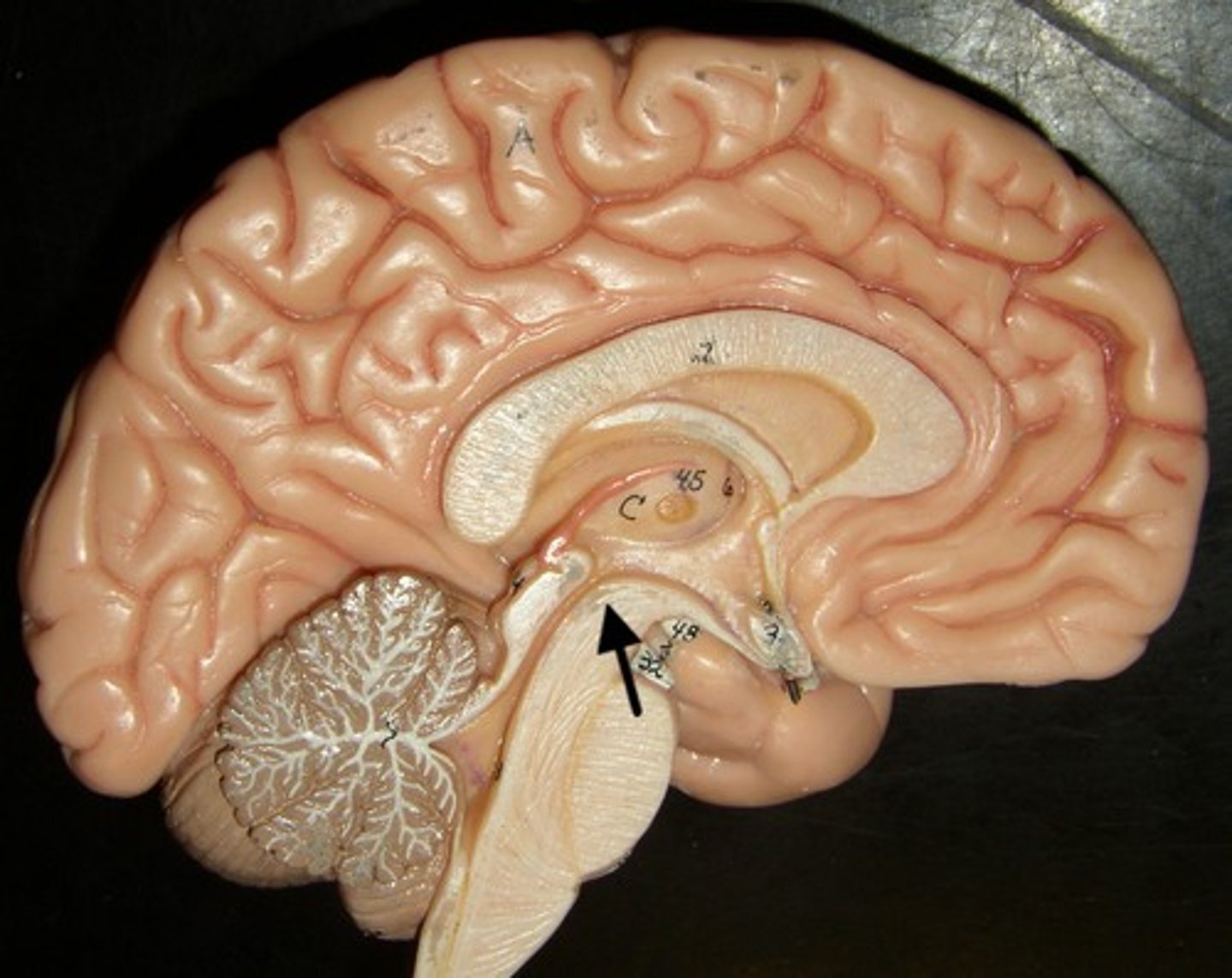
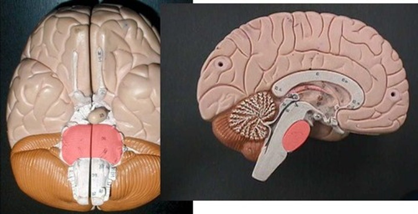
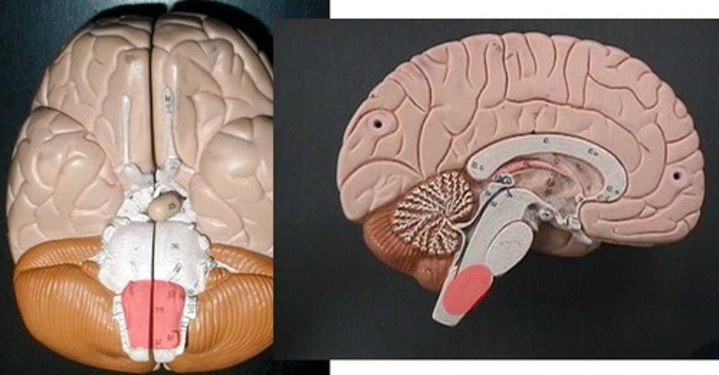
Cerebellum midsagittal section
continued from a different view
Superior colliculus
Inferior colliculus
Arbor vitae (Tree living)
When cut in sagittal section, gray matter can be observed on the exterior, with deeper white matter.
Cerebellar cortex
The outer layer of gray matter.

Pons

Medulla oblongata

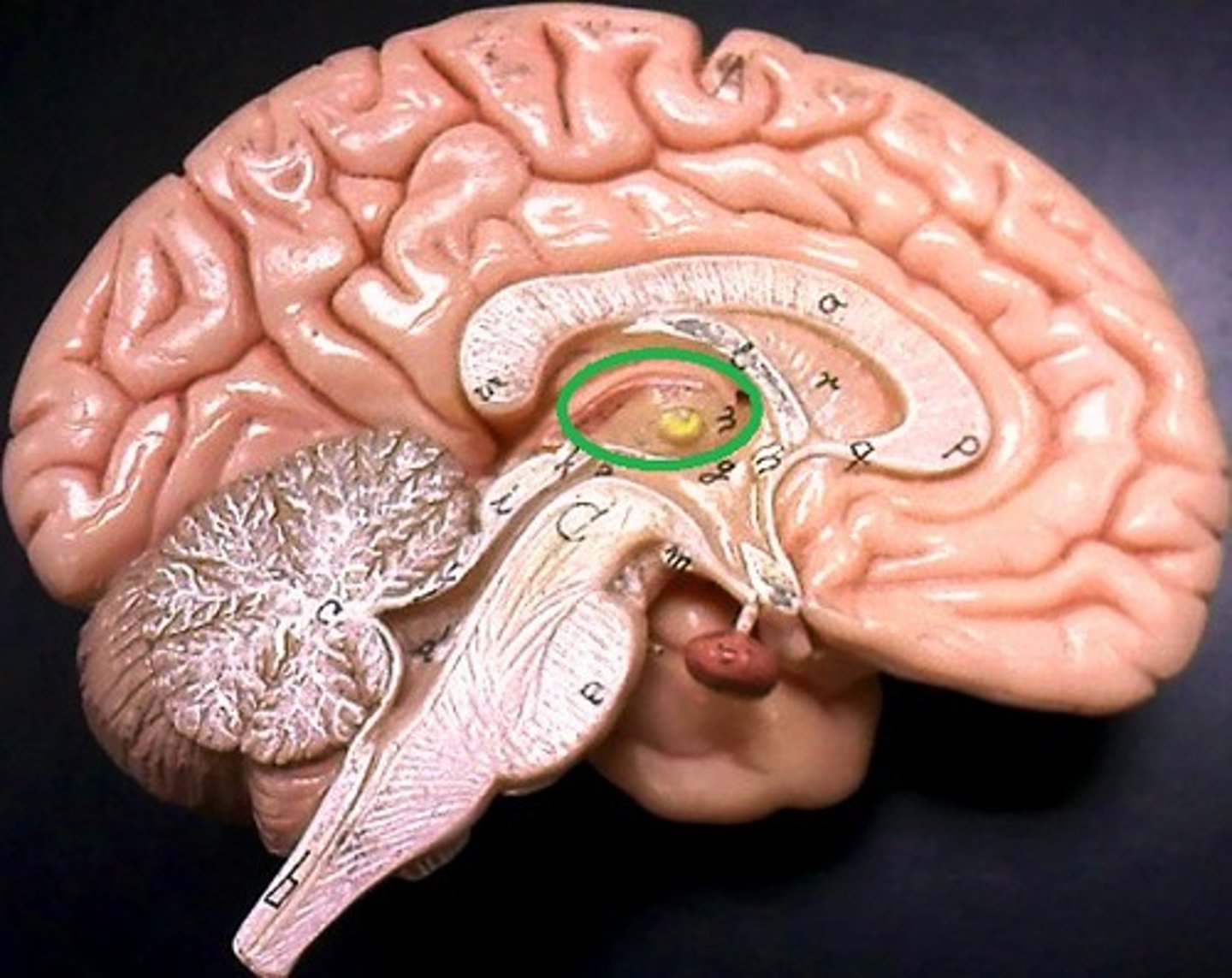
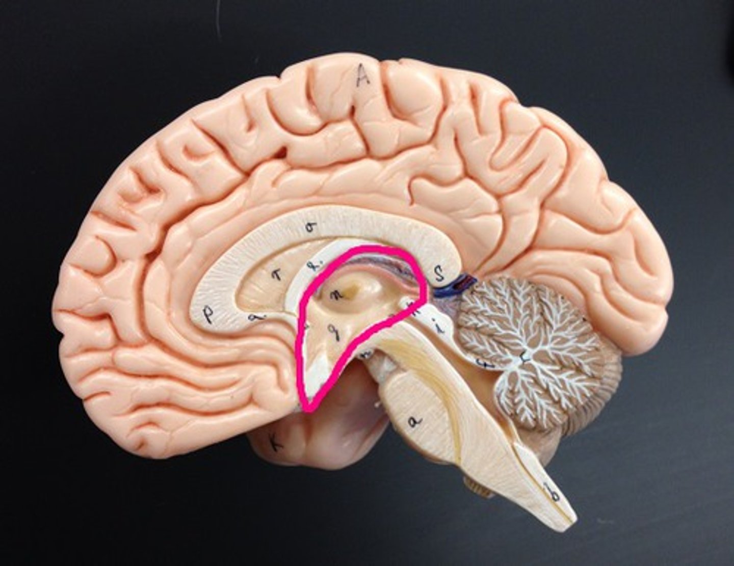
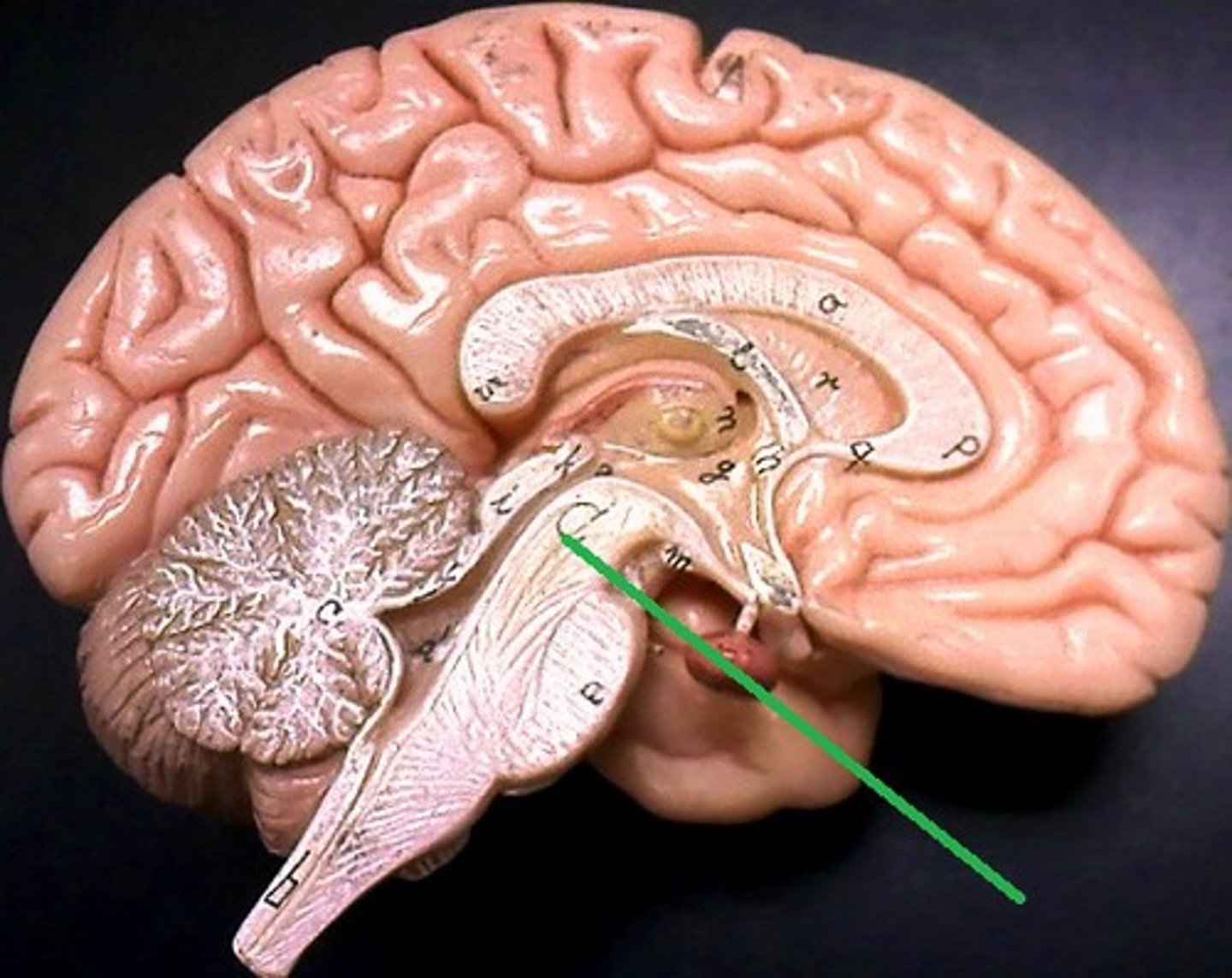
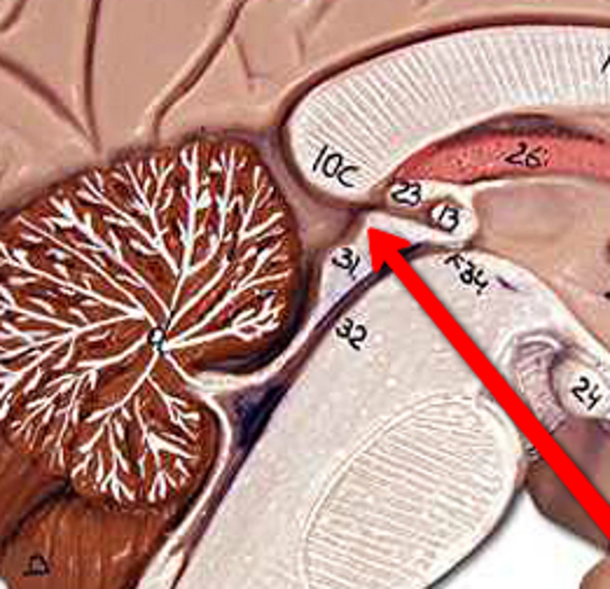
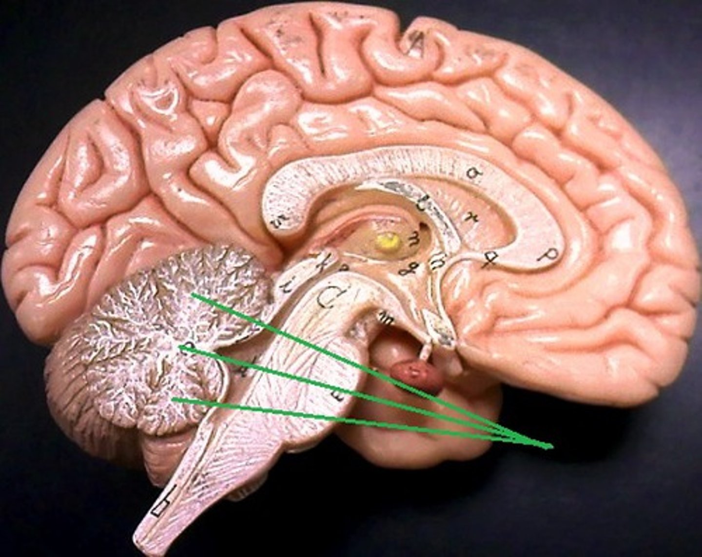
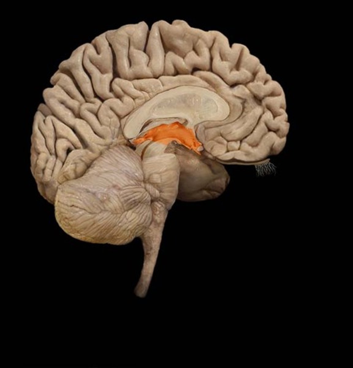
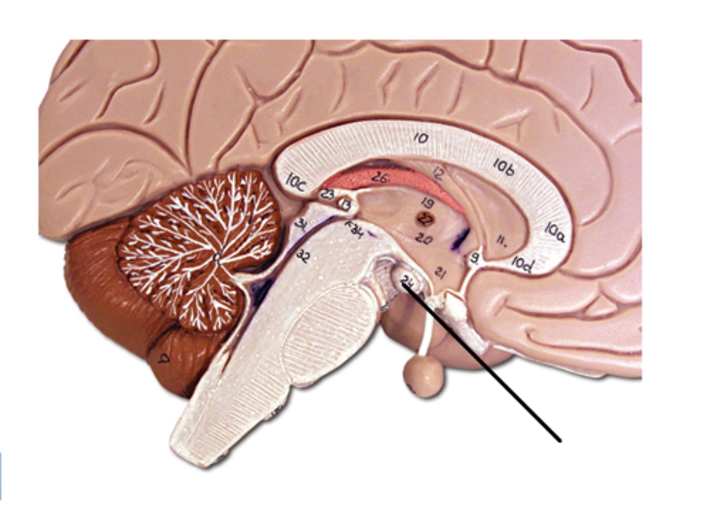
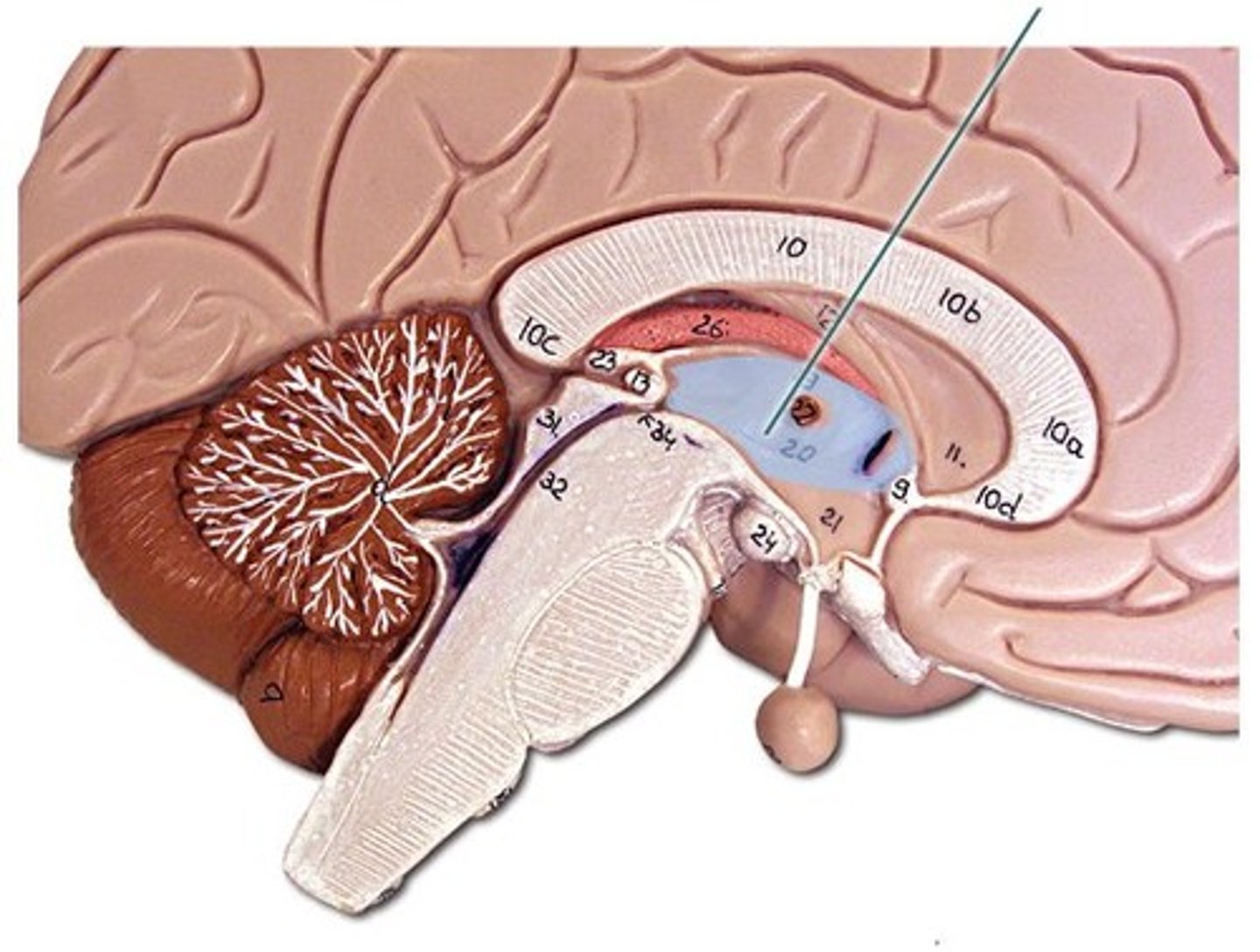
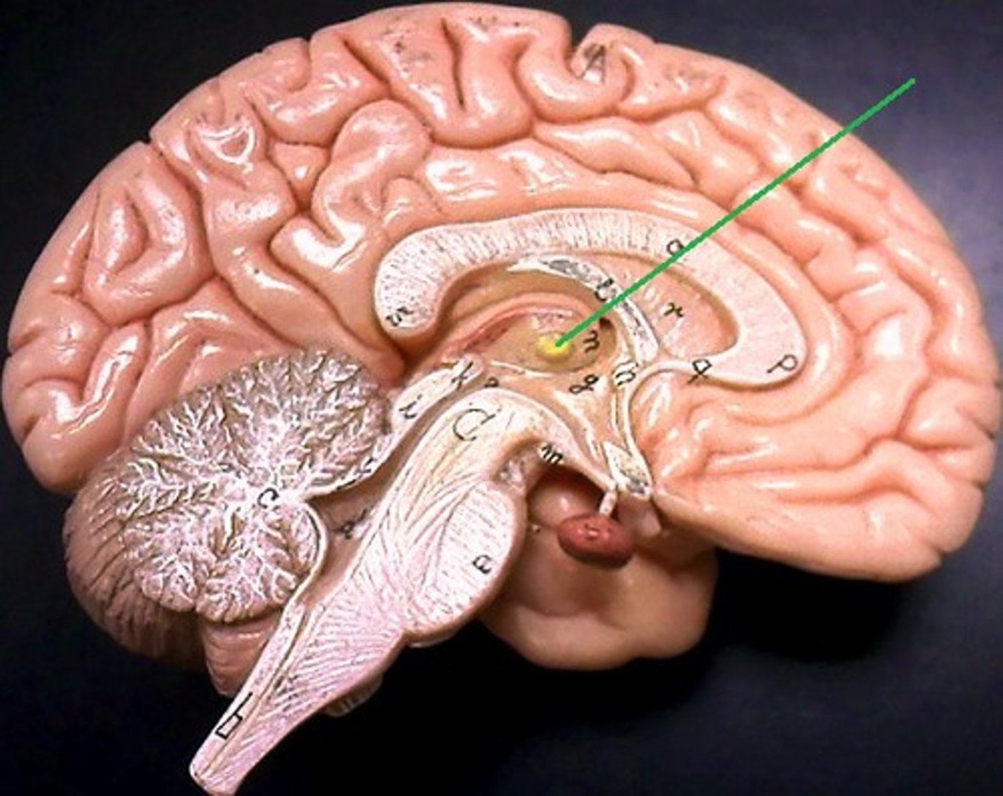
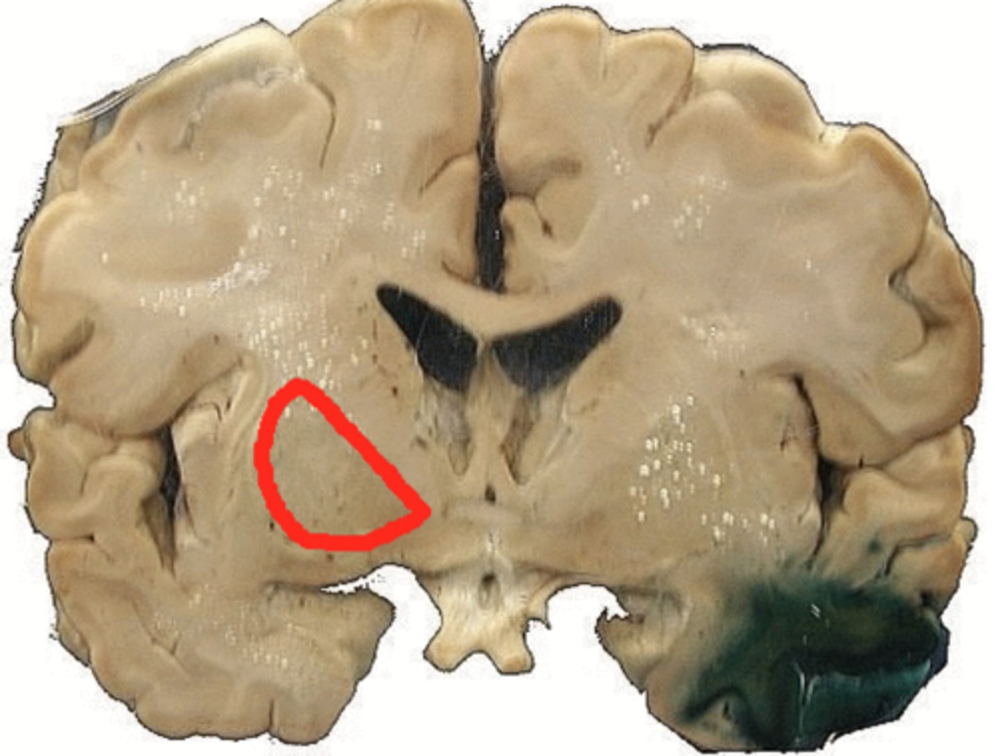
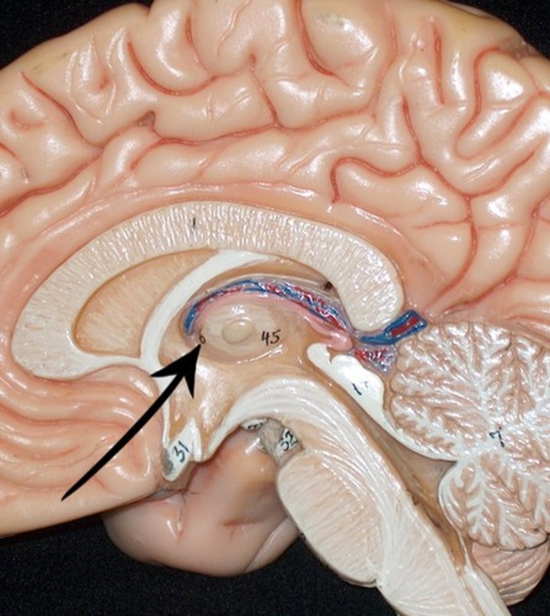
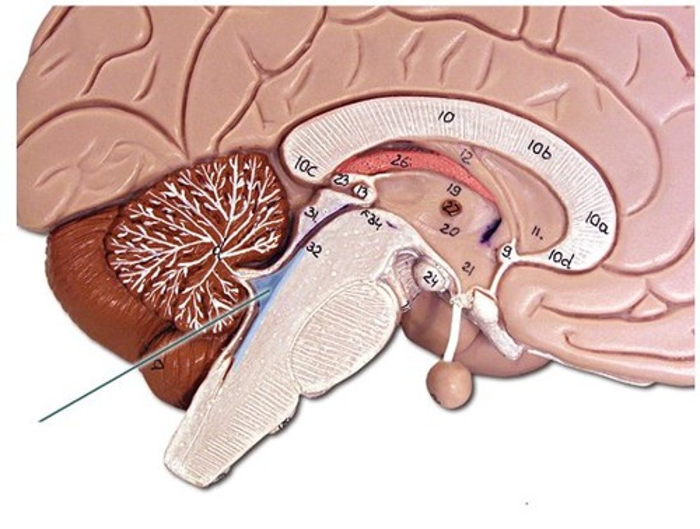
The Diencephalon (Two brain) sagittal section, medial view
Located in the brain's central area and has 3 main regions.
Hypothalamus (Below inner chamber)
Located below the thalamus and is a quadrangular-shaped structure.
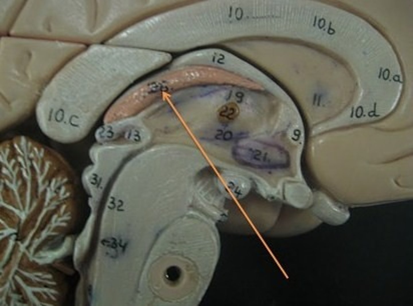
Thalamus (Inner chamber)
Composed of paired, egg-shaped bodies centrally located in the diencephalon and makes up approximately 80% of this structure.
Pineal gland
A small endocrine gland that secretes the hormone melatonin.
Diencephalon
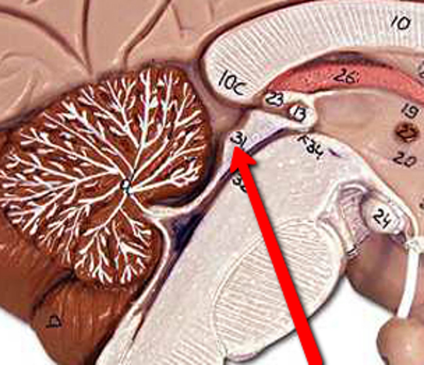
The Diencephalon continued
Sagittal section, magnified view
Mammillary body
Two small, round masses located just posterior to the infundibulum are relay stations for smell and taste reflexes.
Pineal gland
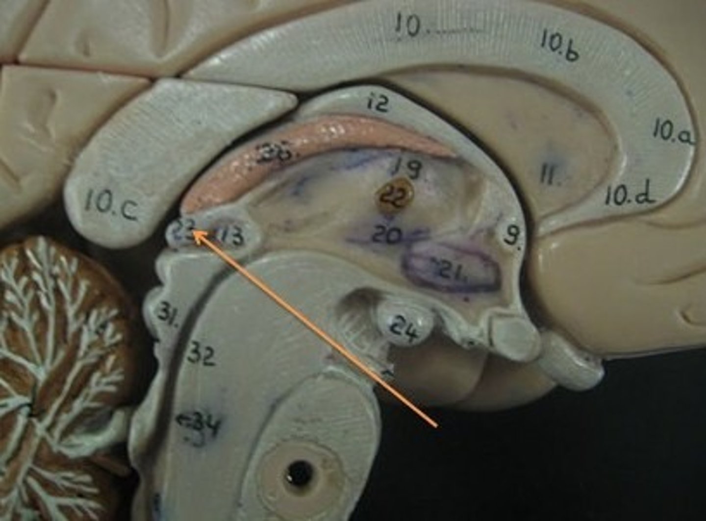
Thalamus
Intermediate mass of thalamus
A small bridge that connects each cerebral hemisphere that contains half of the thalamus.
Hypothalamus
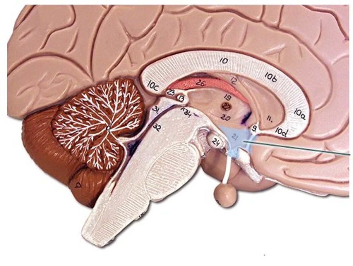
Infundibulum
A stalk that connects the pituitary gland to the hypothalamus.
Pituitary gland
Looks like a large pea and is attached to the end of the infundibulum. The hypothalamus control it.
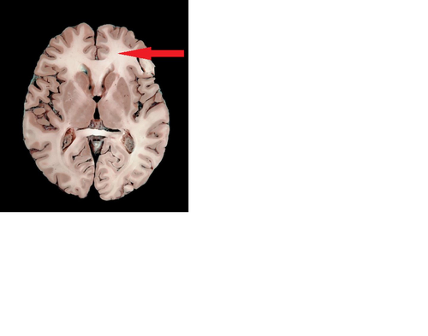
The Diencephalon continued
Frontal section view
Lateral ventricles brain
Thalamus
Third ventricles
Hypothalamus
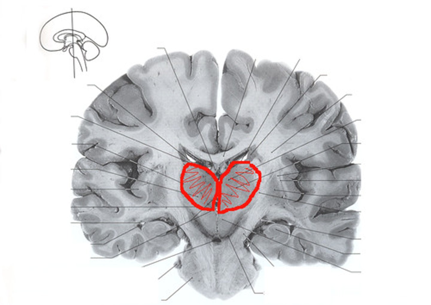
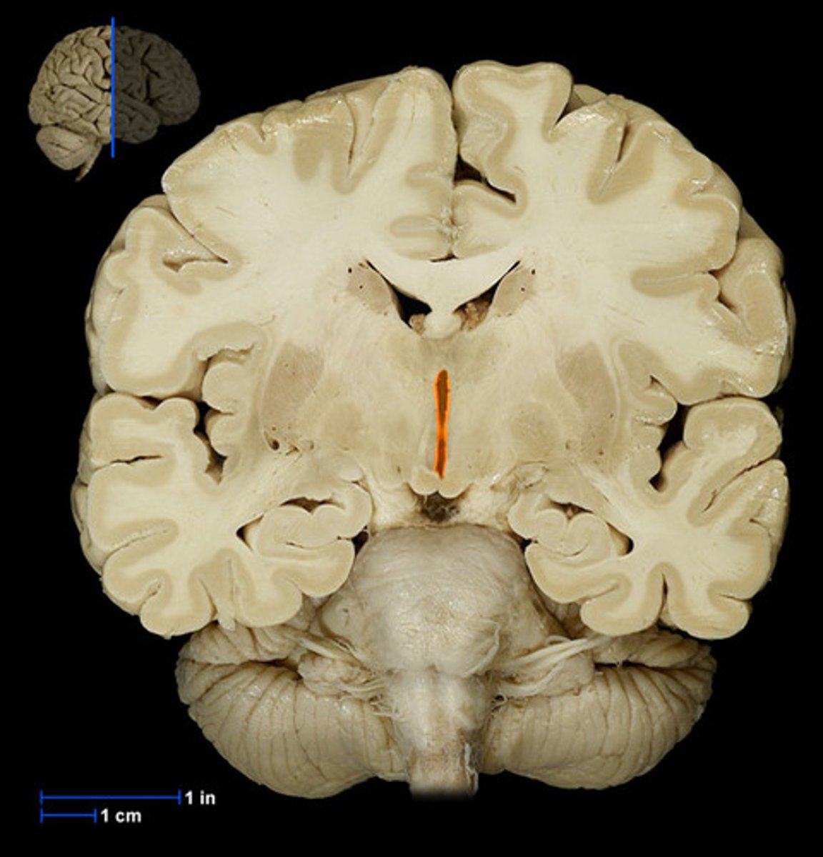
Gray and White matter in the cerebrum
Continued frontal section view
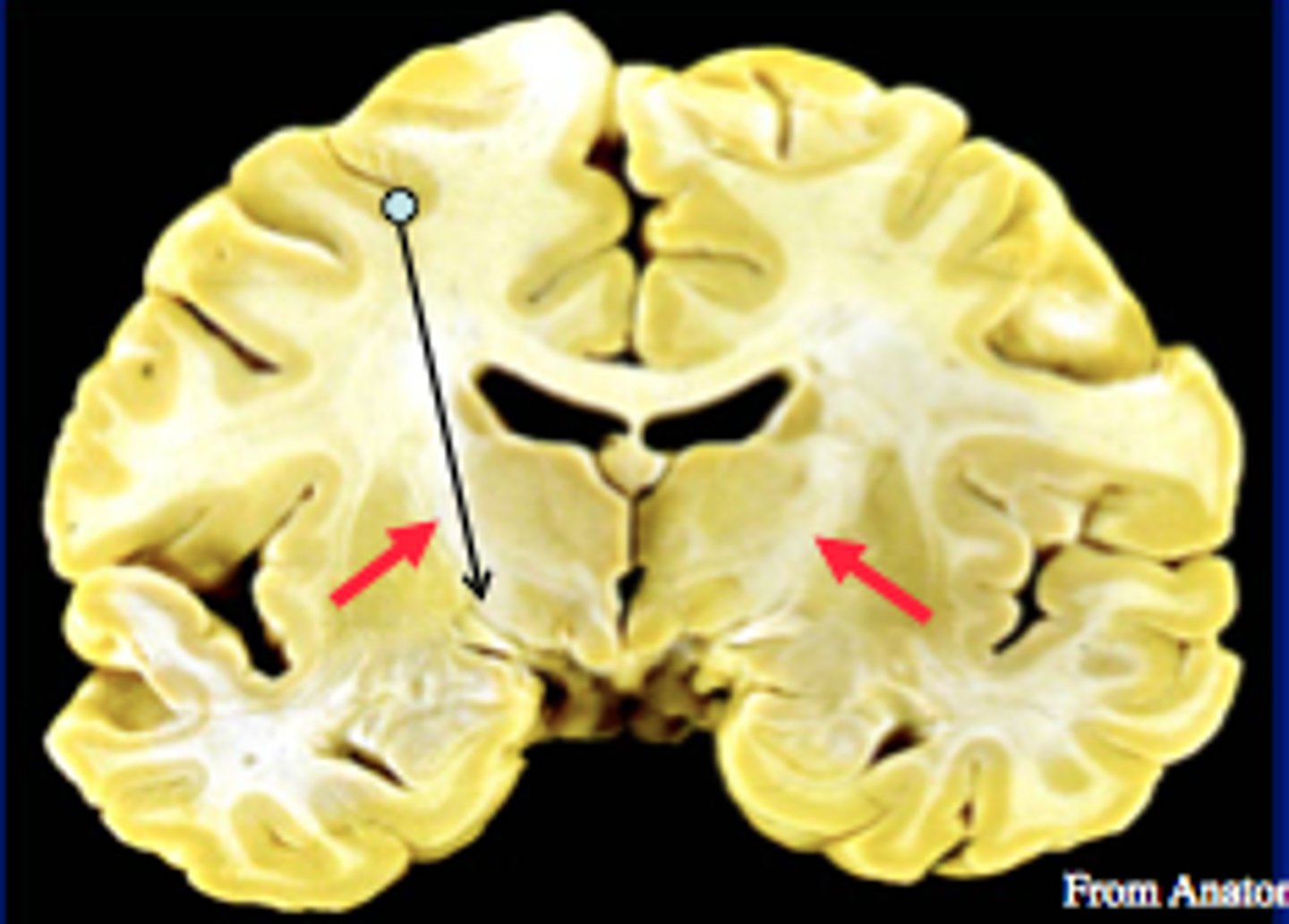
Basal nuclei (Grey matter)
Areas of cerebral gray matter composed of paired nuclei (clusters of neuron cell bodies in the CNS) that are found deep within each cerebral hemisphere.
Cerebral cortex (Grey matter)
The superficial cerebral gray matter on the exterior of the cerebrum composed of nerve cell bodies and dendrites.

Cerebral white matter
Lies deep to the outer cortex and is composed mostly of myelinated axons that give it the white appearance.
Corpus callosum (Commissural fibers)
A prominent commissural fiber tract that is readily observable in midsagittal sections of the brain, connects the two cerebral hemispheres.
Fornix (Association fibers)
Looks like a group of commissural fibers but is actually a tract of arched association fibers.
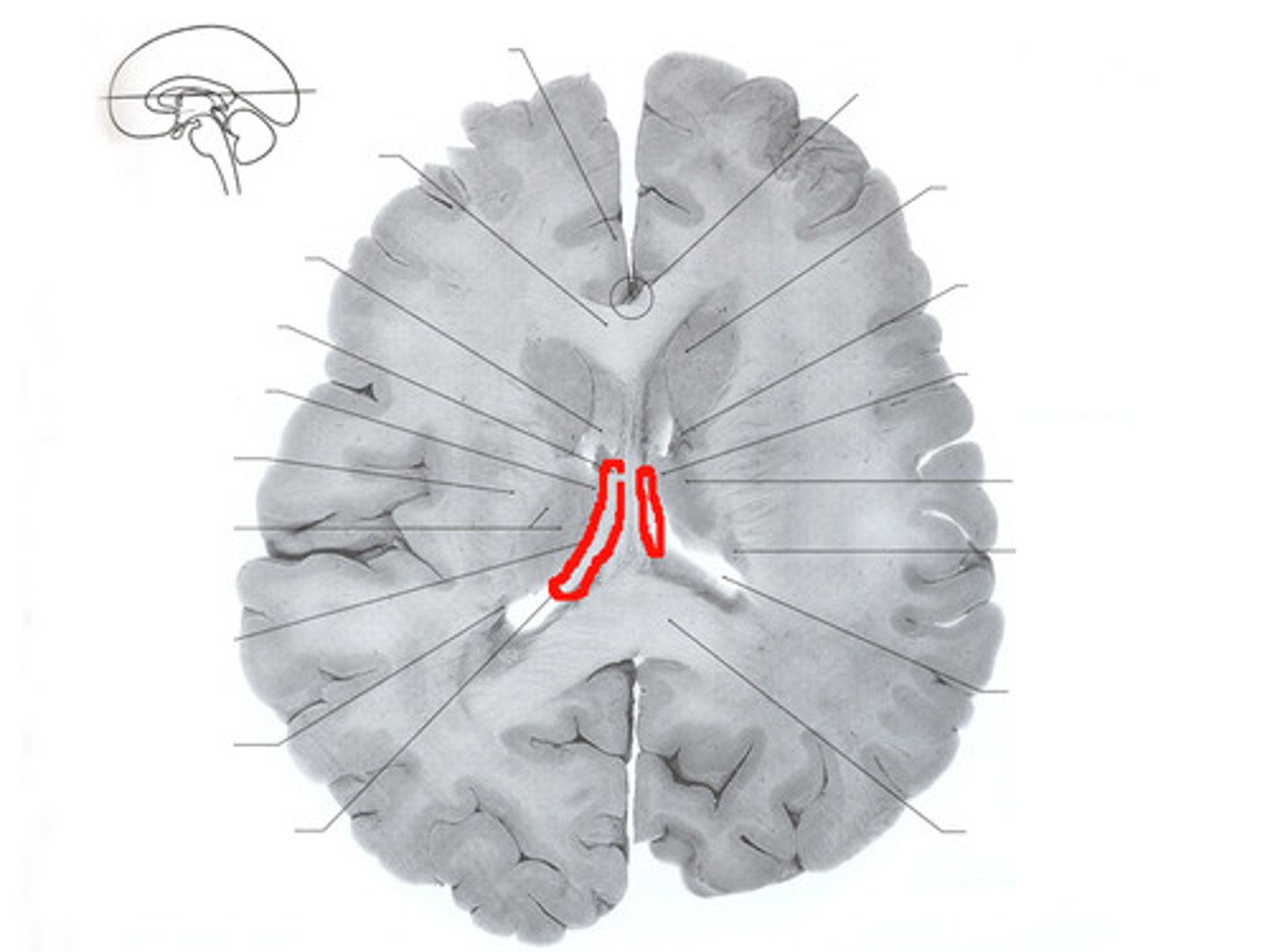
Internal capsule (Projection fibers)
A large group of projection fibers,
contains sensory and motor tracts that connect the cerebral cortex to the brain stem and spinal cord.
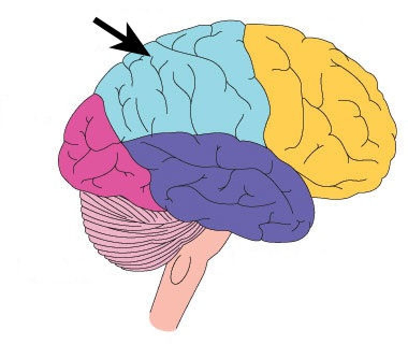
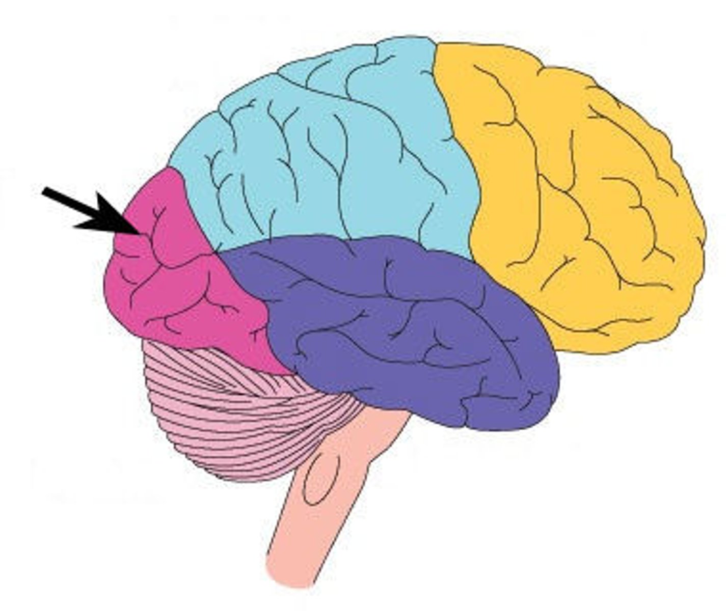
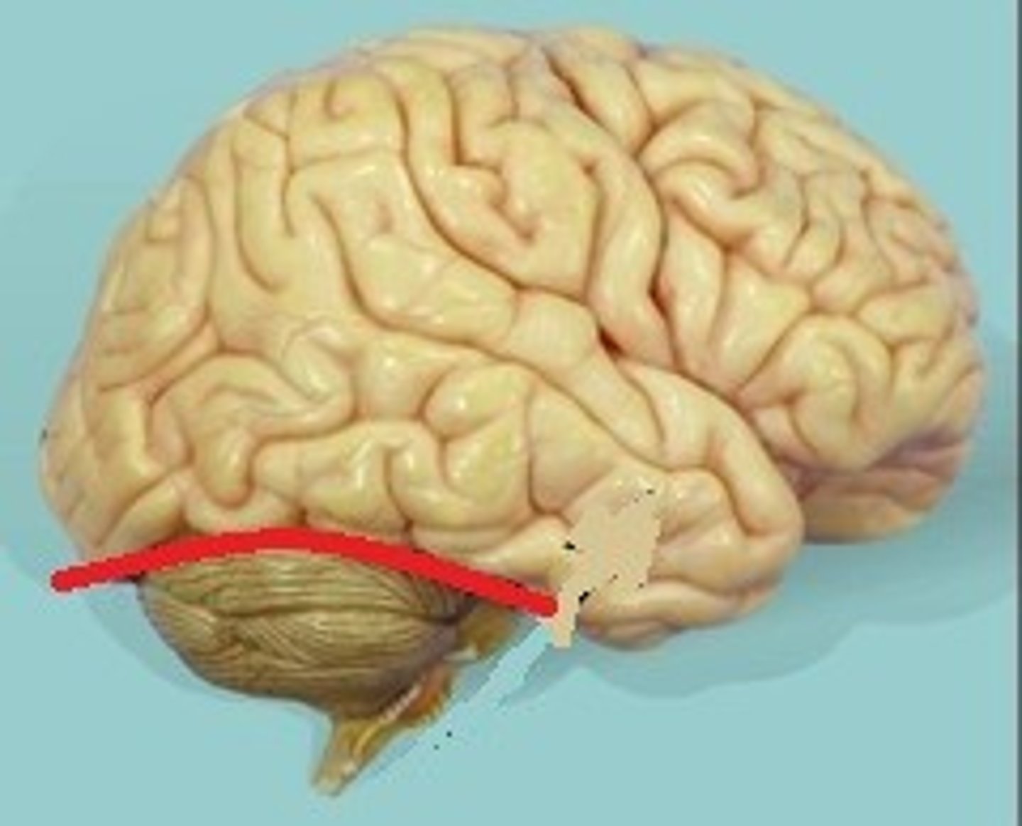
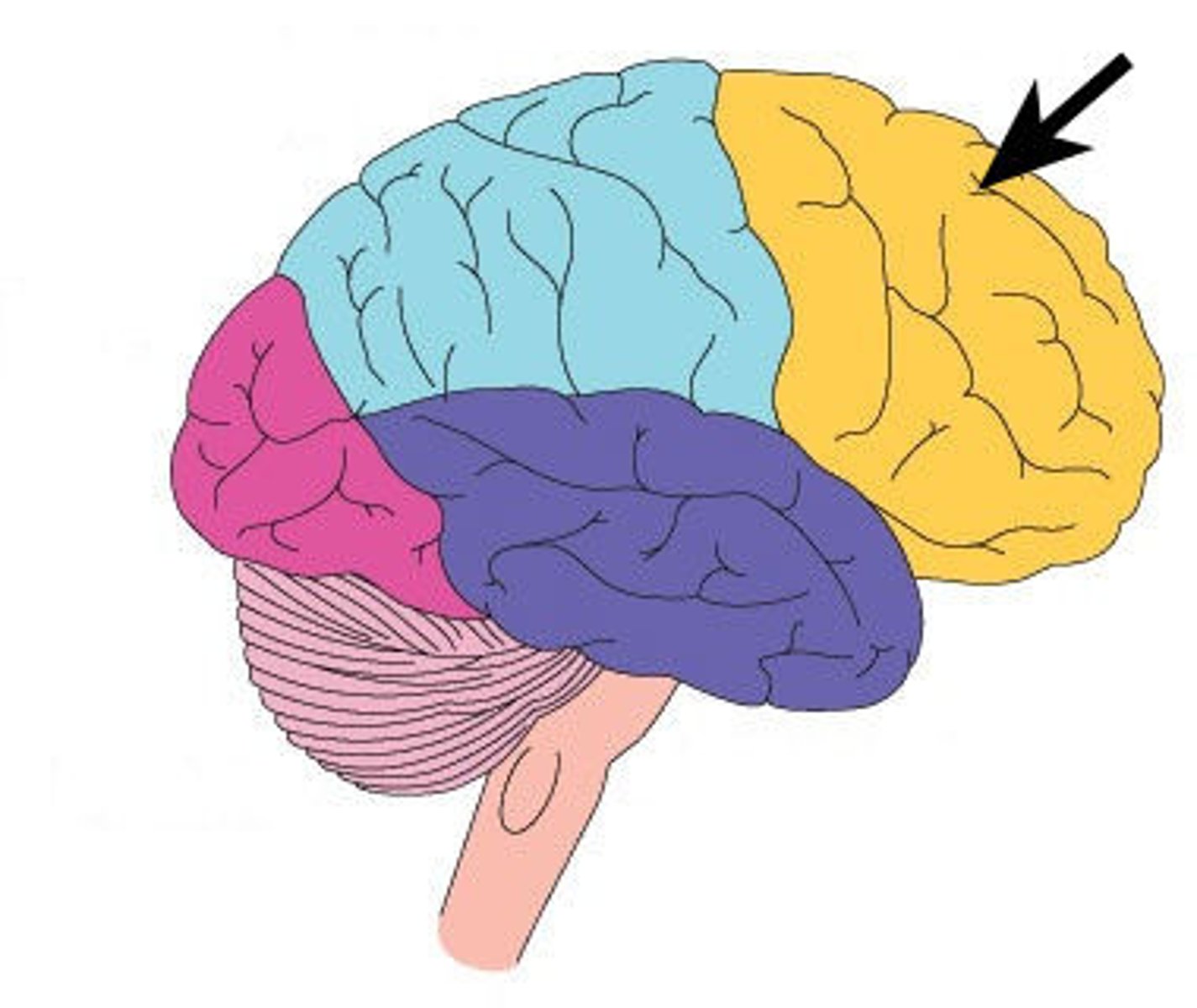
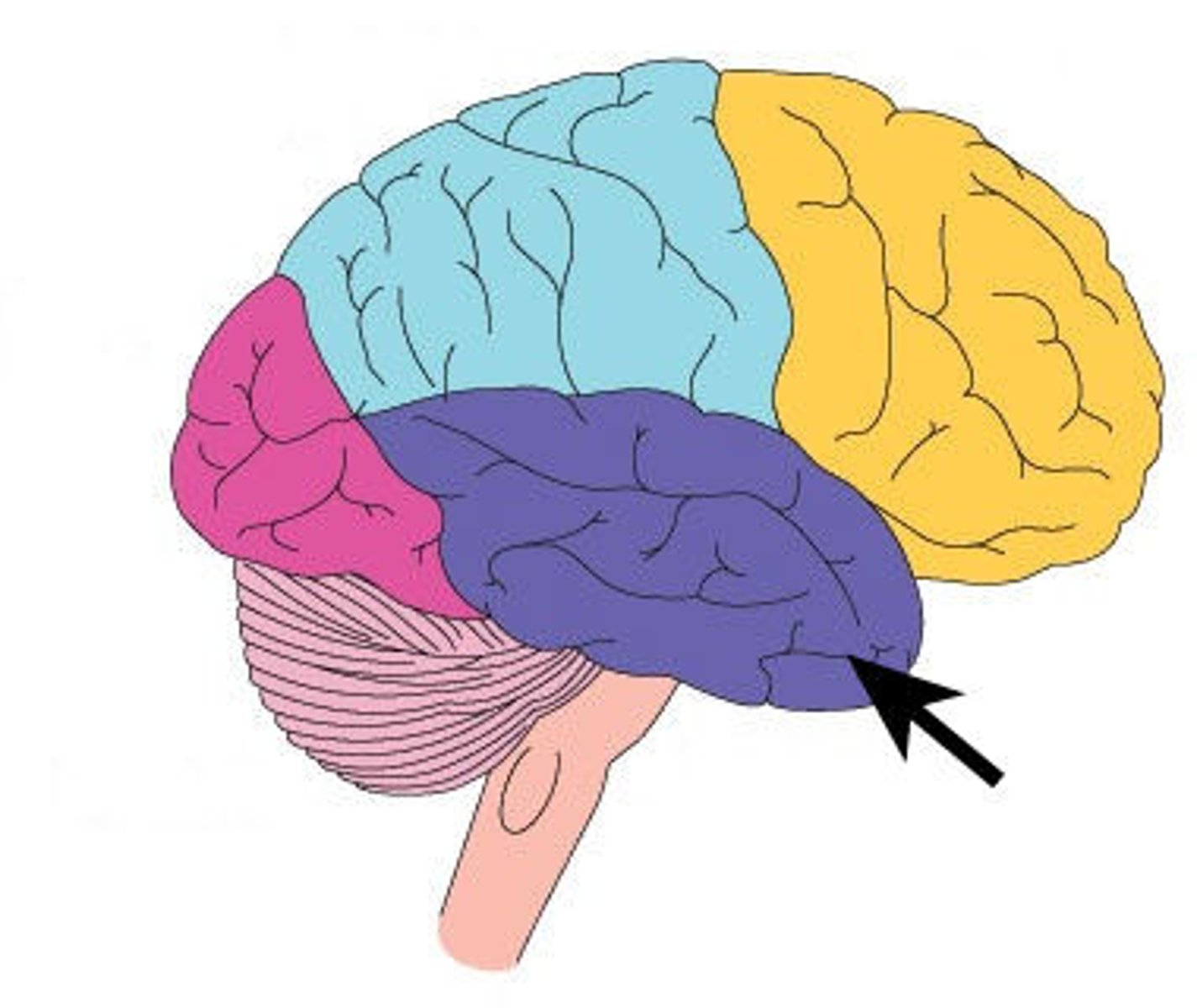
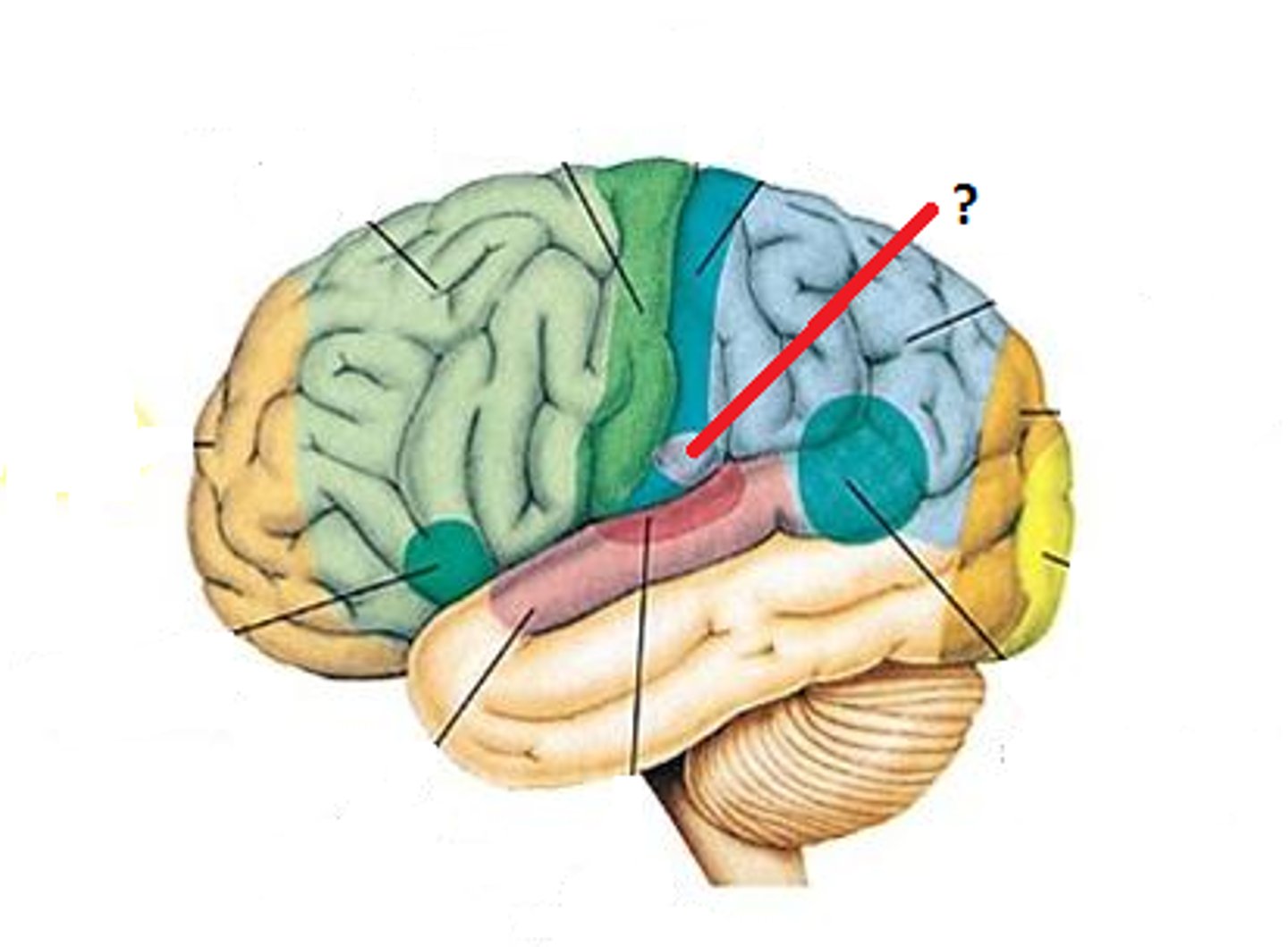
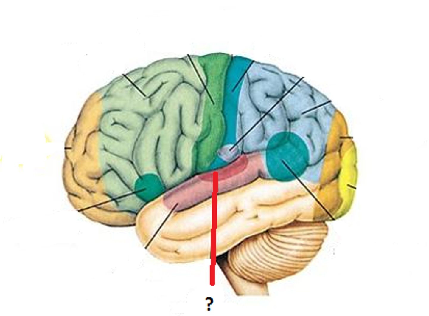
External features of the Cerebrum, right lateral view with temporal lobe cut away
It is composed of 4 lobes which are mainly named for the overlying cranial bones.
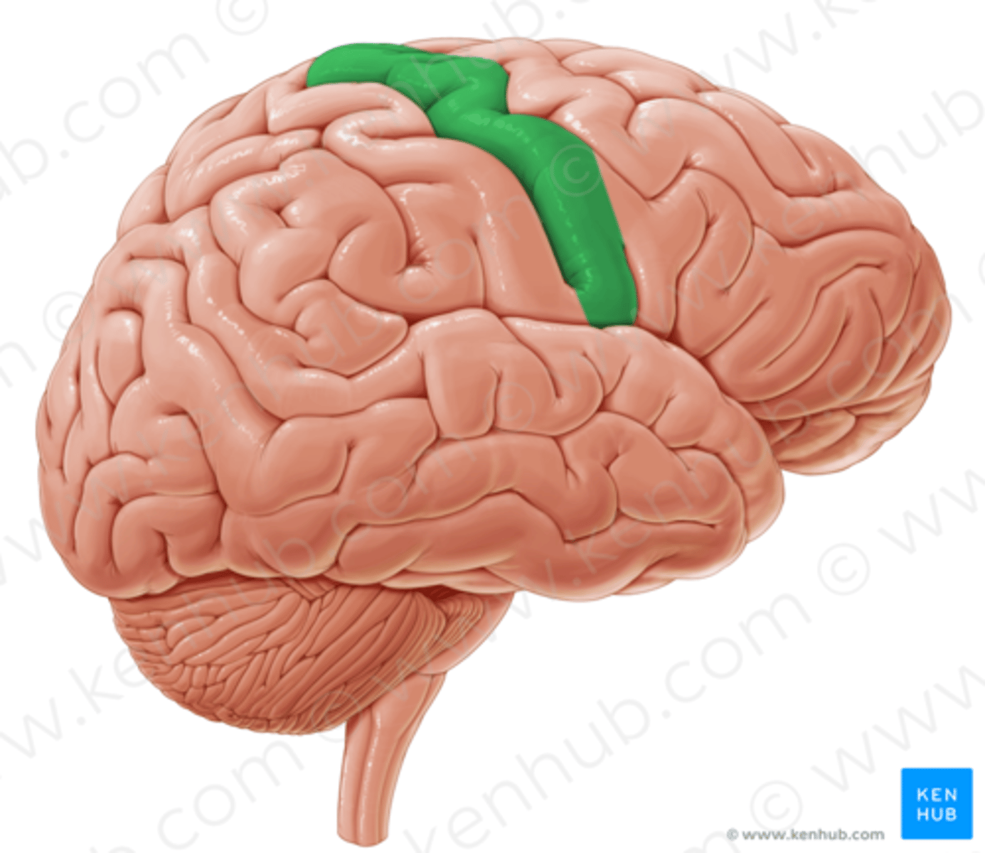
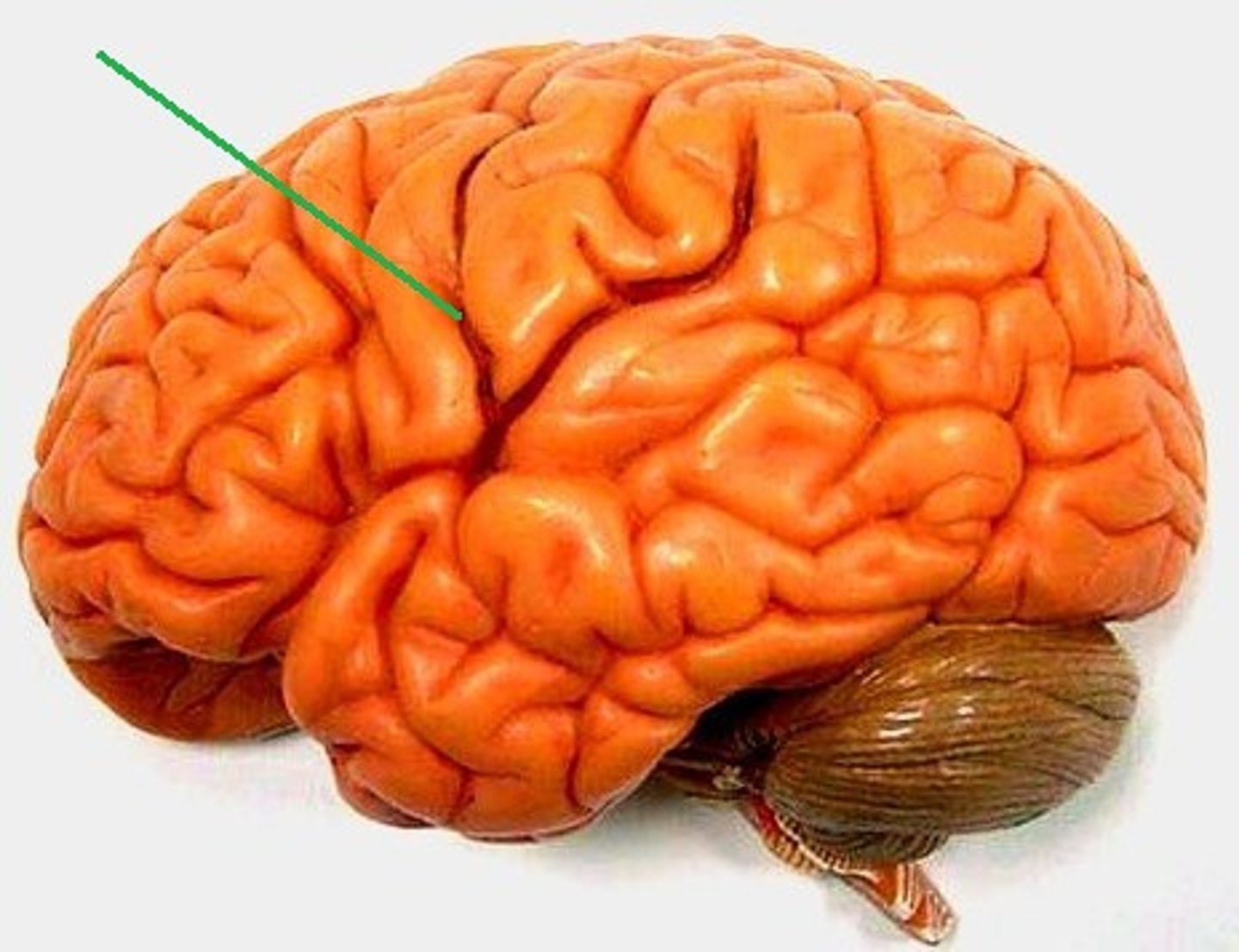
Central sulcus (furrow)
Shallow groove separating frontal lobe from parietal lobe.
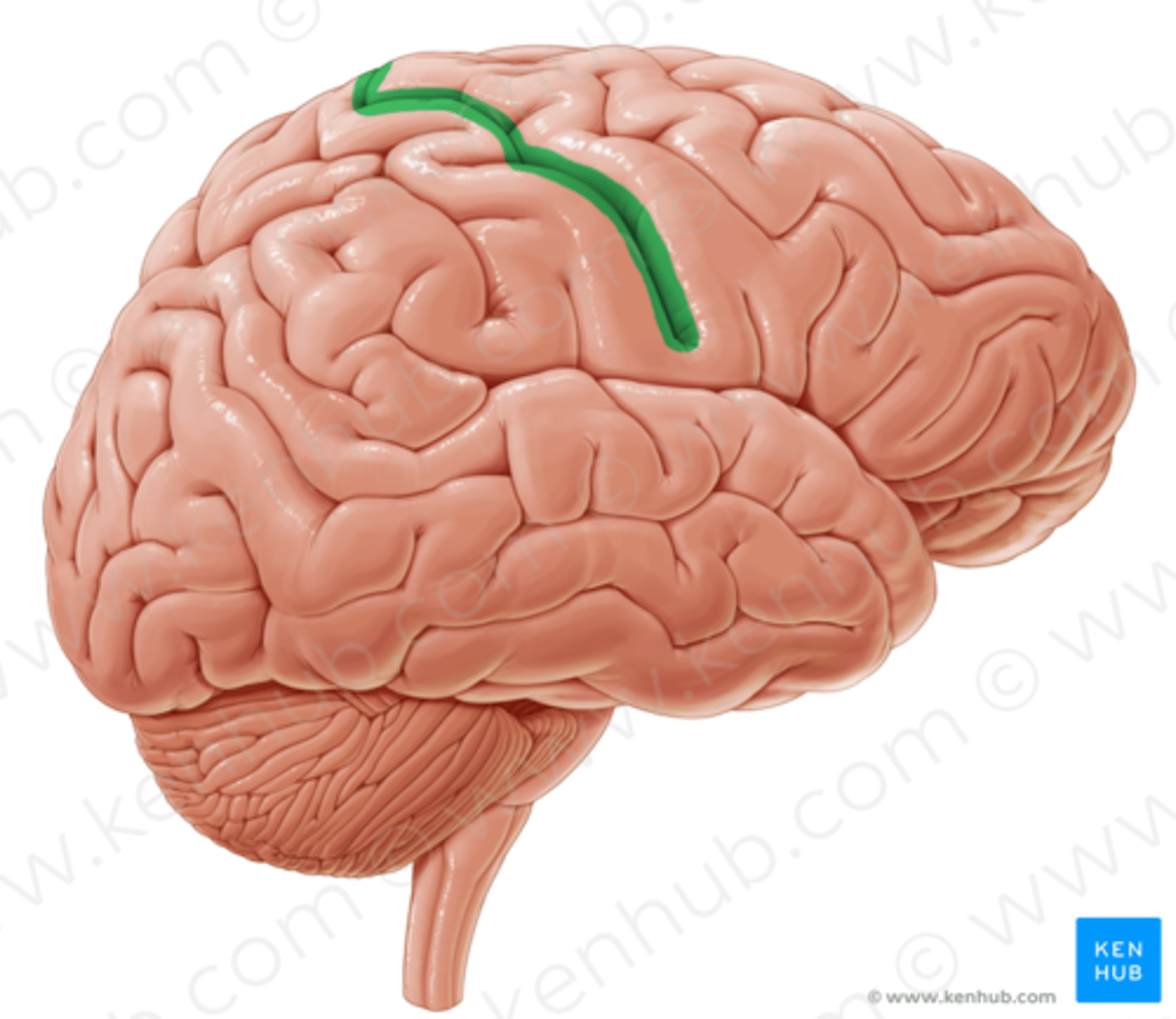
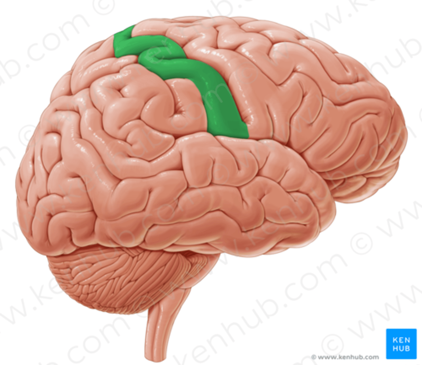
Postcentral gyrus (circle)
Elevation located just posterior to the central sulcus.
Parietal lobe
Occipital lobe
Transverse fissure
Deep groove separating the cerebrum from the cerebellum in the posterior/inferior part of the brain.
Precentral gyrus (circle)
Elevation located just anterior to the central sulcus.
Frontal lobe
Insula
An inner lobe that lies deep to the lateral cerebral fissure and is not visible from the exterior.
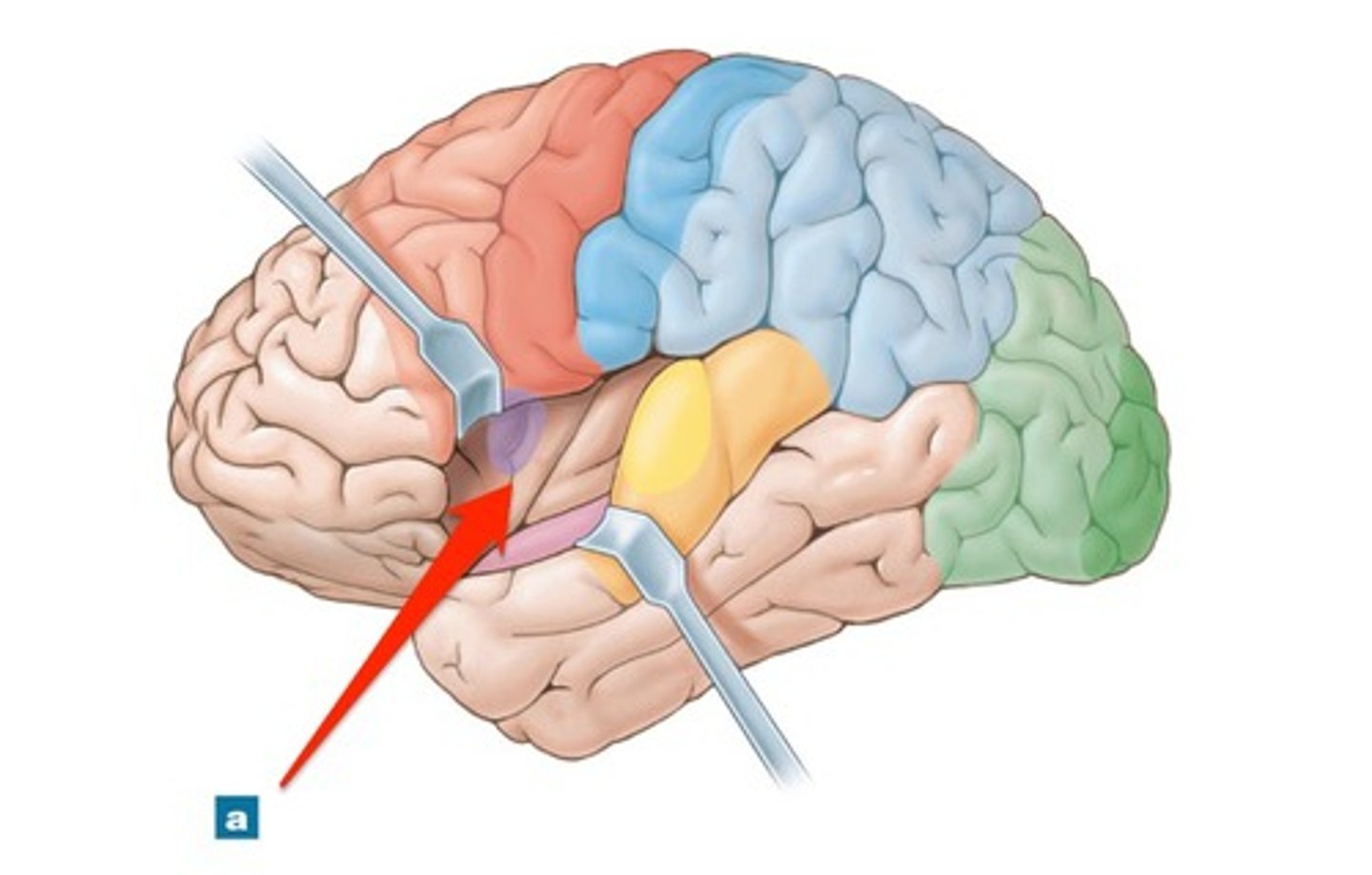
Temporal lobe
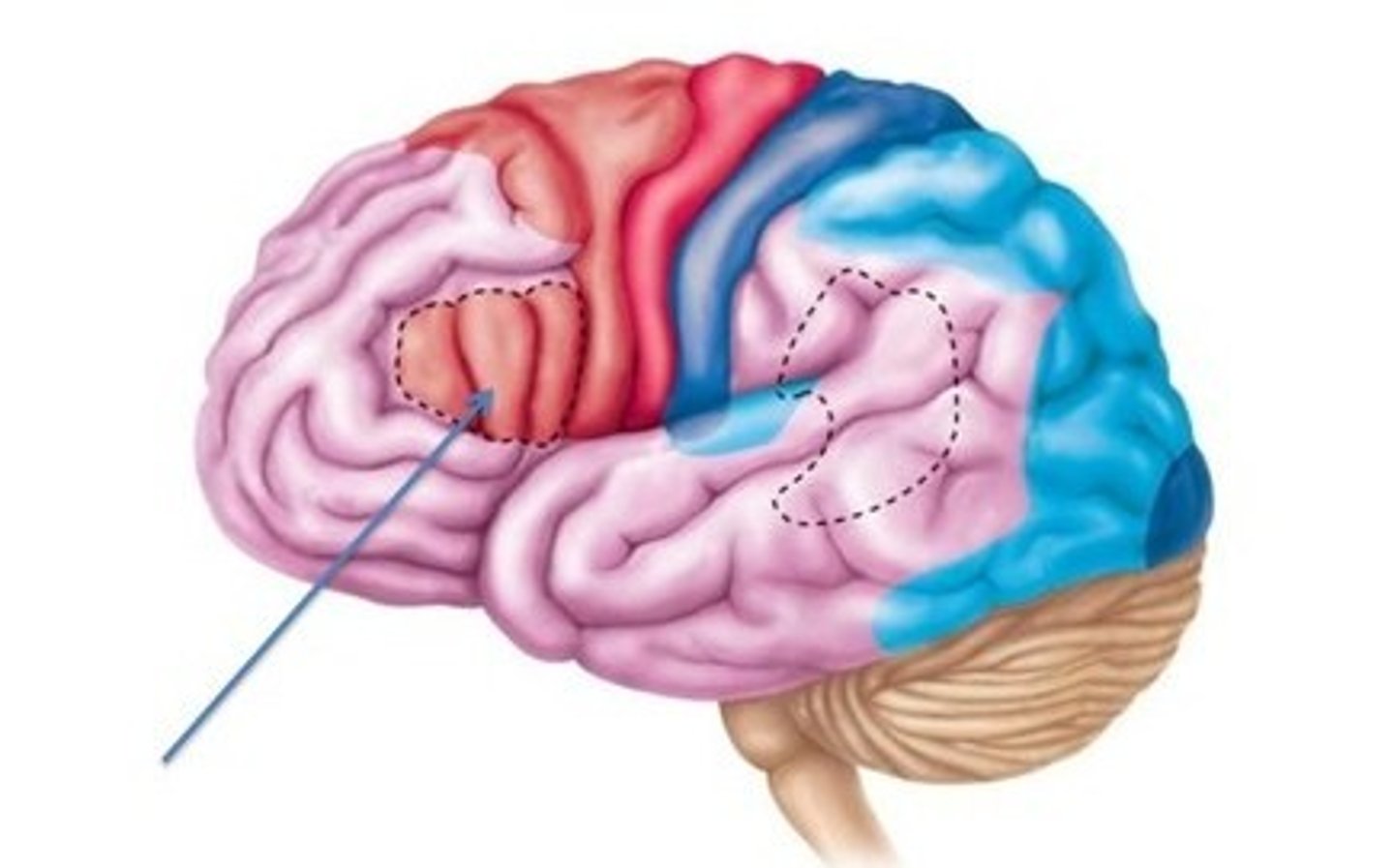
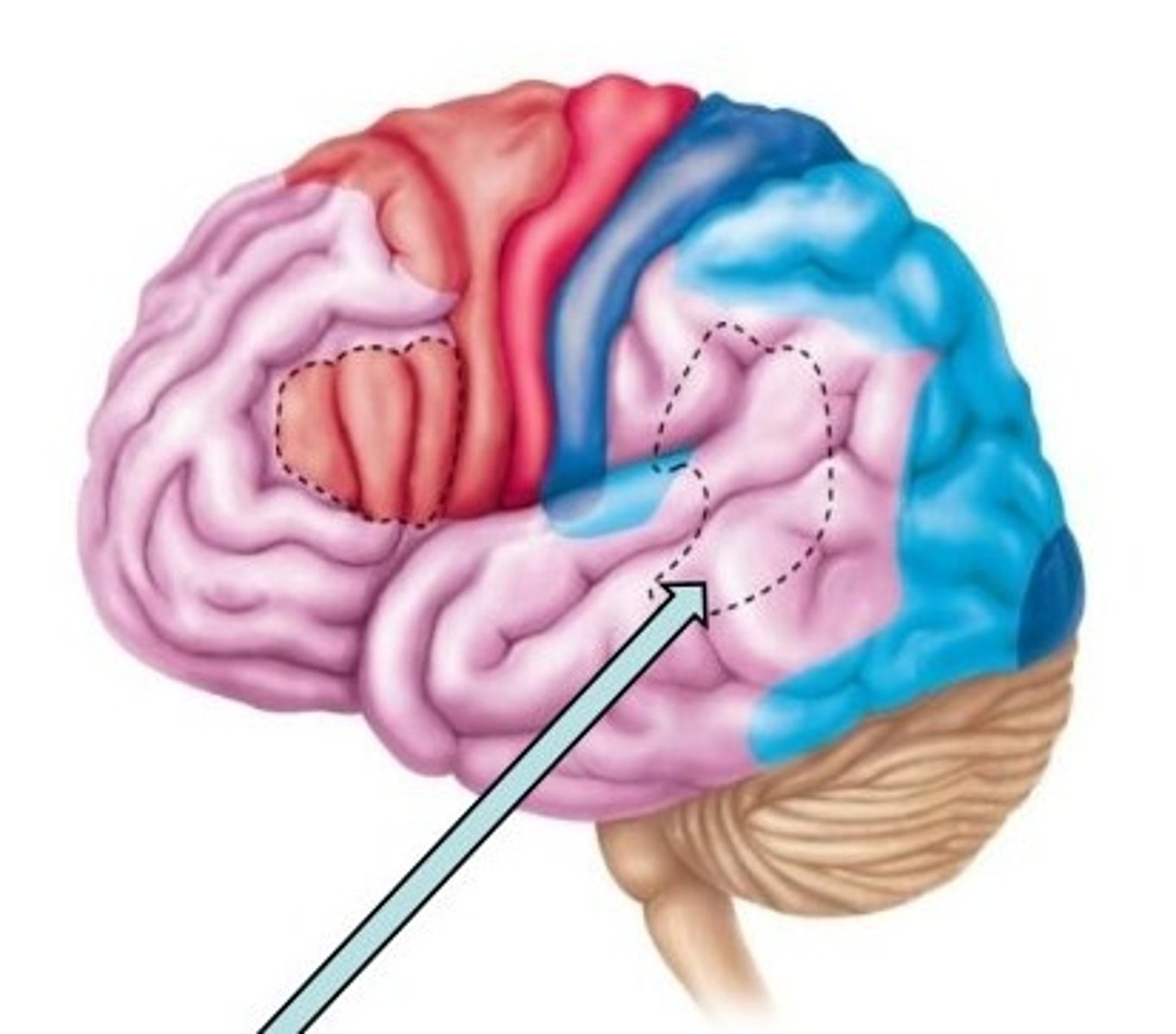
Functional areas of the Cerebral cortex, left lateral view
It is composed of 3 types of functional areas; motor, sensory, and association areas.
Broca's speech area
Located superior to the lateral sulcus and anterior to the primary motor cortex, usually in the left hemisphere; initiates impulses that result in speech. (Motor area)
Primary gustatory area
In each postcentral gyrus, just superior to the lateral sulcus; receives impulses when the taste buds are stimulated. (Sensory area)
Primary motor area
Located in the precentral gyrus of each frontal lobe; initiates impulses to skeletal muscles. (Motor area)
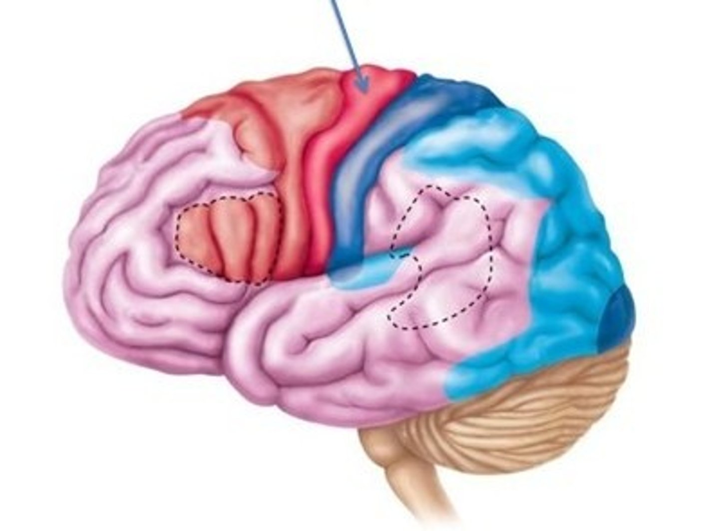
Central sulcus
Primary somatosensory area
Located in the postcentral gyrus of each parietal lobe; receives nerve impulses for touch, proprioception, pain, and temperature. (Sensory area, Selected Association area)
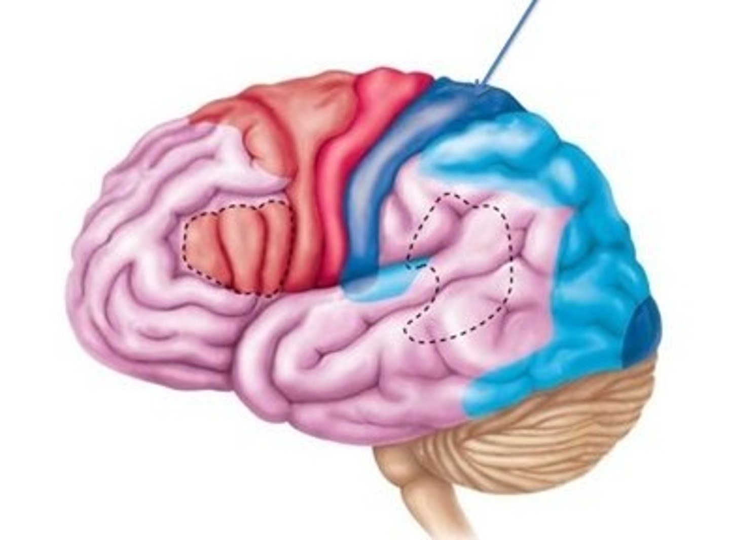
Primary visual area
In the posterior occipital lobe; receives impulses from the thalamus when the retina is stimulated. (Sensory area, Selected Association area)
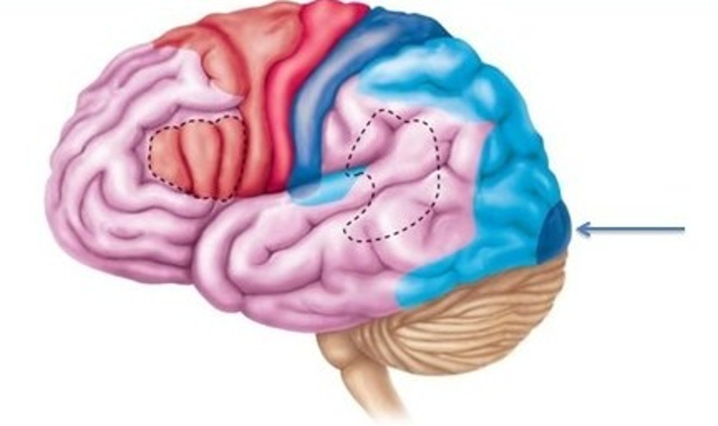
Wernicke's area
Located in left temporal and parietal lobes; recognizes spoken words, translates words into thoughts, and possibly helps us sound out strange or new words. (Selected Association area)
Primary auditory area
Located in each temporal lobe across the lateral sulcus from the gustatory area; receives impulses when the auditory receptors of the ear are stimulated. (Sensory area, Selected Association area)
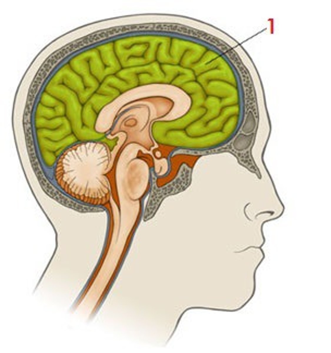
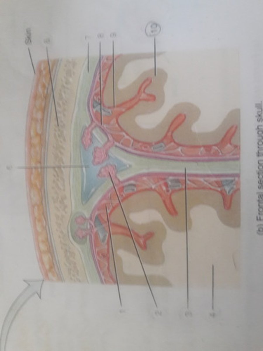
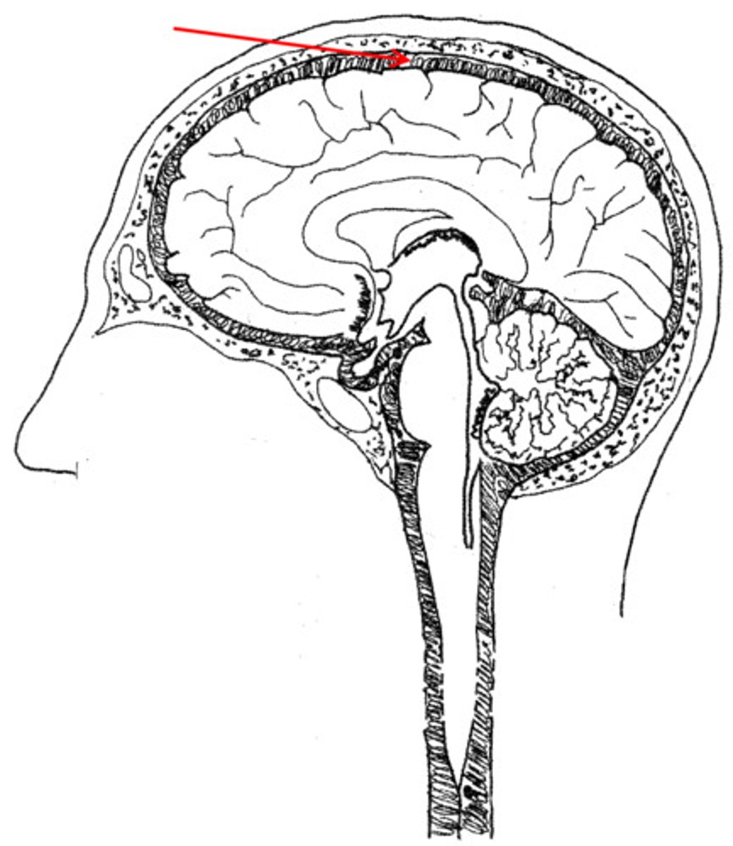
Cranial meninges, frontal section through skull
There are 3 that are made of connective tissue membranes; the dura mater, arachnoid mater, and pia mater.
Subarachnoid space
Between the arachnoid and pia.
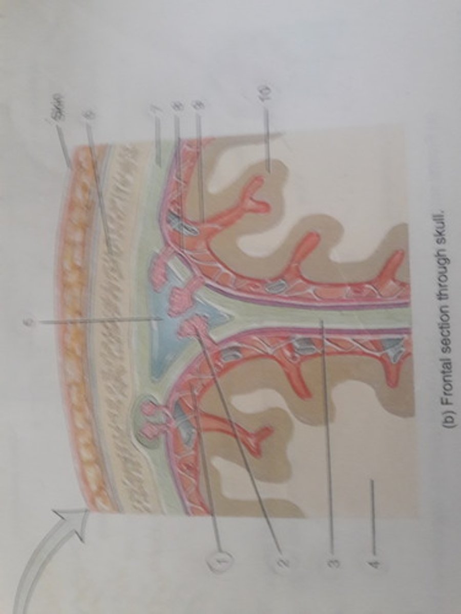
Arachnoid villus (Spider-like tiny projections)
Projections of the arachnoid mater into the dural sinuses.
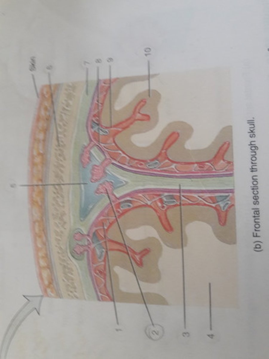
Falx (sickle-shaped)cerebri
The double-layered dura mater extends deep into the longitudinal fissure.
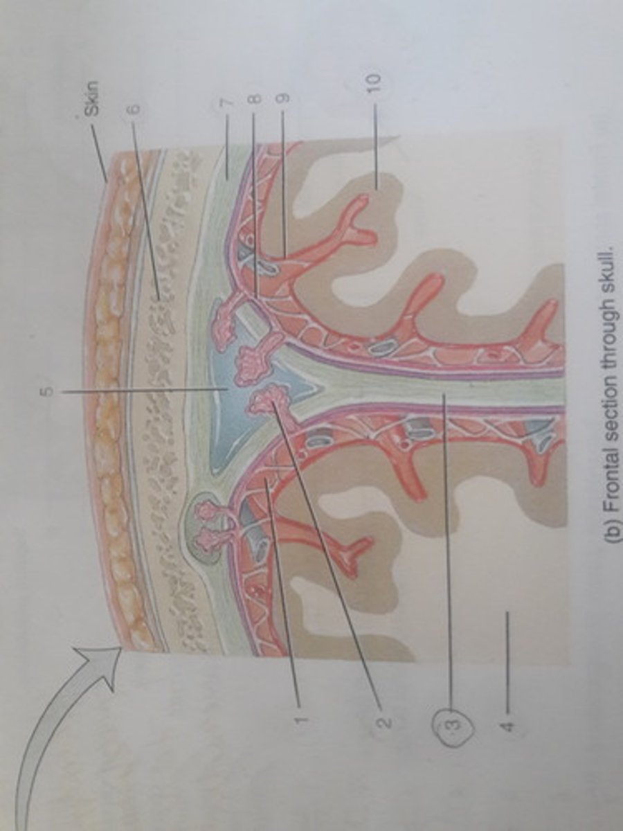
White matter
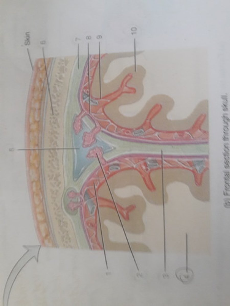
Superior sagittal sinus
Located superior to the longitudinal fissure, is one of the main dural sinuses.
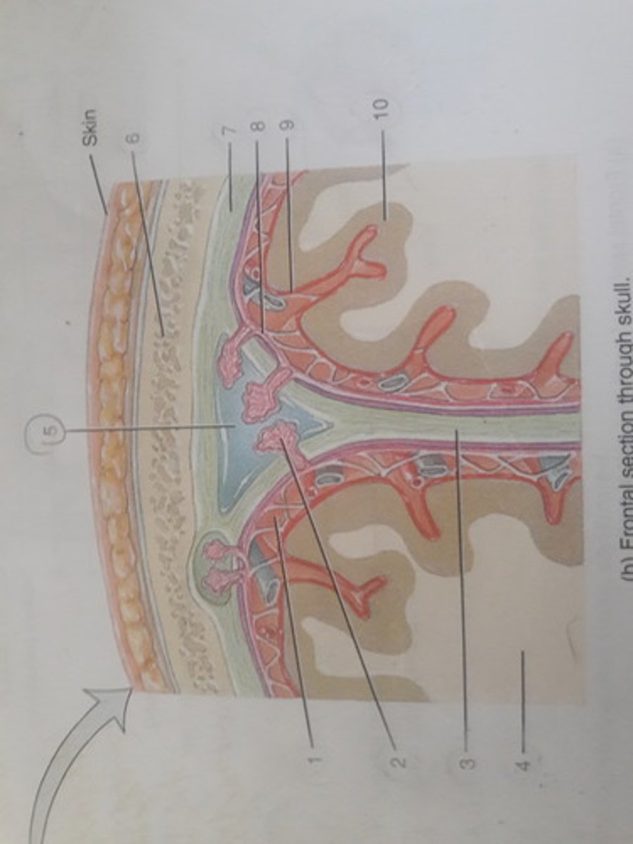
Parietal bone
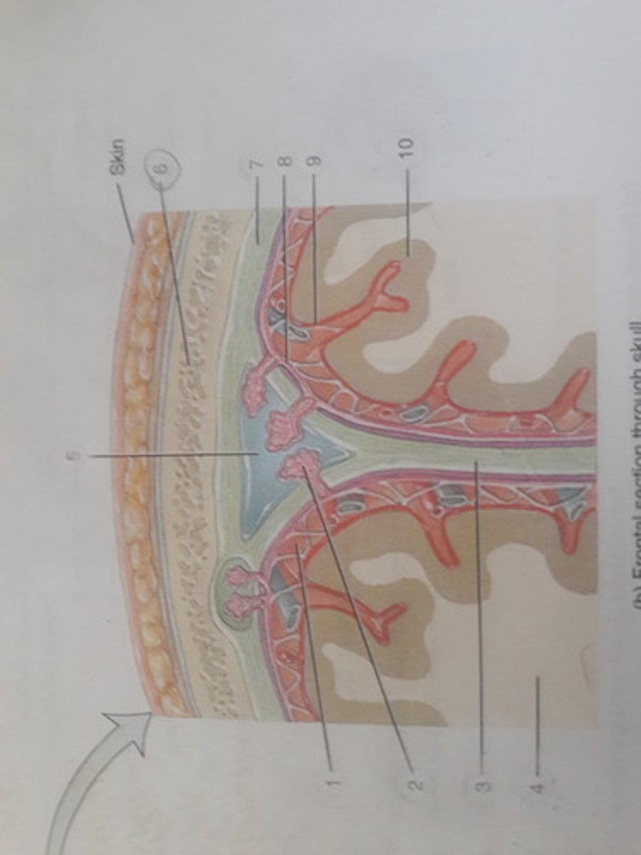
Dura mater (Hard mother)
The first meninx (sing) located deep to the cranial bones.
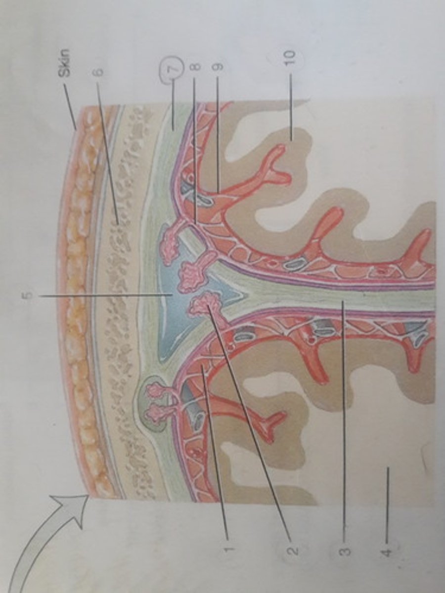
Arachnoid mater (Spider-like mother)
The 2nd meninx (sing) located deep to the dura mater.
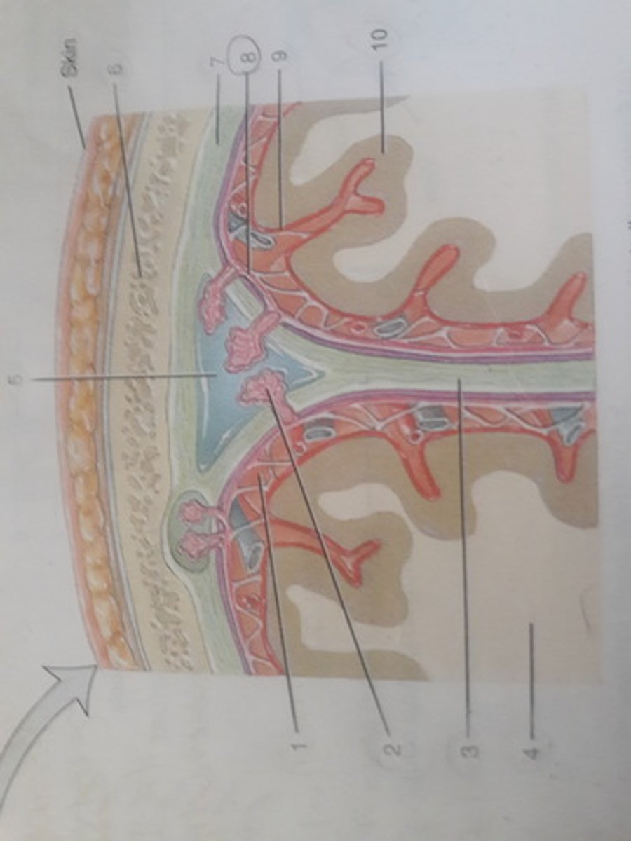
Pia mater (Delicate mother)
The thin, inner meninx (sing). It hugs and overlays the cerebral cortex, following each gyrus and sulcus.
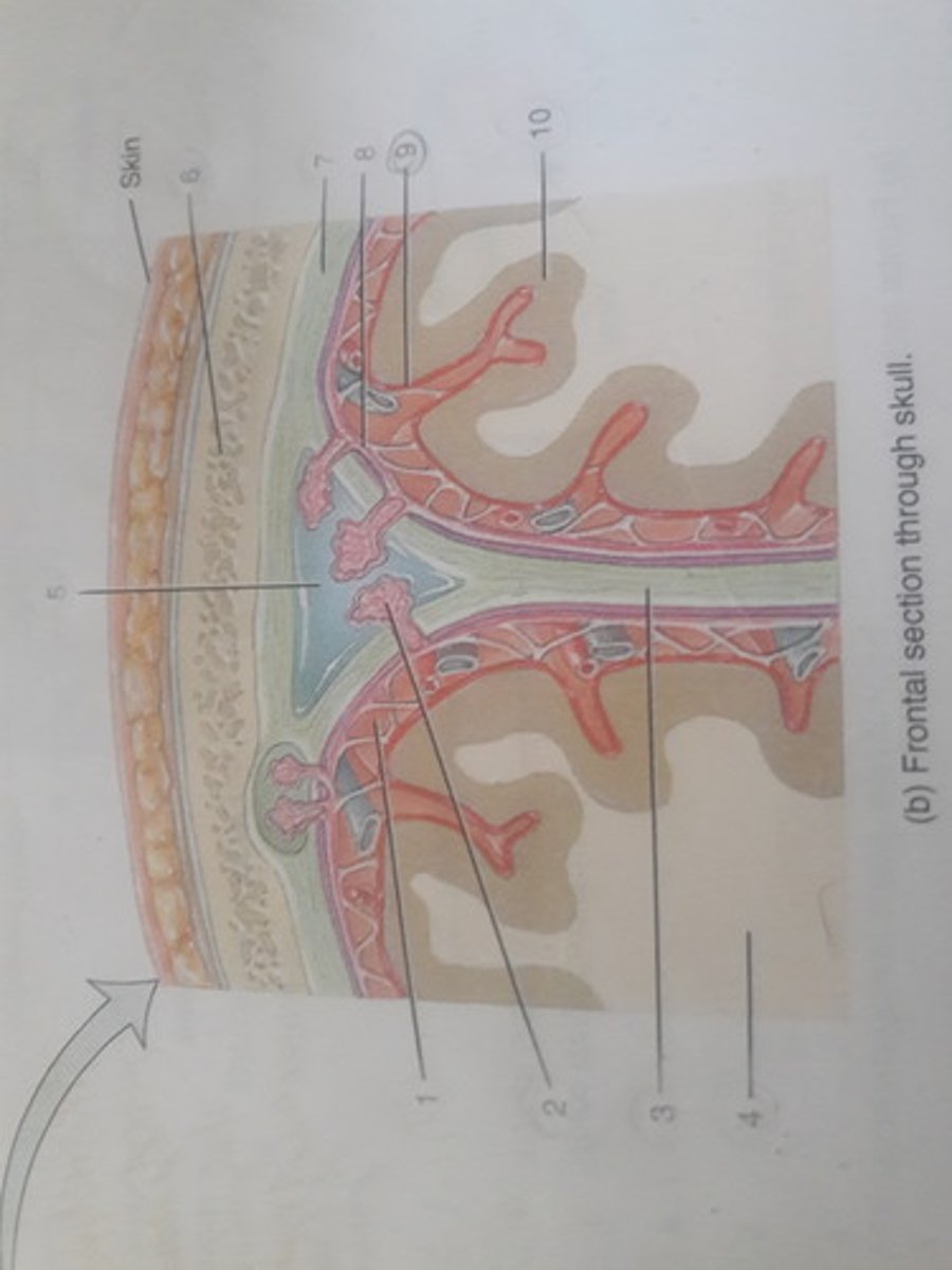
Cerebral cortex
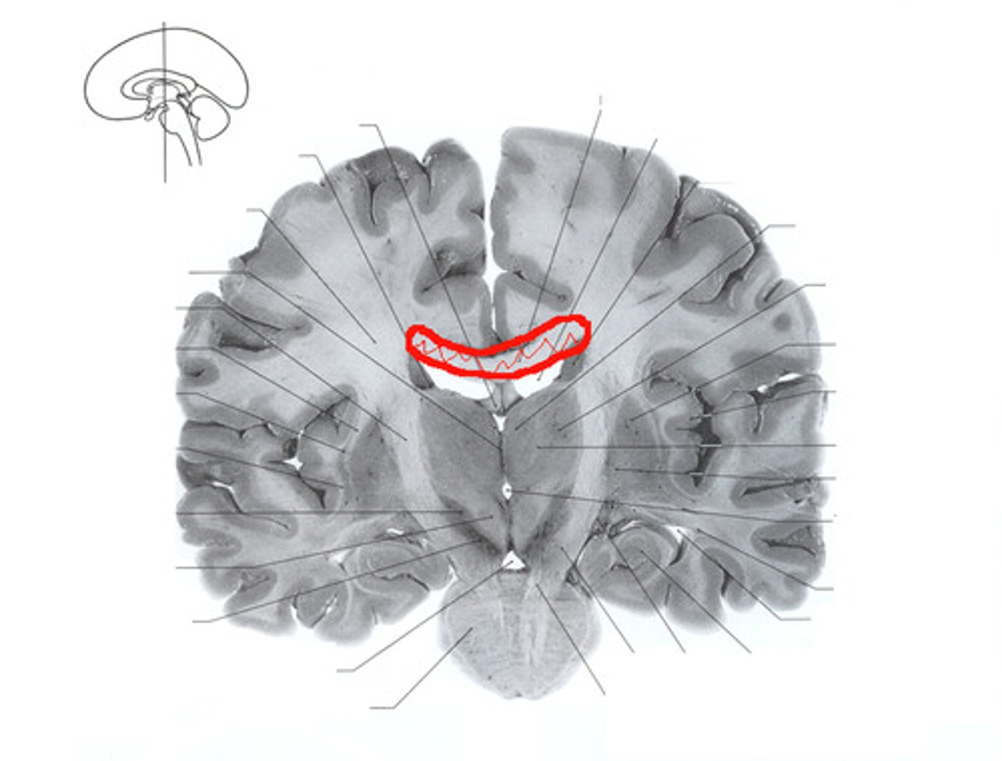
Cerebral spinal fluid (CSF) circulation,frontal section of brain and spinal cord
Constantly bathes the brain and spinal cord with oxygen, nutrients, and vital chemicals.
Lateral ventricle
Located in each cerebral hemisphere with a thin membrane.
Choroid plexus (Membrane-like pleated)
Passes through ependymal cells into 4 small brain cavities or ventricles.
Cerebral aqueduct (waterway)
A thin tube that is connecting the third ventricle to the fourth ventricle.
Fourth ventricle
Located between the pons and the cerebellum.
Third ventricle
Medially located between the paired masses of the thalamus and is narrower and smaller than the other ventricle.
CSF circulation continued
Right lateral view (ventricles superimposed)
Third ventricle
Cerebral aqueduct (Waterway)
Fourth ventricle
Central canal of the spinal cord
CSF circulation continued
Sagittal section of the brain and spinal cord
Superior sagittal sinus
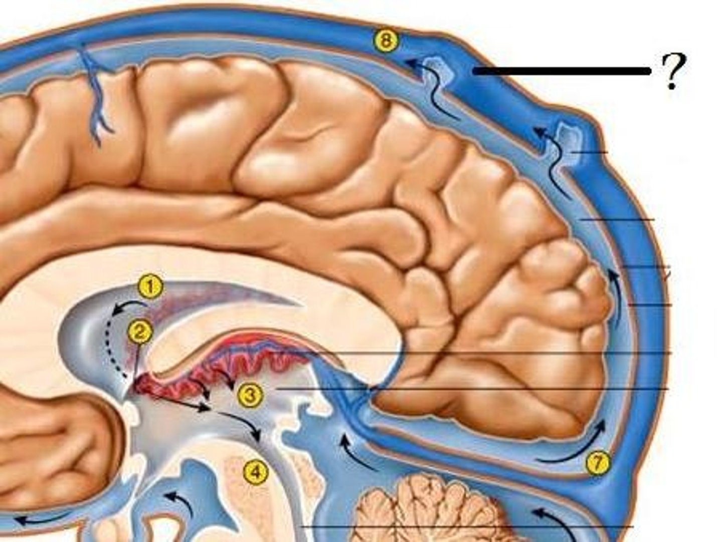
Arachnoid villus
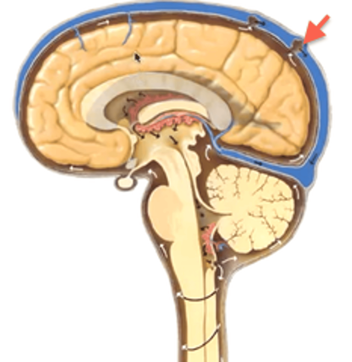
Subarachnoid space
Lateral ventricle
Choroid plexus
Third ventricle