Neuroanatomy Cerebral Cortex & Ventricular System.
1/42
There's no tags or description
Looks like no tags are added yet.
Name | Mastery | Learn | Test | Matching | Spaced | Call with Kai |
|---|
No analytics yet
Send a link to your students to track their progress
43 Terms
What structures make up the cerebrum?
Cerebral cortex (gray matter)
Underlying white matter
Deep cerebral structures
What is the cerebral cortex?
The outer gray matter layer of the cerebrum.
-gyrus
-sulcus (fissure)
Why we have gyri and sulci?
(Why not a smooth surface brain?
Increasing the cortical area
for the total number of neurons
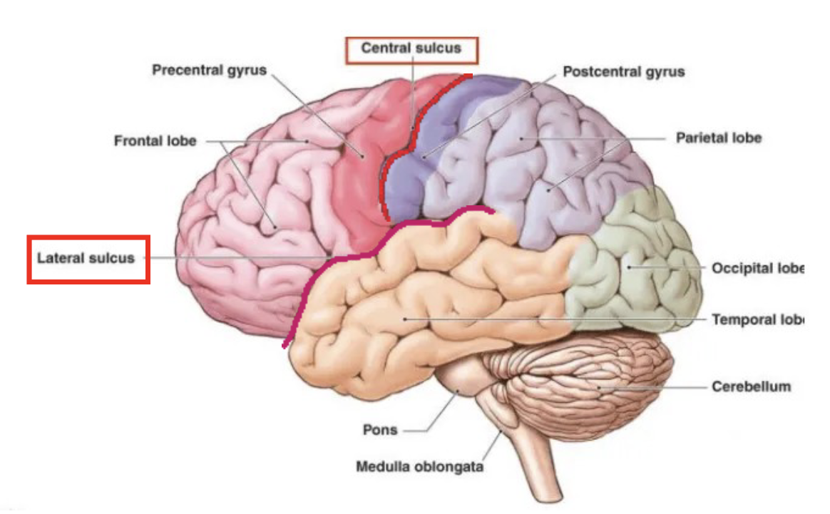
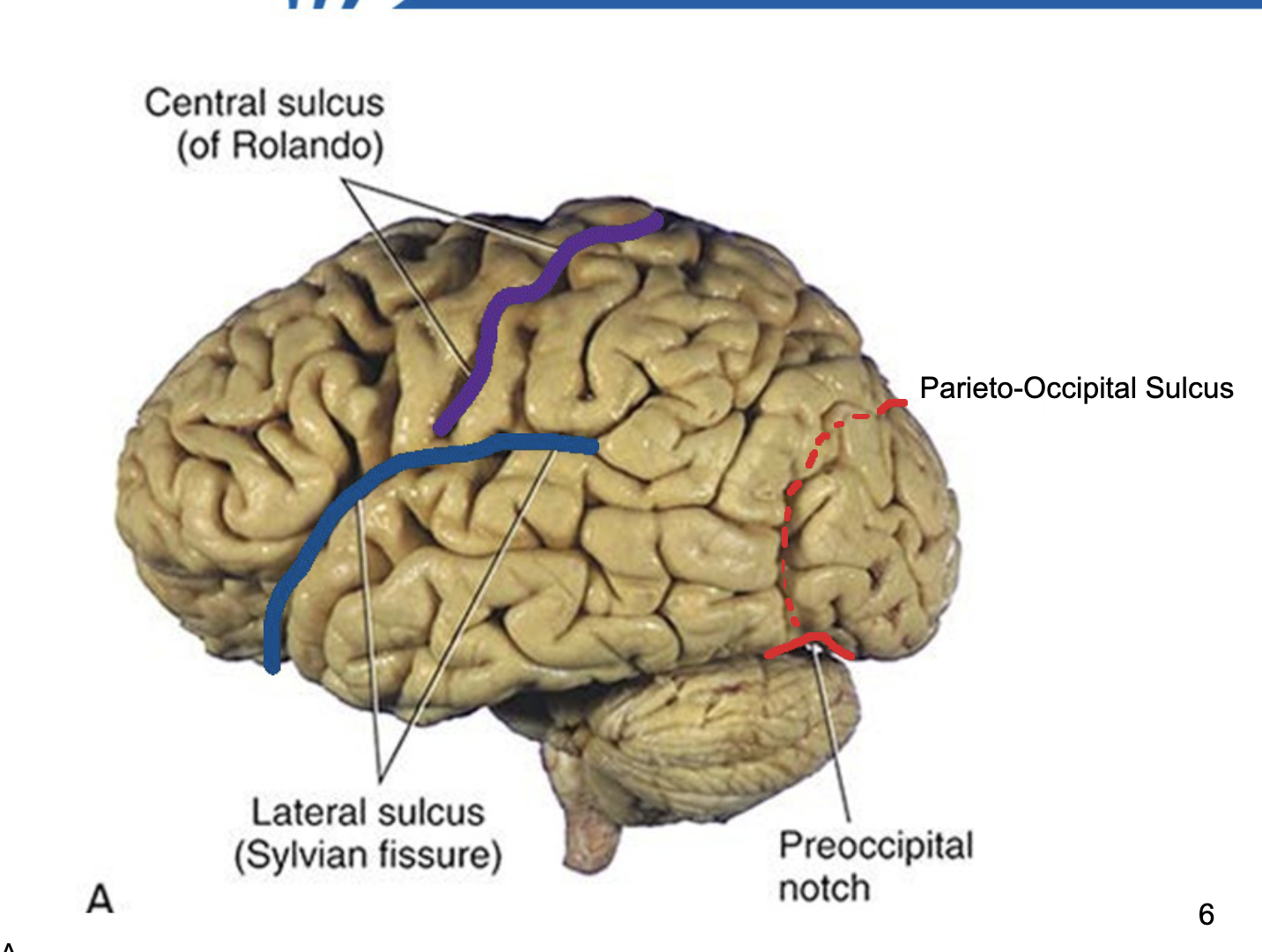
Structures of Major Sulci
Longitudinal fissure
Central sulcus (Rolando sulcus)
Lateral sulcus (Sylvian fissure)
Parieto-occipital sulcus
Preocciptital notchus
Memory trick: LCLP
(Longitudinal → Central → Lateral → Parieto-occipital)
What does the central sulcus separate?
Frontal lobe (anterior)
Parietal lobe (posterior)
What does the lateral sulcus separate?
Frontal
parietal
temporal
What does the longitudinal fissure separate?
Left and right cerebral hemispheres.
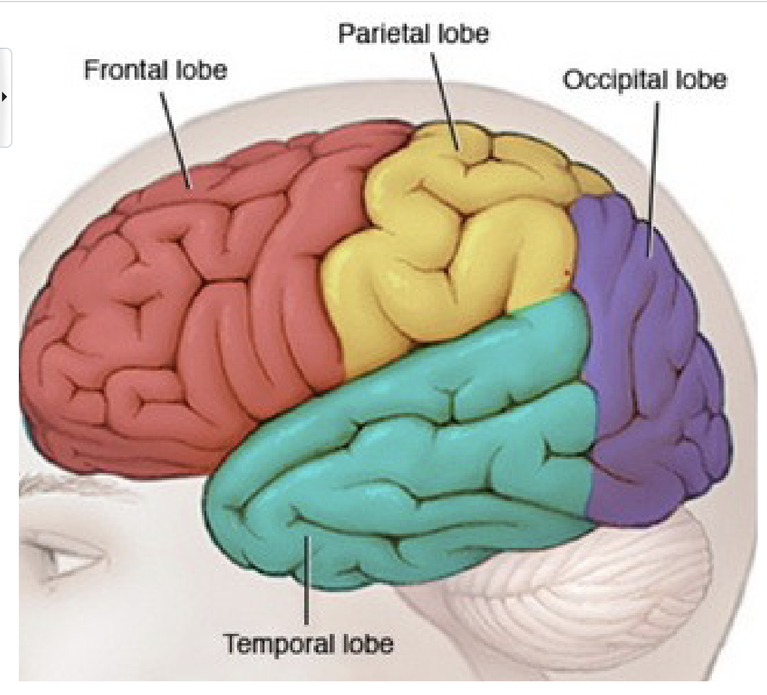
4 main lobes
Frontal
parietal
occipital
temporal
For Major Gyri what is the precentral gyrus?
Primary motor cortex
Part of the frontal lobe
For Major Gyri what is the postcentral gyrus?
Primary Somatosensory Cortex
Part of the Parietal Lobe
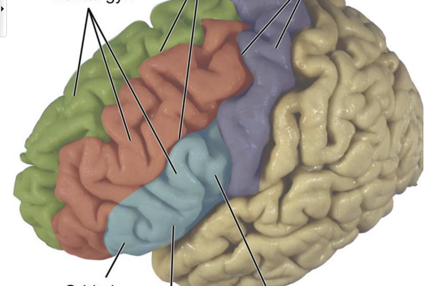
Label Frontal Lobe
Lateral View
Frontal Pole
Precentral gyrus
Precentral Sulcus
Superior frontal sulcus
Inferior frontal sulcus
Superior frontal gyrus
Middle frontal gyrus
Inferior frontal gyrus : divided into 3 parts Orbital part, triangular part, Opercular part
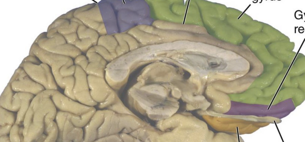
Label Frontal Lobe
Medial View
Anterior Paracentral Lobule
Orbital gyri
Olfactory Sulcus
Gyrus Rectus
Frontal Lobe Functional Areas
Primary Motor Cortex (M1): Pre-central gyrus: Sends out motor commands to your muscles
Secondary Motor Areas: In frontal of pre-central gyrus: Motor planning planning
-Supplementary Area (SMA)
-Premotor Cortex (PMC)
Broca’s Area: Controls your language motor output. around the inferior frontal gyrus, specifically the triangular part, opercular.
What is Motor homunculus?
Map that allows you to know on this primary motor cortex
KNEE TO TONGUE Look slide 15

Frontal lobe
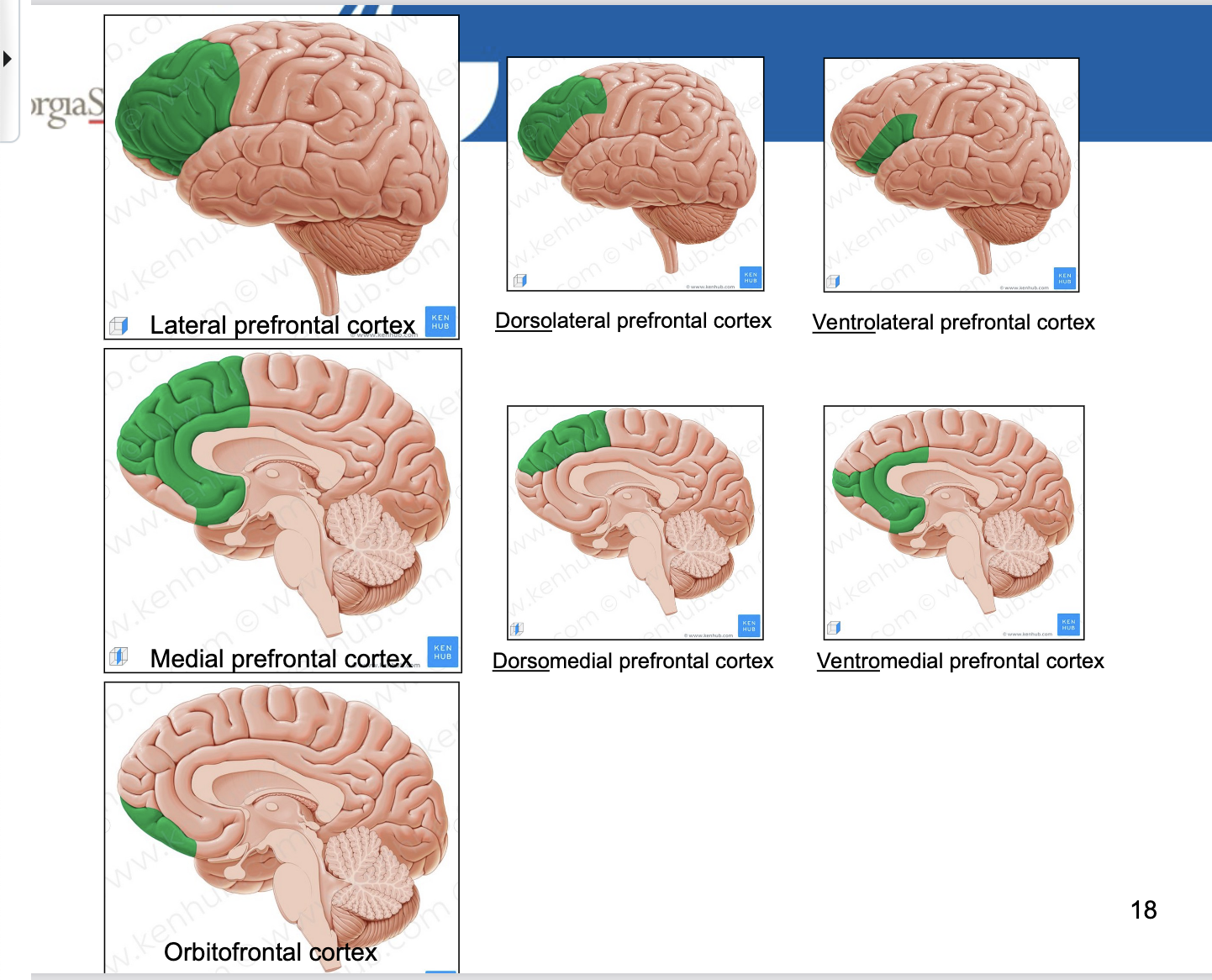
Functional area PREFONTAL CORTEX parts
Lateral part: DORSOlateral Prefrontal Cortex, VENTROlateral Prefrontal Cortex
Medial part: DORSOmedial Prefrontal Cortex, VENTROmedial Prefrontal Cortex
Orbitofrontal Cortex

Label Parietal Lobe
Lateral View
Postcentral Gyrus
Postcentral Sulcus
Intraparietal Sulcus
Superior Parietal Lobule
Inferior Parietal Lobule
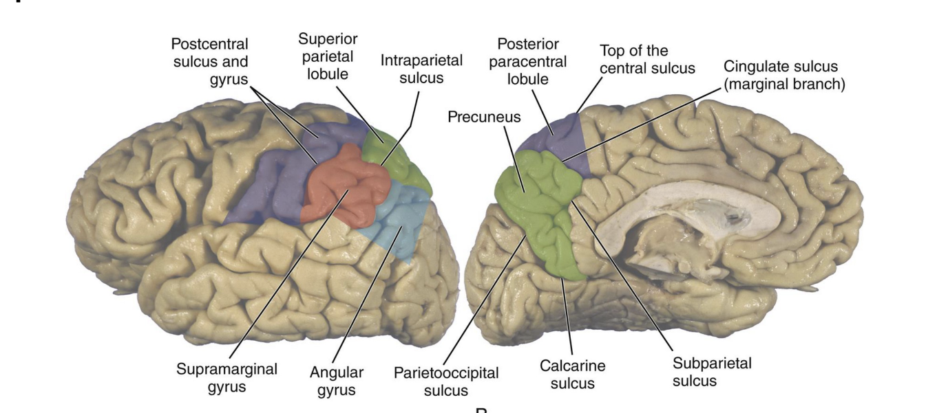
Label Parietal Lobe
Medial View
Posterior Paracentral Lobule
Precuneus
Marginal branch of the Cingulate Sulcus
Subparietal Sulcus

Parietal Lobe Functional Areas
Primary Somatosensory Cortex: Sensory info
Secondary Somatosensory Area: analyze sensory Info
Look at sensory homunculus page 25!

Label Temporal Lobe
Lateral View
Temporal Pole
Superior Temporal Sulcus
Inferior Temporal Sulcus
Superior Temporal Gyrus
Middle Temporal Gyrus
Inferior Temporal Gyrus
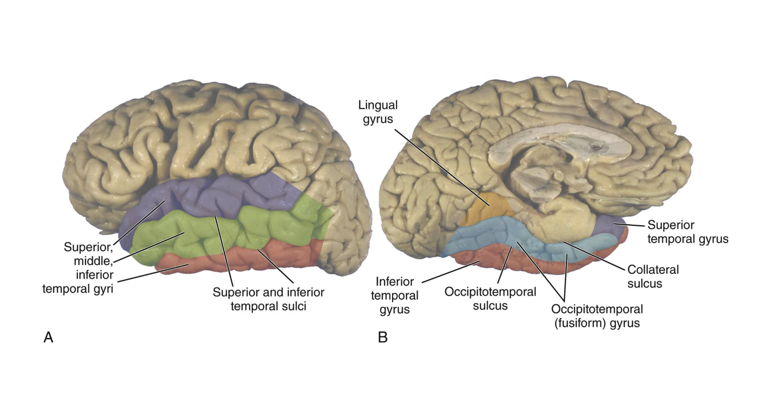
Label Temporal Lobe
Medial View
Occipitotemporal Sulcus
Occipitotemporal gyrus (parietal)
Collateral sulcus

Temporal Lobe Functional Areas
Primary Auditory Cortex: Identify auditory info
Secondary Auditory Area: further analyze auditory info
Wernicke’s Area: Understand language
Rest parts:
-Middle Temporal Gyrus: Memory
-Inferior Temporal Gyrus: Process higher levels of visual info

Occipital Lobe
Lateral view
Lateral Occipital Gyri
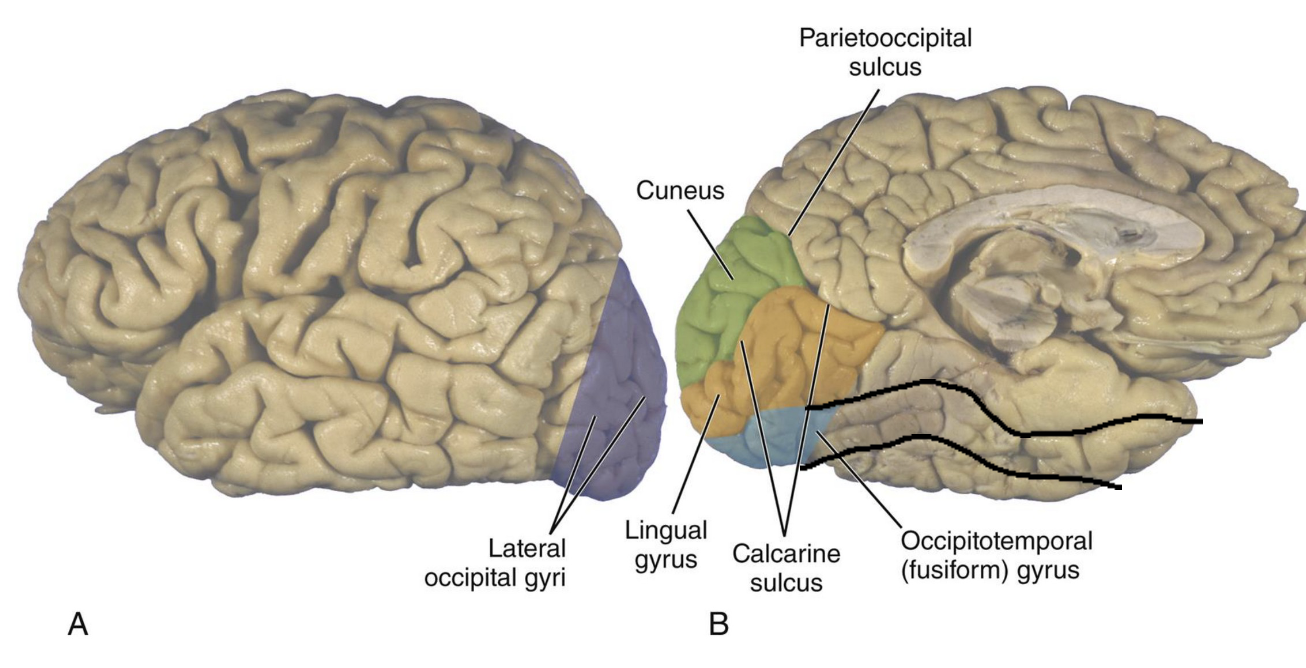
Label Occipital Lobe
Medial View
Occipitotemporal Gyrus (parietal)
Lingual Gryus
Calacrine sulcus: separates rest of occipital lobe into 2 areas
Cuneus

Occipital Lobe
Functional Areas
Primary Visual Cortex : Idenify & recieve visual info from eye nerve
Seconday Visual Area: further analyze Visual info
Limibic Lobe is primarily composed of
Cingulate Gyrus : Isthmus
Parahippocampal Sulcus
Limbic Lobe is part of the what System
Limbic System; which contains hippocampus and amygdala
What is not part of the limbic lobe
Corpus Callosum
Callosal Sulcus
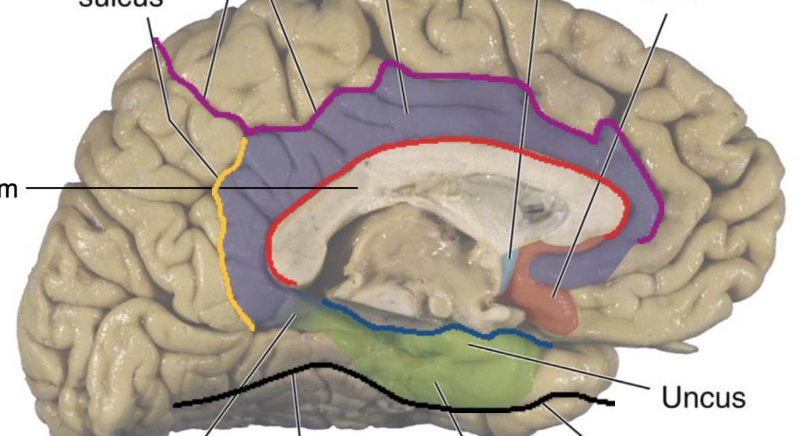
Label Limbic lobe
Subparietal sulcus
Cingulate sulcus and gyrus
paraterminal gyrus
subcallosal area
Corpus Callosum
Uncus
Rhinal sulcus
Parahippocampal Gyrus
Collateral sulcus
Isthmus
Where is the insula? and function
Deep inside the lateral fissure.
Interception (conscious awareness of body and sensation)
insula is covered by?
Frontal operculum
Parietal operculum
Temporal operculum
Lateral ventricles shape and parts
Long C-shaped cavities
5 parts
Anterior Horn (frontal horn)
Body
Posterior Horn( occipital horn)
Atrium (trigone)
Inferior horn (temporal horn)
Key Anatomical Relationship
Roof of the anterior horn and body: CORPUS CALLOSUM
Medial wall of anterior horn: SEPTUM PELLUCIDUM
Floor and medial wall of the inferior horn : HIPPOCAMPUS
Third Ventricle shaped?
Narrow, slit -shaped cavity
Third Ventricle located where?
Located in the midline of the diencephalon
What connects the lateral ventricles to the third ventricle?
Interventricular foramina.
Third ventricle contains what space?
Interthalamic adhesion
Cerebral aqueduct is what?
Narrow Channel connecting the 3rd and 4th ventricles
Located behind the midbrain
most common site of ventricular obstruction
What surrounds the fourth ventricle?
Anterior:
Pons
Medulla
Posterior:
Cerebellum
Fourth Ventricle “Open “ Cavity that communicates with
Subarachniod space through 1 MEDIAN APERTURE and 2 LATERAL APERTURES
What is the central canal?
Continuation of the fourth ventricle into the spinal cord.
Cerebropsinal Fluid
-Clear, colorless fluid
150 ml of CSF is present in adult CNS
25 ml is located within ventricles (only little in ventricles ). REMAINDER occupies the subarachnoid space
Produced at 500 mL/day ( turned 3-4 times daily)
Cerebrospinal Fluid Functions
Provides buoyancy (SPATIAL BUFFERING) and supports for the CNS
Assists in the transport of nutrients and removal of waste products
Cerebrospinal Fluid all ventricles contain
highly vascular, folded structures called CHOROID PLEXUS( in all 4 ventricular), which produce CSF