Veterinary Systems Unit 1
 Call Kai
 Learn
Learn Practice Test
 Spaced Repetition
 Match
Match Flashcards
 Knowt Play
1/34

There's no tags or description
Looks like no tags are added yet.
Name | Mastery | Learn | Test | Matching | Spaced | Call with Kai | Chat |
|---|
No analytics yet
Send a link to your students to track their progress
35 Terms
Why do we stain tissue?
To add color, contrast, and differentiation
Hematoxylin
plant dye combined with metal ion to become a blue cationic dye
Eosin
A fluorescent anionic red dye derived from fluorescein
What does hematoxylin stain?
nucleic acids, nuclei, nucleoli, ribosome-rich cytoplasm, rough ER, and hyaline cartilage matrix
what does eosin stain?
mitochondria, cytoskeleton, extracellular protein matrix proteins (collagen and elastin), many secretory granules, protein-rich cytoplasm, muscle fibers, and red blood cells
basophilic
loves base, binds to hematoxylin
eosinophilic
loves eosin
plane of section
orientation of cut
level of section
where along the structure the cut is made
what we see in a microscope depends on
3d organization, stain used, and plane and level of section
sectioning makes a 3d image
into a 2d image
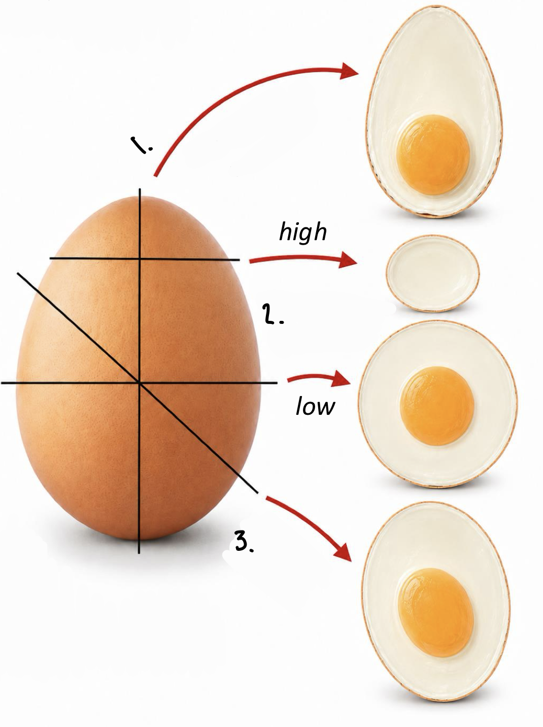
longitudinal section 2. transverse sections 3. oblique section
longitudinal section
cut parallel to long axis of structure
transverse/cross section
cut perpendicular to long axis of structure
oblique section
cut at an angle between longitudinal and transverse sections
tangential section
cut barely passes through structure
nucleus
stores DNA and regulates gene expression
nucleolus
produces rrna and assembles ribosomes
ribosomes
synthesize proteins from mrna
rough endoplasmic reticulum (RER)
synthesizes and secretes membrane proteins
golgi apparatus
modifies, sorts, and packages proteins
secretory granules
store and release secreted products
plasma membrane
controls transport and cellular signaling
smooth endoplasmic reticulum (SER)
synthesizes lipids, detoxifies, and stores calcium
mitochondria
generate ATP through oxidative phosphorylation
cytoskeleton
maintains shape and enables cellular movement
lysosomes
digest and recycle cellular materials
lipid droplet
stores lipids for energy
why are some secretory granules basophilic?
they contain acidic anionic glycoproteins
euchromatin
being used, so more dispersed and lighter staining
heterochromatin
not being used, so more condensed and darker staining
poorly stained H&E structures
lipid droplets, golgi, glycogen, mucus, plasma membrane
why are lipid droplets and golgi not stained well?
hydrophobic and H&E are water based dyes
why are glycogen and mucus not well stained?
poorly preserved
why is plasma membrane poorly stained?
too thin to see