Chapter 1 - Anatomical Kinesiology
 Call Kai
 Learn
Learn Practice Test
 Spaced Repetition
 Match
Match Flashcards
 Knowt Play
1/161

There's no tags or description
Looks like no tags are added yet.
Name | Mastery | Learn | Test | Matching | Spaced | Call with Kai | Chat |
|---|
No analytics yet
Send a link to your students to track their progress
162 Terms
What is anatomical kinesiology?
Study of the human musculoskeletal system and musculotendinous system.
what is biomechanics?
Application of mechanical physics to human motion.
What is kinetics?
Study of the forces involved that cause movement to occur.
What is kinematics?
Study of motion without regard to cause (ignores forces).
An athletic trainer observes athletes performing a squat jump. What method of study is being used?
Kinematics – study of motion without regard to forces.
under anatomical kinesiology
A researcher records ground reaction forces during jump squats. What method of study is being used?
Kinetics – study of forces involved in movement.
under biomechanics
Anatomical position
Standard reference position used for all body descriptions: standing upright, facing forward, feet parallel and close, arms at sides, palms facing forward. This reference applies regardless of body orientation (standing, supine, prone).
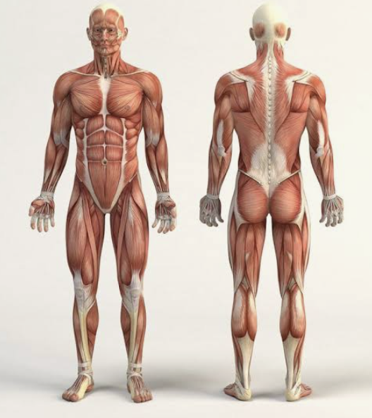
Fundamental position
Similar to anatomical position, but arms are at the sides with palms facing the body instead of forward.

What does lateral mean in anatomical terminology?
Farther from the midline relative to another structure.
What does medial mean in anatomical terminology?
Closer to the midline relative to another structure.
What does median mean?
Located at the midline itself (on the midsagittal plane).
How should anatomical directional terms be stated correctly?
Structure A in reference to Structure B (directional terms are relative).
Where is the mid-sternal line?
Vertical line running down the midline of the sternum on the anterior chest.
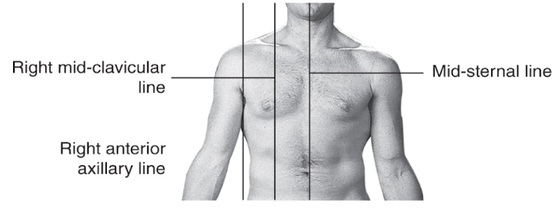
Where is the mid-clavicular line?
Vertical line passing through the midpoint of the clavicle on the anterior torso
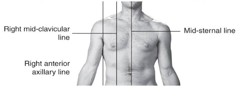
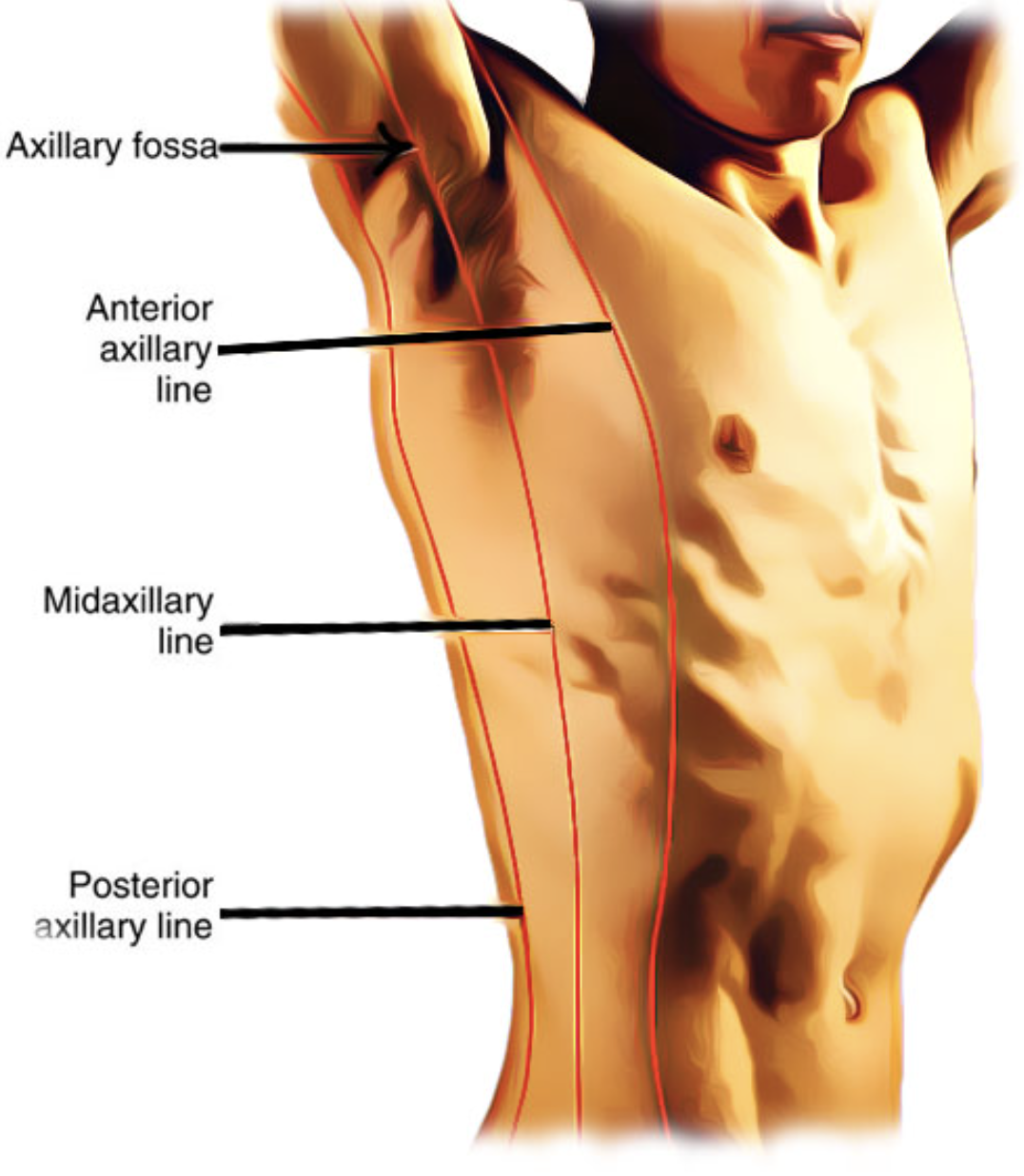
Where is the anterior axillary line?
Vertical line along the anterior border of the axilla (front of armpit).
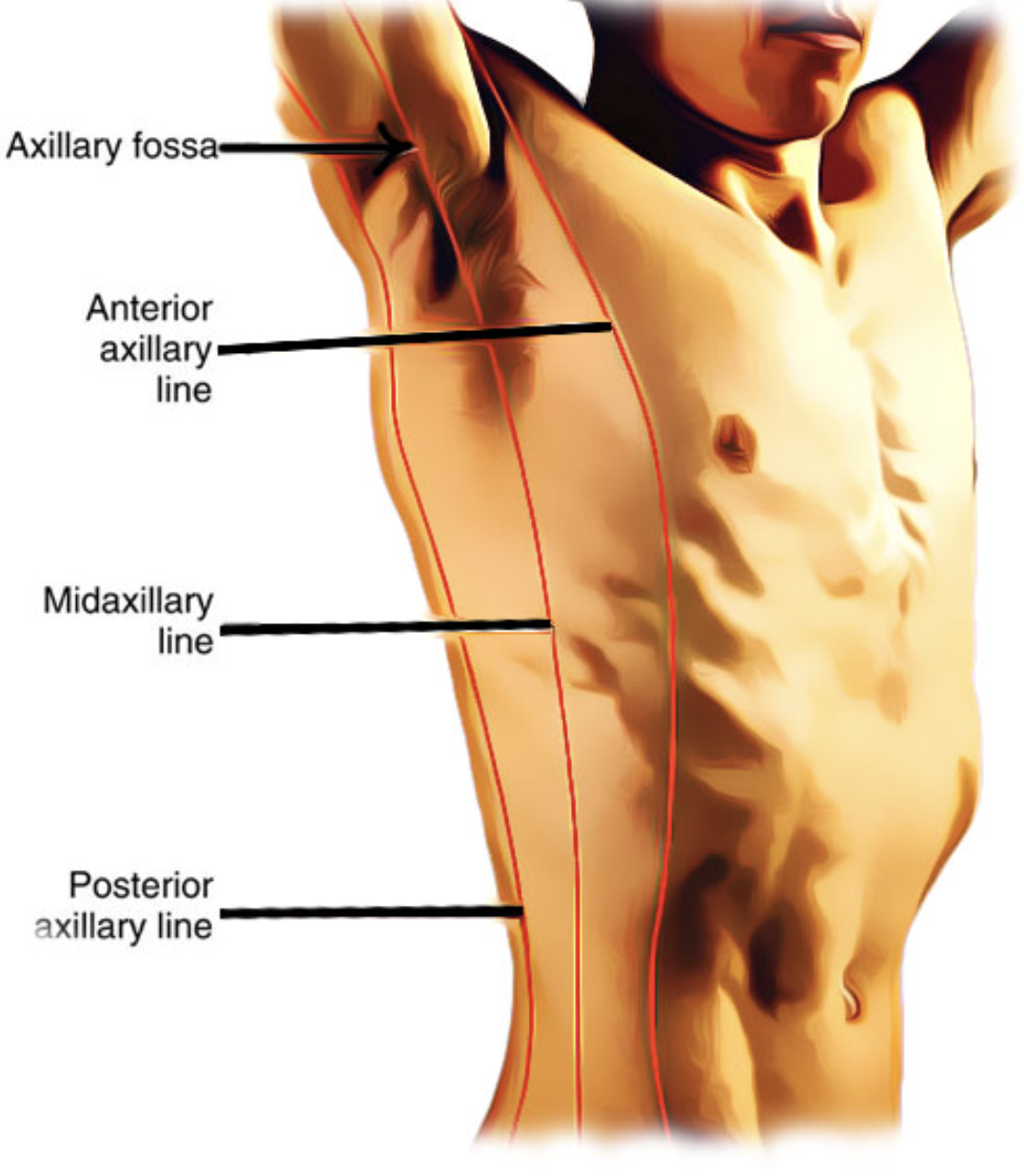
Where is the mid-axillary line?
Vertical line running down the center of the axilla (middle of armpit).

Where is the posterior axillary line?
vertical line along the posterior border of the axilla (back of armpit).
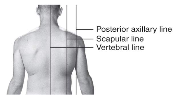
Where is the scapular line?
Vertical line passing through the inferior angle of the scapula on the posterior torso.
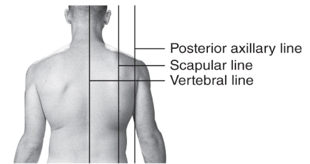
Where is the vertebral line?
Vertical line running along the spinous processes of the vertebral column.
Where is the mid-inguinal point?
Midpoint between the ASIS and the pubic symphysis; landmark for the femoral artery.

Complete the statement correctly: “The shoulder is ___ to the sternum.”
Lateral.
Complete the statement correctly: “The sternum is ___ to the shoulder.”
Medial.
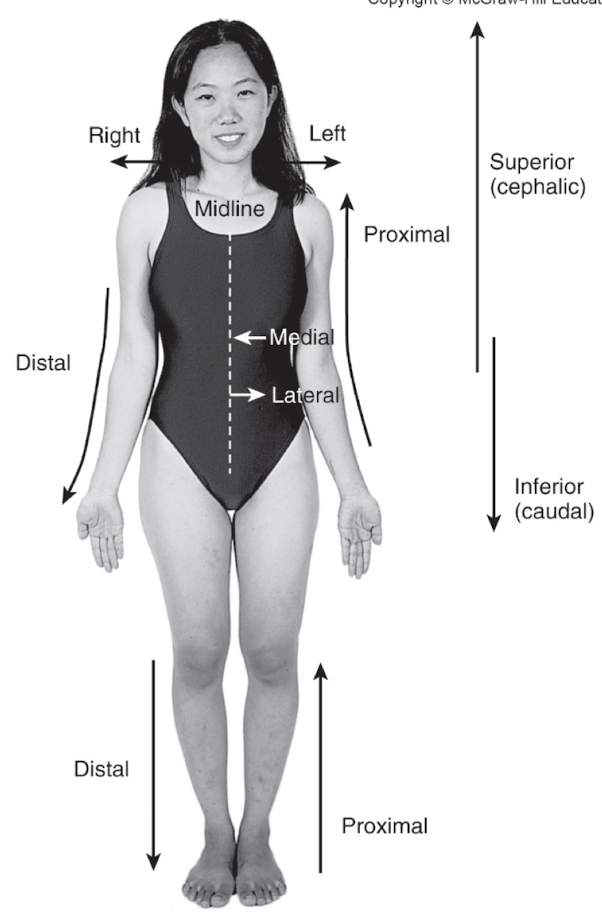
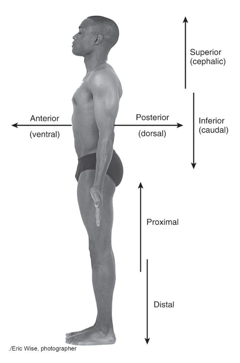
Anatomical directional terms: superior, inferior, cephalic, caudal — what do they mean?
Superior (supra) = above another structure; higher; toward the head
Inferior (infra) = below another structure; lower; toward the feet
Cephalic = toward the head; synonymous with superior
Caudal = toward the tail/feet; synonymous with inferior
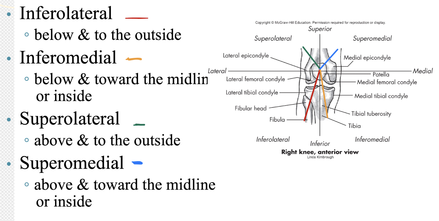
Using the patella as a reference, explain inferolateral, inferomedial, superolateral, and superomedial.
Inferolateral: Fibular head is inferolateral to the patella (below + outside)
Inferomedial: Tibial tuberosity is inferomedial to the patella (below + toward midline)
Superolateral: Lateral femoral condyle is superolateral to the patella (above + outside)
Superomedial: Medial femoral condyle is superomedial to the patella (above + toward midline)
Anatomical directional terms: deep vs superficial — what do they mean?
Deep = beneath or farther from the surface; describes relative depth of a structure in a circumferential manner (not directional like medial/lateral).
Superficial = near the surface; describes the relative depth of a structure circumferentially.
Example: The triceps muscle is superficial to the humerus; the humerus is deep to the triceps.
Anatomical directional terms: anterior, anteroinferior, anterosuperior — what do they mean?
Anterior = in front of another structure.
Anteroinferior = in front of and below another structure.
Anterosuperior = in front of and above another structure.
How do combined anatomical directional terms (e.g., anteroinferolateral) work?
They stack multiple directions at once (front/back + up/down + medial/lateral) to give a more precise relative location. All terms are relative to a reference structure.
What does anteroinferolateral mean? Give a shoulder example.
Anteroinferolateral = in front, below, and toward the outside.
Example: The anteroinferolateral shoulder capsule is anteroinferolateral to the glenoid fossa.
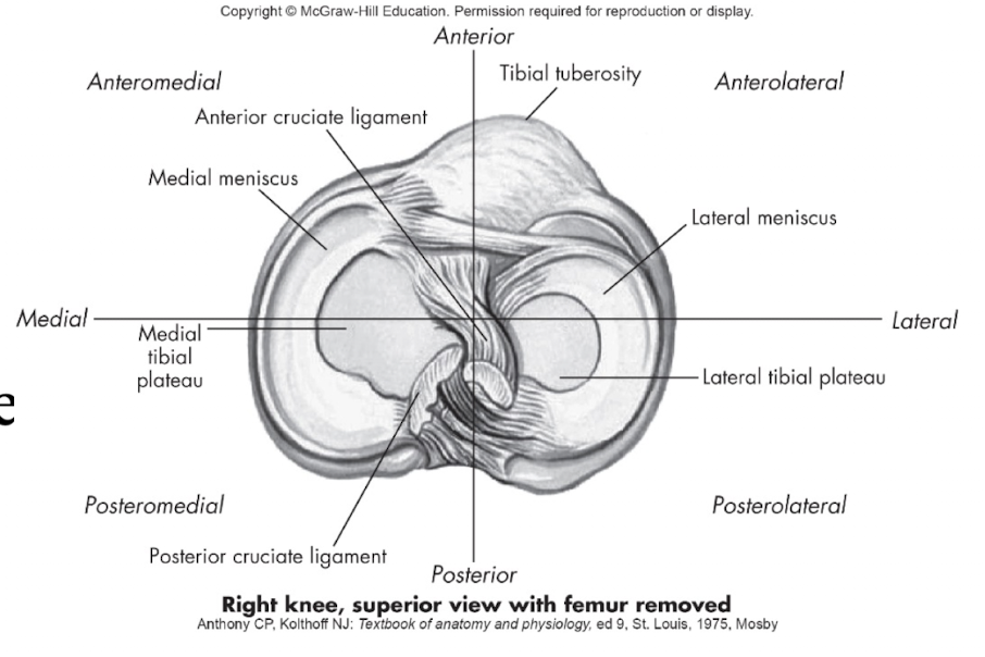
What does anterolateral mean, and where is an example of this in the right knee (superior view)?
Anterolateral = in front and toward the outside.
The lateral meniscus and lateral tibial plateau are anterolateral relative to the posterior cruciate ligament (PCL).
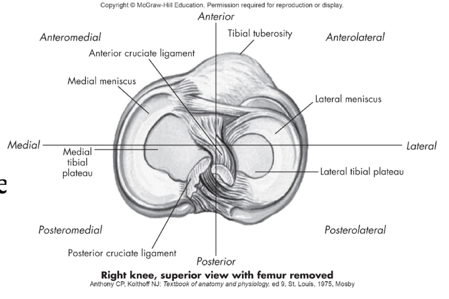
What does anteromedial mean, and where is an example of this in the right knee (superior view)?
Anteromedial = in front and toward the midline.
Example: The medial meniscus and medial tibial plateau are anteromedial relative to the PCL.
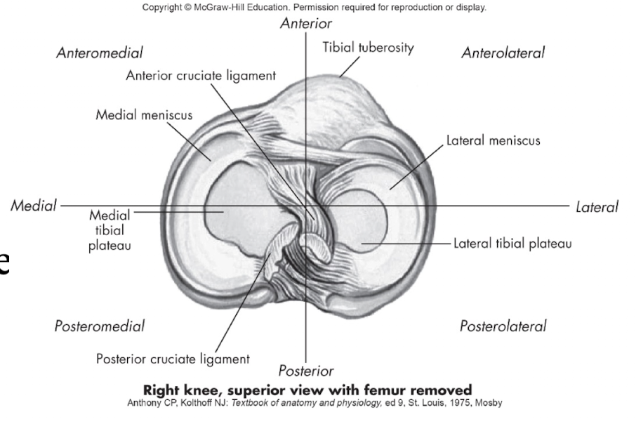
What does anteroposterior mean, and how is it shown in the right knee (superior view)?
Anteroposterior = relating to the front-to-back direction.
Example: The knee joint has an anteroposterior orientation from the tibial tuberosity (anterior) to the posterior cruciate ligament (posterior).
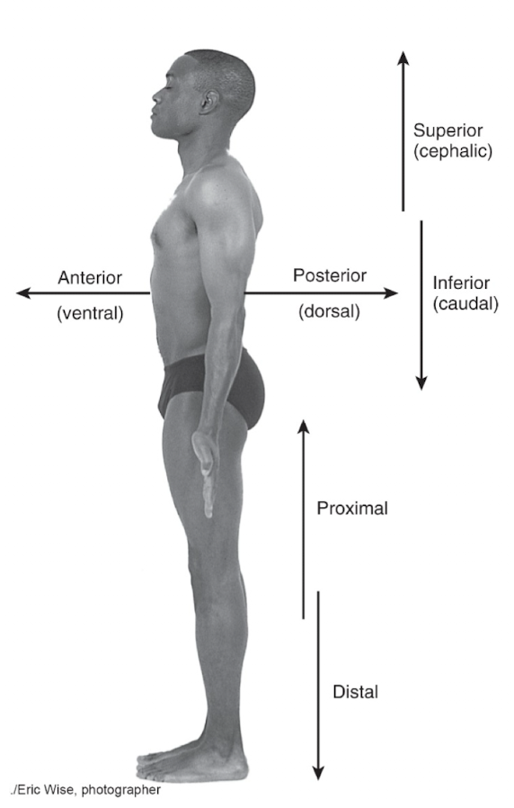
What do posterior, posteroinferior, and posterolateral mean, and where is an example of each on the body?
Posterior = behind or in the back.
Example: The gluteal region is posterior to the abdomen.Posteroinferior = behind and below.
Example: The gluteal region is posteroinferior to the lumbar spine.Posterolateral = behind and toward the outside.
Example: The posterolateral shoulder (posterior deltoid region) is posterolateral to the sternum.
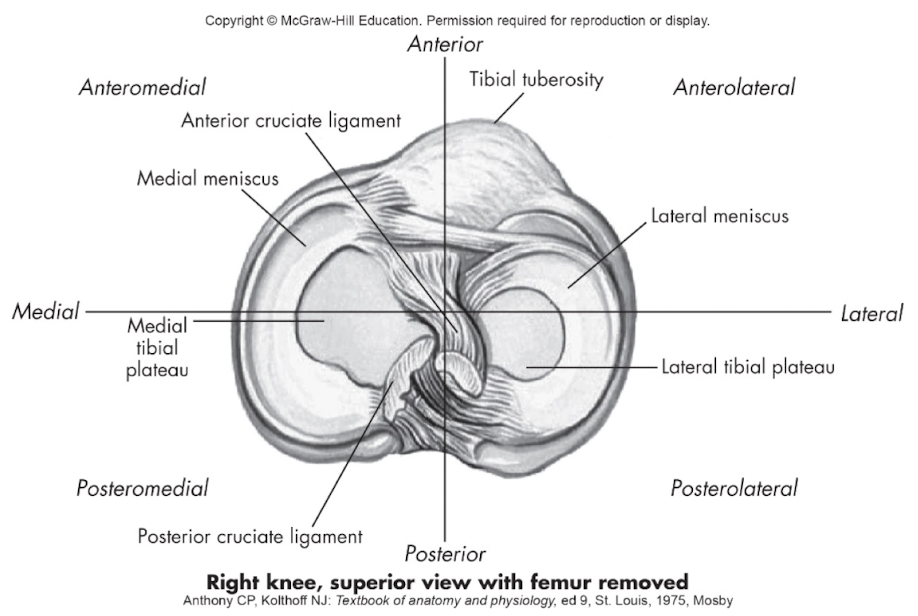
What does posteromedial mean, and where is an example of this in the right knee (superior view)?
Posteromedial = behind and toward the midline.
Example: The posterior cruciate ligament (PCL) lies posteromedial relative to the anterior cruciate ligament (ACL); the posteromedial meniscal region is posteromedial relative to the center of the knee.
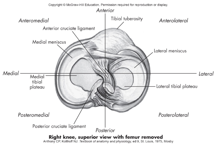
What does posterosuperior mean, and where is an example of this in the right knee (superior view)?
Posterosuperior = behind and above.
Example: The posterior portion of the tibial plateau is posterosuperior relative to more anterior tibial structures (e.g., tibial tuberosity region).
What does contralateral mean?
Contralateral = on the opposite side.
Example: The right hand is contralateral to the left foot.
What does ipsilateral mean?
Ipsilateral = on the same side.
Example: The right arm is ipsilateral to the right leg.
What does bilateral mean?
Bilateral = involving both sides.
Example: Both shoulders moving at the same time is a bilateral movement.
What do proximal and distal mean, and what are they always referenced to?
Proximal = closer to the trunk or point of origin.
Distal = farther from the trunk or point of origin.
Unlike other directional terms, proximal and distal are always referenced to the trunk, NOT to another structure.
Example:
The elbow is proximal to the wrist (closer to the trunk).
The fingers are distal to the elbow (farther from the trunk).
What do prone and supine mean?
Prone = lying face down (stomach down), palms down.
Supine = lying face up (on the back), palms up.
What do dorsal and ventral mean? what are they for the food and hand.
Dorsal = relating to the back or posterior surface.
• Examples: back of the body, back of the hand, top (dorsum) of the foot
Ventral = relating to the belly/abdomen or anterior surface.
• Examples: front of the body, palm of the hand, sole (plantar surface) of the foot
What do palmar, volar, and plantar mean?
Palmar: Relating to the palm or volar aspect of the hand.
Volar: Relating to the palm of the hand or sole of the foot.
Plantar: Relating to the sole or undersurface of the foot.
What does fibular mean in anatomical directional terminology?
Fibular = relating to the lateral (outer) side of the lower extremity.
Example: The fibula is on the outer side of the leg.
Memory trick: Fibula = outside of the leg
What does tibial mean in anatomical directional terminology?
Tibial = relating to the medial (inner) side of the lower extremity.
Example: The tibia (shin bone) is on the inner side of the leg.
Memory trick: Tibia = inner shin
What does radial mean in anatomical directional terminology?
Radial = relating to the lateral (thumb) side of the forearm or hand (in anatomical position).
Example: The thumb is on the radial side of the hand.
Memory trick: Radial = thumb side
What does ulnar mean in anatomical directional terminology?
Ulnar = relating to the medial (pinky) side of the forearm or hand (in anatomical position).
Example: The pinky finger is on the ulnar side of the hand.
Memory trick: Ulnar = pinky side
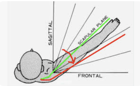
What is the scapular plane?
The scapular plane is the plane of movement aligned with the natural resting position of the scapula on the posterior rib cage.
It lies approximately 30–45° anterior to the frontal plane, so movements in this plane occur in line with the scapula, not directly frontal.
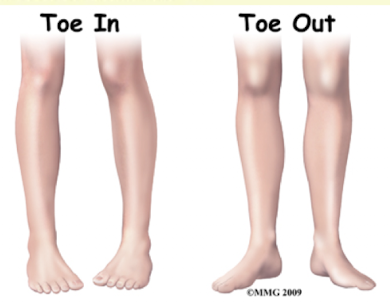
What do anteversion and retroversion mean in alignment variation terminology?
Anteversion = abnormal or excessive forward rotation of a structure.
• Common example: Femoral anteversion → hip internally rotated → toes in, knees point inward.Retroversion = abnormal or excessive backward rotation of a structure.
• Common example: Femoral retroversion → hip externally rotated → toes out.
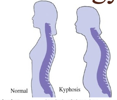
What is kyphosis?
Kyphosis = increased outward/backward curvature of the spine in the sagittal plane.
Common description: hunchback.
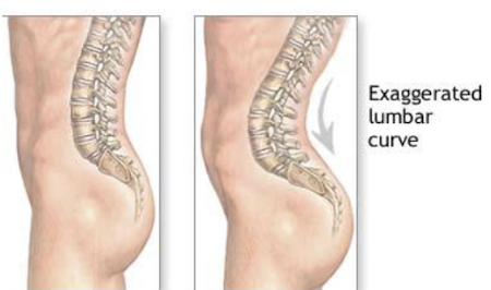
What is lordosis?
Lordosis = increased inward/forward curvature of the spine in the sagittal plane.
Common description: exaggerated lumbar curve or “swayback.”
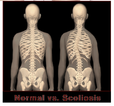
What is scoliosis?
Scoliosis = lateral (side-to-side) curvature of the spine.
Occurs in the frontal (coronal) plane.
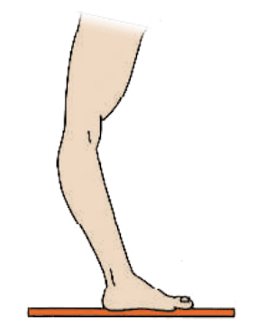
What is recurvatum?
Recurvatum = backward bending of a joint.
Example: Genu recurvatum = knee hyperextension.
Memory hook: Re-curve = curve backward
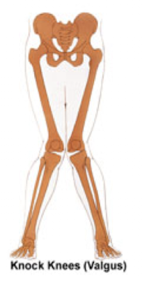
What is valgus alignment?
Valgus = outward angulation of the distal segment of a bone or joint.
Example: Knock-knees (knees move medially, ankles stay apart).
Load direction: lateral → medial.
Memory hook: ValGUS = knees go “in”
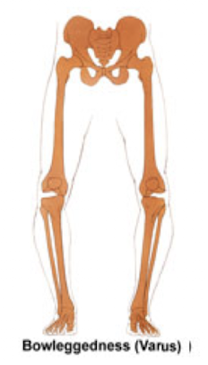
What is varus alignment?
Varus = inward angulation of the distal segment of a bone or joint.
Example: Bowlegs (knees stay apart, ankles move medially).
Load direction: medial → lateral.
Memory hook: VaRUS = knees go “out”
What is a plane of motion, and how does it relate to its axis?
A plane of motion is an imaginary 2D surface through which a body segment moves.
Motion in a plane occurs around an axis.
The plane and its axis are perpendicular (90°) to each other.
memorize:
Movement occurs IN a plane and AROUND an axis.
And the plane and axis are always perpendicular (90°) to each other.
Plane = WHERE the movement happens
Axis = the imaginary line you rotate AROUND
The key idea is that the axis sticks through the plane at 90° (perpendicular).
For example, when you do a biceps curl:
Your arm moves in the sagittal plane
Your elbow rotates around a mediolateral axis (an imaginary line going side-to-side through your elbow)
That axis is 90° to the sagittal plane
A good analogy is a door:
The path/area the door moves through = plane
The hinges it rotates around = axis
What are the 3 cardinal planes of motion?
Sagittal (anteroposterior) plane
Frontal (lateral/coronal) plane
Transverse (horizontal) plane
What is the sagittal (anteroposterior) plane?
Divides the body into right and left halves.
Example: Sit-up (flexion/extension occurs in the sagittal plane).

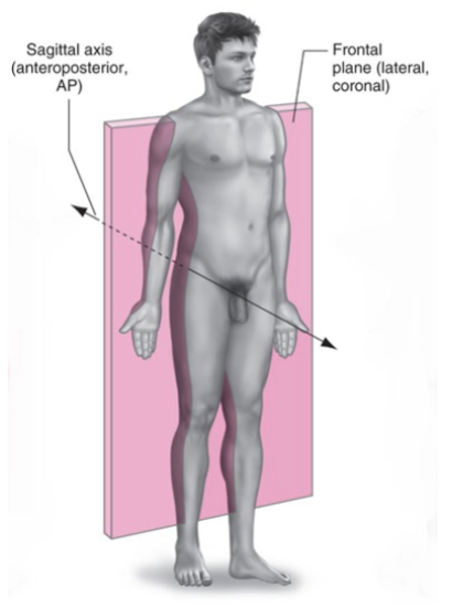
What is the frontal (lateral/coronal) plane?
Divides the body into anterior (front) and posterior (back) halves.
Example: Jumping jacks (abduction/adduction occurs in the frontal plane).
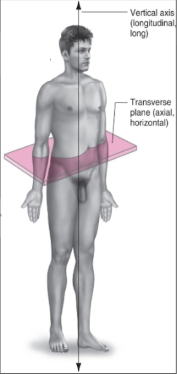
What is the transverse (axial/horizontal) plane?
Divides the body into superior (top) and inferior (bottom) halves.
Example: Spinal rotation to the left or right.

What movements occur in the high diagonal plane?
Upper-limb movements at the shoulder, especially overhand skills.
Example: Baseball pitch.

What movements occur in the low diagonal plane?
Upper extremity (UE): Upper limbs at the shoulder → underhand skills
Example: Discus throwLower extremity (LE): Lower limbs at the hip
Example: Kicking/punting
What are axes of rotation?
For movement to occur in a plane, the body/segment must rotate around an axis.
Axes are named based on their orientation.
The single sentence I'd memorize is:
An axis is an imaginary line around which a body part rotates; it is named according to the direction the line runs.
when given an axis remember that movement occurs in the perpendicular plane
vertical axis = movment occurs in transverse plane (NEED TO ADD TYPE OF MOMVMENT)
Frontal axis = movement occurs in the sagittal plane
saggital axis = movment occurs in the frontal plane
What is the frontal (coronal/lateral/mediolateral) axis?
Runs side to side (medial ↔ lateral) and is perpendicular (90°) to the sagittal plane.
allows movement on the perpendicular plane (saggttal)
Movements: Flexion and extension.

What is the sagittal (anteroposterior) axis?
Runs front to back (anterior ↔ posterior) and is perpendicular (90°) to the frontal plane.
Movements: Abduction and adduction.

What is the vertical (long/longitudinal) axis?
Runs top to bottom (superior ↔ inferior) and is perpendicular (90°) to the transverse plane.
Movements: Internal and external rotation.
What is the diagonal (oblique) axis?
Also known as the oblique axis.
Runs at a right angle (90°) to the diagonal plane.


What structures make up the axial region (body regions)?
Cephalic (Head):
Cranium
Face
Cervical:
Neck
Trunk:
Thoracic = Thorax
Dorsal = Back
Abdominal = Abdomen
Pelvic = Pelvis

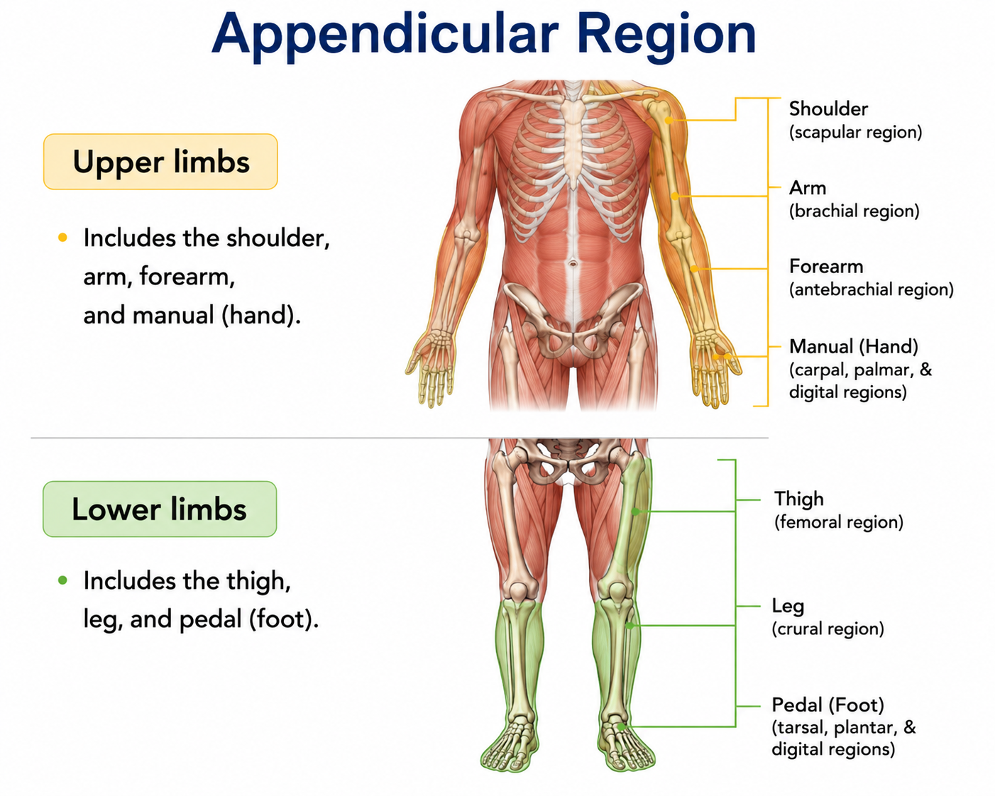
What structures make up the appendicular region (body regions)?
Upper limbs:
Shoulder
Arm
Forearm
Manual = Hand
Lower limbs:
Thigh
Leg
Pedal = Foot
How many bones are in the adult skeleton, and how are they divided?
Adult skeleton = 206 bones
Axial skeleton = 80 bones
Appendicular skeleton = 126 bones
There may be occasional anatomical variations.
What are the 5 main functions of the skeletal system?
Protection – protects organs (heart, lungs, brain)
Support – maintains posture
Movement – muscle attachment and acts as levers
Mineral storage – calcium and phosphorus
Hemopoiesis – blood cell formation in red bone marrow
hat are the 5 types of bones and examples of each?
Long bones – humerus, fibula
Short bones – carpals, tarsals
Flat bones – skull, scapula
Irregular bones – pelvis, ethmoid, ear ossicles
Sesamoid bones – patella
What are the characteristics of long bones?
Long cylindrical shaft with relatively wide, protruding ends
Shaft contains the medullary canal
Examples: phalanges, metatarsals, metacarpals, tibia, fibula, femur, radius, ulna, humerus

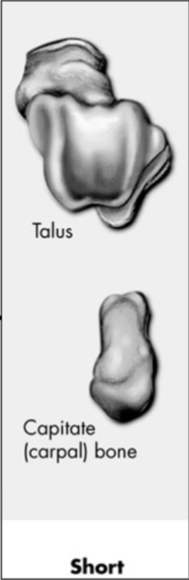
What are the characteristics of short bones?
Small, cubical-shaped solid bones
Have proportionally large articular surfaces to articulate with multiple bones
Examples: carpals and tarsals
What are the characteristics of flat bones?
Usually have a curved surface
Vary from thick where tendons attach to very thin
Examples: ilium, ribs, sternum, clavicle, and scapula

What are the characteristics and examples of irregular and sesamoid bones?
Irregular bones:
Complex-shaped bones
Examples: vertebrae, ischium, pubis, maxilla
Sesamoid bones:
Small bones embedded within tendons
Provide protection and improve mechanical advantage
Examples: patella, 1st metatarsophalangeal, 1st metacarpophalangeal

What are the typical bony features of a long bone?
Diaphysis: Long cylindrical shaft
Cortex: Hard, dense compact bone forming the walls of the diaphysis
Periosteum: Dense, fibrous membrane covering the outer surface of the diaphysis
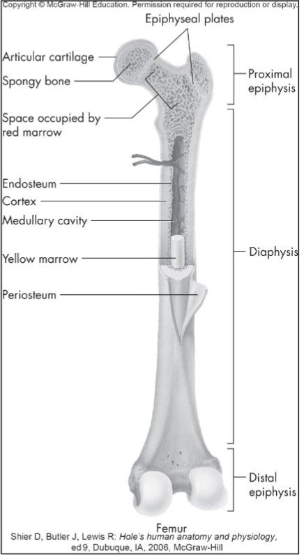
What are the endosteum and medullary cavity?
Endosteum: Fibrous membrane that lines the inside of the cortex
Medullary (marrow) cavity: Space within the diaphysis that contains yellow (fatty) marrow
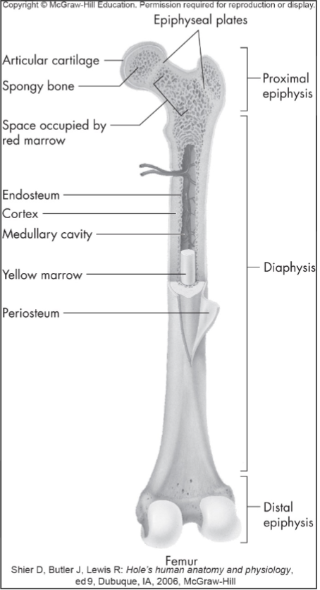
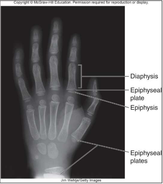
What are the epiphysis and epiphyseal plate?
Epiphysis: Ends of long bones composed of cancellous (spongy/trabecular) bone
Epiphyseal plate (growth plate): Thin cartilage plate that separates the diaphysis from the epiphysis
What is articular (hyaline) cartilage?
Articular cartilage: Covers the epiphysis of a bone to provide cushioning and reduce friction at joints.

What are endochondral bones and how do they develop?
Endochondral bones:
Develop from hyaline cartilage
Begin as hyaline cartilage models during the embryonic stage
Cartilage is gradually replaced by bone through ossification

When does longitudinal bone growth occur and stop?
Longitudinal bone growth continues while the epiphyseal (growth) plates are open.
After adolescence → epiphyseal plates close/ossify → longitudinal bone growth stops.
Bone grows at the diaphysis
Memory: Open growth plate = ↑ length; closed growth plate = no more length growth.
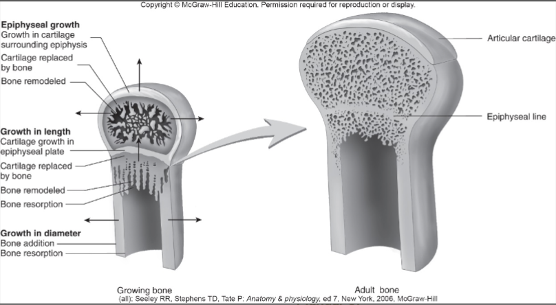
How does bone growth change with age?
Epiphyseal plates → most close by ~18, but some remain open until ~25 → longitudinal growth stops when they close
Appositional growth → increases bone diameter → continues throughout life

How does appositional bone growth increase bone diameter?
Periosteum → osteoblasts add new bone to the outside
Medullary cavity → osteoclasts resorb bone on the inside
Result → bone increases in diameter while the medullary cavity enlarges
Osteoblasts = build bone
Osteoclasts = break down/resorb bone

What gives bone its properties?
Calcium carbonate + calcium phosphate → hardness/rigidity (60–70% of bone weight)
Collagen → flexibility + tensile strength
Water → 25–30% of bone weight
Aging → ↓ collagen → ↑ brittleness
Compare cortical vs. cancellous bone.
Cortical bone → outer bone, low porosity (5–30%), stiffer, withstands greater stress but less strain
Cancellous bone → underneath cortical, spongy/high porosity (30–90%), withstands greater strain before fracturing
Cortical = stronger/stiffer
Cancellous = more deformable
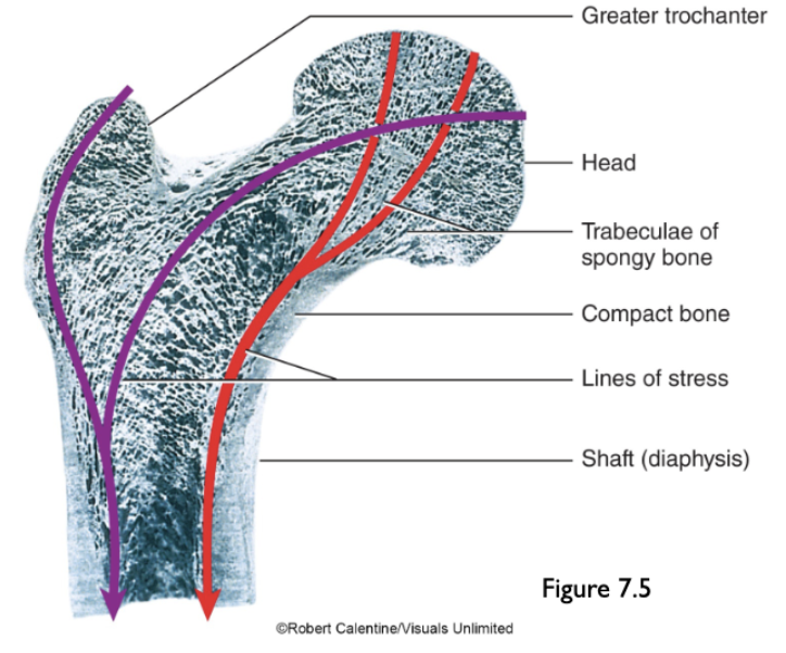
What is Wolff’s Law?
Bone adapts/remodels in response to habitual stress.
↑ stress/loading → ↑ bone mass
Bone size and shape adapt based on the magnitude and direction of forces applied.
What bone processes form joints?
Condyle → rounded articular projection
Facet → small, flat articular surface
Head → rounded, expanded end of a bone
All are processes/projections involved in forming joints.

What bone processes serve as attachment sites for muscles, tendons, or ligaments?
Crest, epicondyle, line, process, spine, suture, trochanter, tubercle, tuberosity
→ All are elevations/projections that serve as attachment sites.

What bone markings are classified as cavities/depressions?
Facet, foramen, fossa, fovea, meatus, sinus, sulcus
→ These are depressions, openings, or grooves in bone.

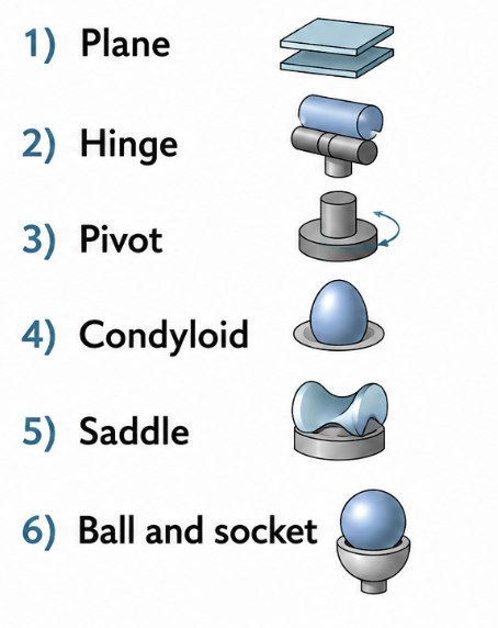
What are the 6 types of (diarthrodial) synovial joints based on the shape of their articular surfaces?
Plane
Hinge
Pivot
Condyloid
Saddle
Ball-and-socket
What is an articulation (arthrosis), and what determines the range of movement at a joint?
Articulation (arthrosis) = connection between bones at a joint, usually allowing movement between bone surfaces.
Joint movement is limited/determined by:
Shape/configuration of the bones
Ligaments
Muscles
Key idea: Humans have similar types of joint movements, but freedom, range, and strength of movement vary based on joint structure and surrounding tissues.
What are the 3 major classifications of joints based on structure and movement characteristics?
Synarthrodial
Amphiarthrodial
Diarthrodial
How do the functional classifications of joints correspond to their structural classifications? (overview)
Synarthrodial (immovable)
Fibrous → Gomphosis, Suture
Amphiarthrodial (slightly movable)
Fibrous → Syndesmosis
Cartilaginous → Symphysis, Synchondrosis
Diarthrodial (freely movable)
Synovial →
Arthrodial
Condyloidal
Enarthrodial
Ginglymus
Sellar
Trochoidal
Key:
Synarthrodial = no movement
Amphiarthrodial = little movement
Diarthrodial = free movement
What are synarthrodial joints and what are the two main types?
Synarthrodial joints = immovable joints
Two types:
Suture → joints between bones of the skull
Gomphosis → teeth anchored into the maxilla or mandible
Both are structurally classified as fibrous joints.

What are amphiarthrodial joints and what are the 3 types?
Amphiarthrodial joints = slightly movable joints → allow a small amount of motion.
3 types:
Syndesmosis
Symphysis
Synchondrosis
What is a syndesmosis joint?
Syndesmosis = slightly movable (amphiarthrodial) joint
2 bones joined by a strong ligament or interosseous membrane
Allows minimal movement
Bones may or may not touch at the actual joint
Examples:
Coracoclavicular joint
Distal tibiofibular joint
What is a symphysis joint?
Symphysis = slightly movable (amphiarthrodial) joint
Bones separated by a fibrocartilage pad
Allows very slight movement between bones
Examples:
Pubic symphysis
Intervertebral discs
What is a synchondrosis joint?
Synchondrosis = slightly movable (amphiarthrodial) joint
Bones separated by hyaline cartilage
Allows very slight movement between bones
Example:
Costochondral joints of the ribs with the sternum