its da final
 Call Kai
 Learn
Learn Practice Test
 Spaced Repetition
 Match
Match Flashcards
 Knowt Play
1/284

There's no tags or description
Looks like no tags are added yet.
Name | Mastery | Learn | Test | Matching | Spaced | Call with Kai | Chat |
|---|
No analytics yet
Send a link to your students to track their progress
285 Terms
Differential Stain
detects differences between organisms through the use of many dyes
and reagents
Example of Differential Stain
Gram Stain
Peptidoglycan
Differentiates bacteria into two groups based on amount of cell wall
Gram Staining: Primary Dye
Crystal Violet
Gram Staining Mordant
Gram’s Iodine
Gram Staining Decolorizer
95% Ethanol
Gram Staining Secondary Dye or Counterstain
Safranin
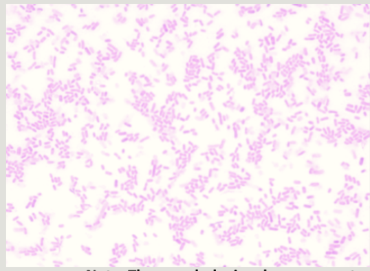
Gram Negative
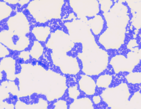
Gram Positive
Cationic Dyes Definition
Basic dyes, positively charged chromophores
Cationic Dyes Examples
Methylene Blue or Crystal Violet
Anionic Dyes Definition
Acidic dyes, negatively charged chromophores
Anionic Dyes Examples
Acid Fuchsin, Congo Red, Nigrosin
Nigrosin
Black Anionic Dye
Dye For Simple Stain
Crystal Violet (Cationic)
Illumination of Microscope
Light Source
Condensor
Directs light towards the objective lens in bright field microscopy
Iris diaphragm
Adjusts the diameter of the cone of light so that it just fills the objective lens
Ocular
10x
Objectives
4X, 10X, 40X and 100X
Ocular x Objective
Total Magnification
Resolution
Smallest distance between two objects which can be seen as separate
Oil Immersion
100x
d(Resolution)
λ/(2NA)
Illumination / λ
Wavelength
NA
Numerical Aperture
Numerical Aperture Definition
Light Gathering Capacity of Lens
Morphology
Bacterial Shapes
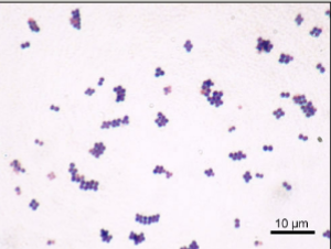
Staphylococcus
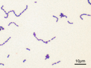
Streptococcus
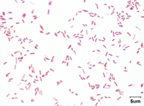
Bacillus (Rods)
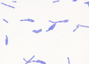
Streptobacillus
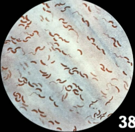
Comma Shaped
Vibrio (Cholerae)
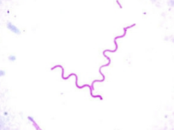
Rigid & Spiral
Spirillum Volutans
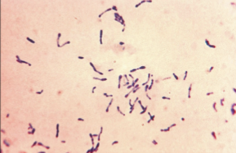
Pleomorphic
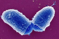
Club Shaped
Acid Fast Staining Type
Differential Stain
Endospore Stain Staining Type
Structural Stain
Structural Stain
Confirm structural characteristics of cells
Acid-Fast/Ziehl-Neelsen Primary Dye
Carbol Fuschin
Acid-Fast/Ziehl-Neelsen Mordant
Steam
Acid-Fast/Ziehl-Neelsen Decolorizer
Acid Alcohol
Acid-Fast/Ziehl-Neelsen Secondary Dye / Counterstain
Methylene Blue
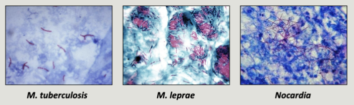
Acid Fast Genera
Mycobacterium
Spore Staining/Schaeffer-Fulton Primary Dye
Malachite Green
Spore Staining/Schaeffer-Fulton Mordant
Steam
Spore Staining/Schaeffer-Fulton Decolorizer
Water
Spore Staining/Schaeffer-Fulton Secondary/Counterstain
Safranin

Green
Free/Endospores
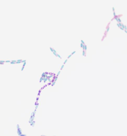
Pink
Vegetative Cells
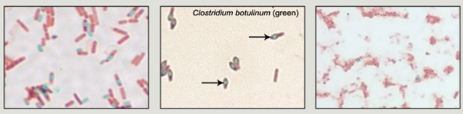
Gram Positive Rods:
Bacillus (Anthracis)
Clostridium (Botulinum, Tetani, Difficile)
Spore Forming Bacteria
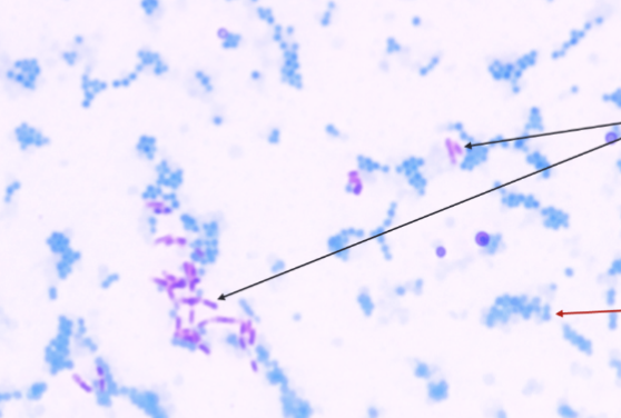
Cerise/Pink/Violet
Acid-Fast Cells (High Mycolic Acid)
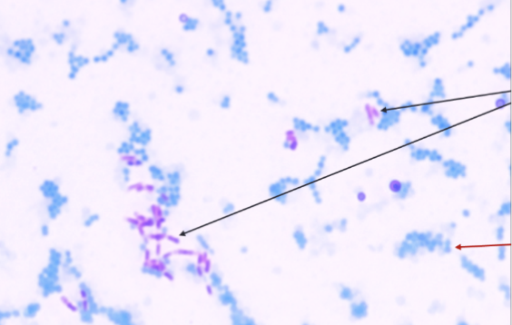
Blue
Acid-Fast Negative
Pure Culture
Single Microbial Species
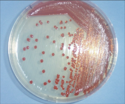
What Streaking is This
Three-Zone
Three Zone Dilutes From
Primary Streak
Subculture from Primary Streak
Secondary Streak
When streaking is considered a pure culture
Secondary Streak
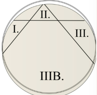
Three-Zone Streak Zones
Support growth of variety of microorganisms
General Purpose Media
Nutrient Agar (NA), Tryptic Soy Agar (TSA), Brain Heart Infusion (BHI)
Example General Purpose Media
Use inhibitors to prevent growth of certain organisms
Selective Media
Phenyl Ethyl Agar (PEA), Eosin Methylene Blue (EMB), MacConkey Agar
Example Selective Media
Allow growth of many microbes, but differentiation is seen by indicators that detect changes that have occurred
Differential Media
EMB, Blood agar, MacConkey Agar
Example Differential Media
Both Selective & Differential
Combination Media
EMB & MacConkey Agar
Example Combination Media
Undefined, exact chemical composition not known
Complex Media
Example Complex Media
BHI, TSA
Known chemical composition for each component
Defined Media
Organisms that have flagella
Motile
Motility Detected by Using
Soft Agar
Organisms that can swim through the soft agar and spread away from the inoculation stab line
Motile
Organisms that remain confined to the path of inoculation by the soft gel of the medium
Non-Motile
Tube Dilution x Plate Dilution
Final Dilution Equation
A / A + B
Dilution Equation
Strong chemical agents used on inanimate surfaces
Disinfectants
Chemical agents that may be safely applied to living tissue
Antiseptics
Compounds that may be administered for systemic circulation
Antibiotics
Kirby Bauer
Standardized antibiotic sensitivity testing method
Zone is measured to determine if an organism is susceptible, resistant, or intermediate to a certain antibiotic
Zone of inhibition
Growth Medium for Kirby-Bauer
Agar Muller-Hinton (MH)
Must be maintained within a narrow range (7.2 to 7.4), pH outside this range can affect the stability and function of the antimicrobials
pH Muller-Hinton (MH)
Some media have factors that can interfere with antimicrobial function
Nutrient Composition Muller-Hinton (MH)
–4 mm, can affect lateral diffusion rate of antibiotics
Agar Depth Muller-Hinton (MH)
Overestimation of bacterial sensitivity
LOW Initial concentration of bacteria inoculated
“Swamp” the antimicrobial, or quickly deplete nutrients, which can affect the growth of the bacteria
HIGH Initial concentration of bacteria inoculated
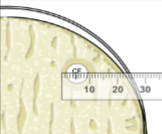
≤14mm
Resistant
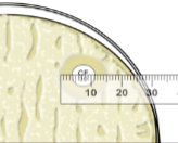
15-17 nm
Intermediate
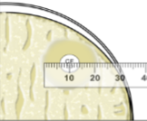
≥18mm
Susceptible
Antimicrobial in the disk start to diffuse into the agar
Diffusion Area
Highest concentration closest to the disk and lower concentrations farther from the disk
Concentration Gradient
Litmus Turns Pink
Acidic pH
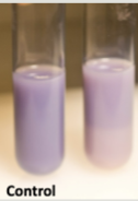
Litmus Result
WEAK Lactose Fermentation Acid
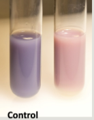
Litmus Result
STRONG Lactose Fermentation Acid
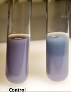
Litmus Result
Alkaline Peptone Deamination
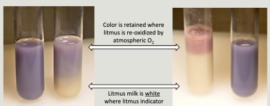
Litmus Result: Color Retention
O2 ReOxidation