BIOL2273: Anatomy and Physiology 1 Exam 2 (Ch. 5,6,7,8 & 9)
1/278
There's no tags or description
Looks like no tags are added yet.
Name | Mastery | Learn | Test | Matching | Spaced | Call with Kai |
|---|
No analytics yet
Send a link to your students to track their progress
279 Terms
What four things does the integumentary system consist of? (Chapter 5)
skin, hair, nails, and glands
What are the five functions of the integumentary system?
1. protection
-against abrasion and harmful effects of UV light
-keeps microorganisms from entering the body
-prevents dehydration by reducing water loss
2. sensation
-has sensory receptors that can detect heat, cold, touch, pressure, and pain
3. temperature regulation
-amount of blood flow through the skin and sweat gland activity regulate body temp.
4. vitamin D production
-important regulator of calcium balance
-skin produces a molecule when exposed to UV light that can be transformed into vitamin D
5. excretion
-waste products are excreted through the skin and glands
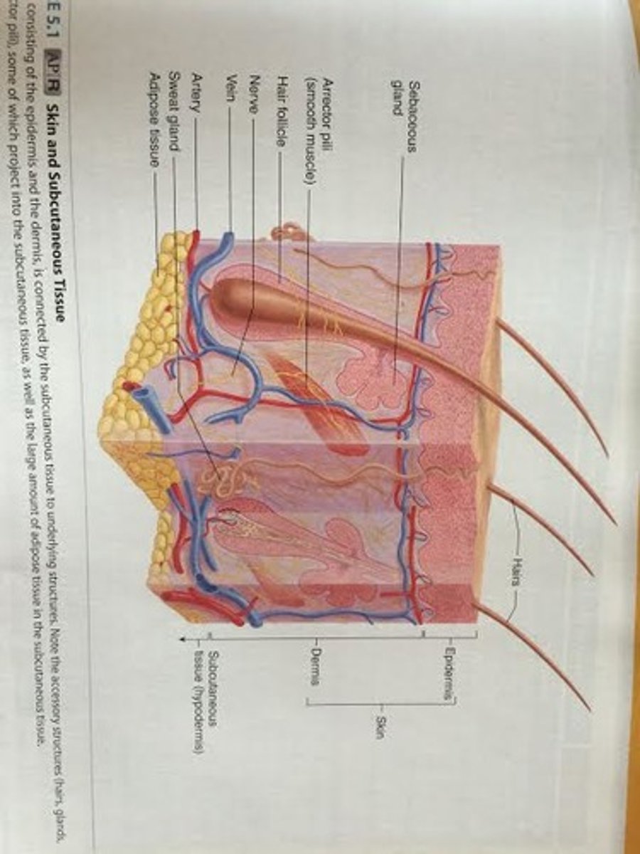
What tissue layers make up the skin? What are they responsible for?
1. epidermis
-superficial layer of the skin
-stratified squamous epithelium
-protection- resists abrasion on skin's surface & reduces water loss
-not as thick as the dermis and contains no blood vessels
-contains the five strata
2. dermis
-connective tissue
-responsible for structural strength of the skin
-contains the papillary layer and reticular layer
*****hypodermis is not a part of the skin
What tissue does the skin rest on?
hypodermis
-subcutaneous tissue-loose connective
-connects the skin to underlying muscles or bone
*****not part of the skin or the integumentary system
What separates the dermis and the epidermis?
a basement membrane
-dermal papilla are in the papillary layer of the dermis
Extra notes of what is contained in the epidermis, dermis, and hypodermis
1. Epidermis
2. Dermis- contains nerve endings, hair follicles, smooth muscles, glands, and lymphatic vessels
3. Hypodermis
The epidermis is mostly composed of what type of cell, what do those cells do? What are three other cells that also compose the epidermis?
keratinocytes
-produce a protein mixture called keratin
-keratin makes cells more durable
-cells protect- resist abrasion and reduce water loss
extra cells:
-melanocytes- contribute to skin color
-Langerhans cells- part of the immune system
-Merkel cells- specialized cells associated with nerve endings that detect light tough and superficial pressure
What is keratinization of cells? What happens during this process? Why is this important?
when the keratinocytes accumulate keratin (fibrous structural protein)
-cells change shape and chemical composition
-happens when cells move from the deeper epidermal layers to the surface
-progression of maturation of keratinocyte
During keratinization, the cells eventually die and produce an outer layer of dead, hard cells that resist abrasion and form a permeability barrier
important because many skin diseases result from malfunctions in this process
-psoriasis-when large scales of epidermal tissues are sloughed off
-by comparing normal and abnormal keratinization, effective therapies can be personally developed for psoriasis
When are new keratinocytes produced?
when keratinocyte stem cells undergo mitosis in the deepest layer of the epidermis
-as new cells form, they push older cells to the surface, where they are sloughed/shed off
-outermost cells protect surface
-deeper replicating cells replace cells lost from the surface
-when the cells move from the deeper epidermal layers to the surface, the cells change shape and chemical composition (keratinization) and they accumulate keratin
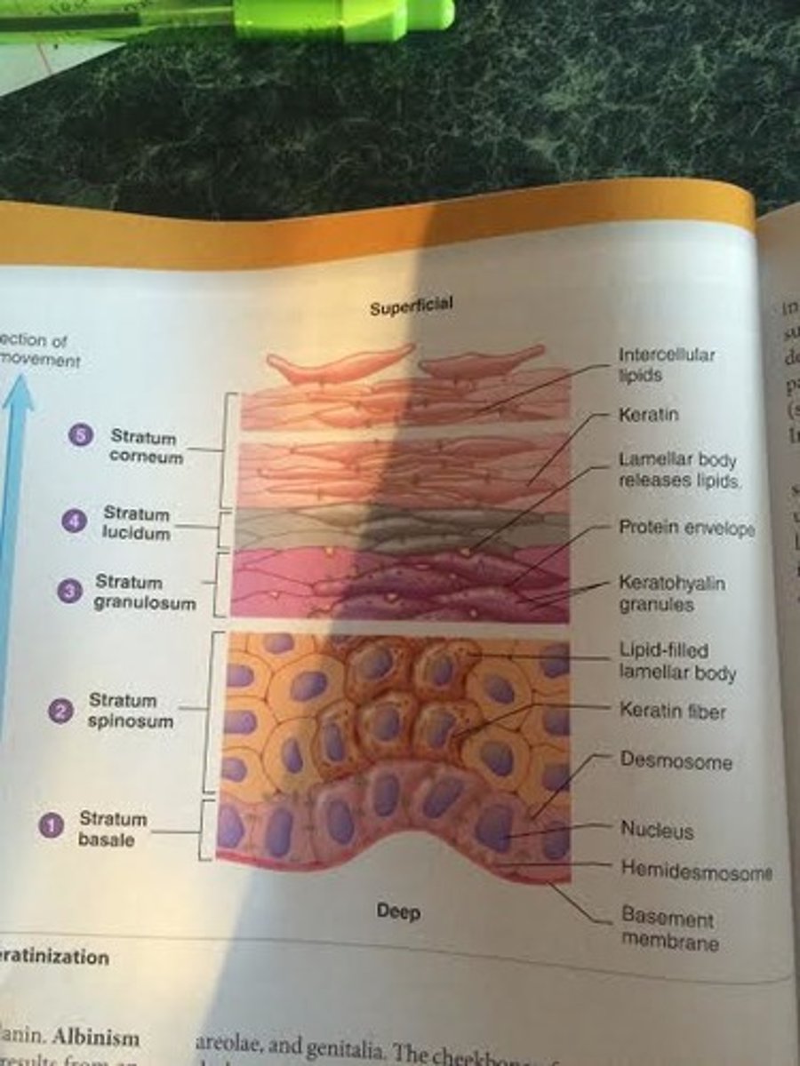
What are the five regions, or strata, of the epidermis, in order from outermost to the innermost layer?
1. Stratum Corneum
2. Stratum Lucidum
3. Stratum Granulosum
4. Stratum Spinosum
4. Stratum Basale
CLGSB
Colby Love Goes Spring Breaking
What does the stratum corneum consist of? How many layers of cells are in this region?
many layers of dead squamous cells
-most superficial layer of skin
-Carotene goes here and adipocytes when in excess
What does the stratum lucidum consist of? How many layers of cells are in this region?
3-5 layers of dead, transparent cells
-keratohyalin (granules in stratum granulosum) has dispersed around keratin fibers to make it look clear
-present in thick skin, absent in most thin skin
Lucidum-Liquid-clear
What does the stratum granulosum consist of? How many layers of cells are in this region?
2-5 layers of flattened cells
-cells filled with granules of keratohyalin, accumulate in the cytoplasm
-hard protein envelope forms beneath the plasma membrane
-lamellar bodies release lipids and the cells die
GRANULes of keratohyalin in stratum GRANULosum
What is keratohyalin?
nonmembrane-bound proteins
-does not deteriorate when pushed through the layers, along with keratin fibers, unlike the other organelles and the nucleus
-accumulates in the cytoplasm of the keratinocytes
-lipid soluble that help to form a waterproof barrier that prevents water loss from the body
-in the form of granules (small compact particles) in stratum granulosum
-dispersed in stratum lucidum
What does the stratum spinosum consist of? How many layers of cells are in this region?
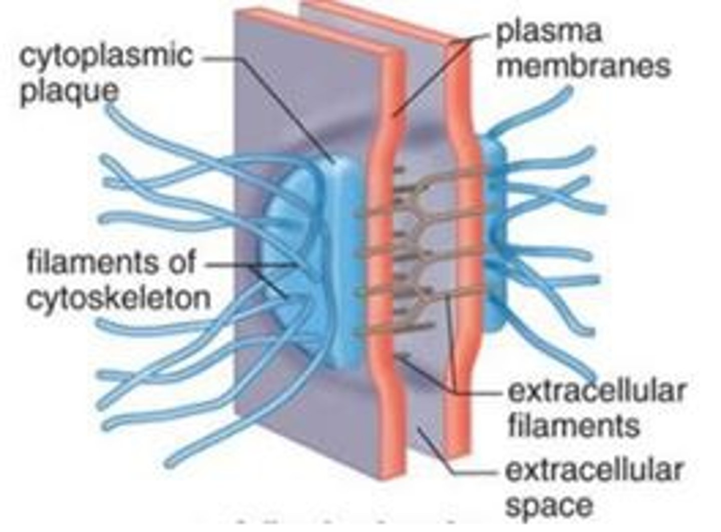
several layers of cells held together by many desmosomes
-additional keratin fibers and lamellar bodies (membrane-bound organelles that release lipids) form inside the keratinocytes
-melanocytes in between here and the stratum basale
What do the lamellar bodies do as they move to the stratum granulosum from the stratum spinosum?
In the stratum granulosum, they move to the plasma membrane and release their lipid contents into the extracellular space. Inside the cell, a protein envelope forms beneath the plasma membrane (makes the dead cells in the stratum corneum have this hard protein envelope). In the most superficial layers of the stratum granulosum, the nucleus and other organelles degenerate, and the cell dies. However, the keratin fibers and keratohyalin granules do not die.
-lamellar bodies are lipid-filled, membrane-bound organelles
What does the stratum basale consist of? How many layers of cells are in this region?
consists of keratinocytes, which produce the cells of the more superficial strata
-single layer of cuboidal and columnar cells
-melanocytes in between here and the stratum spinosum
-deepest portion of the epidermis
Extra notes on the direction of movement of cells in the epidermal layers
5. In the stratum corneum, the dead cells have a hard protein envelope, containing keratin, and are surrounded by lipids.
4. In the stratum lucidum, the cells are dead and contain dispersed keratohyalin.
3. In the stratum granulosum, keratohyalin granules accumulate, and a hard protein envelope forms beneath the plasma membrane; lamellar bodies release lipids; cells die.
2. In the stratum spinosum, keratin fibers and lamellar bodies accumulate.
1. In the stratum basale, cells divide by mitosis, and some of the newly formed cells become the cells of the more superficial strata.
What is the difference between soft keratin and hard keratin?
soft keratin- found in the skin
hard keratin- found in nails and external part of the hair
-cells containing hard keratin are more durable and do not shed
What is the difference between thick and thin skin? Where are each of them found?
1. thick skin- has all five epithelial strata
-found in palms of hands and soles of feet
2. thin skin- contains fewer cell layers per stratum, and the stratum lucidum is usually absent
-hair is found only in thin skin
-only refers to the epidermis only, not for total skin thickness
What two things can develop on thick and thin skin when subjected to friction or pressure?
1. callus- number of layers of stratum corneum increase to produce a thicker area
2. corn- over bony areas- can be very painful
What are the three factors that determine skin color?
1. pigments in the skin
2. blood circulating through the skin
3. thickness of the stratum corneum
What is the group of pigments that are responsible for skin, hair, and eye color?
Melanin
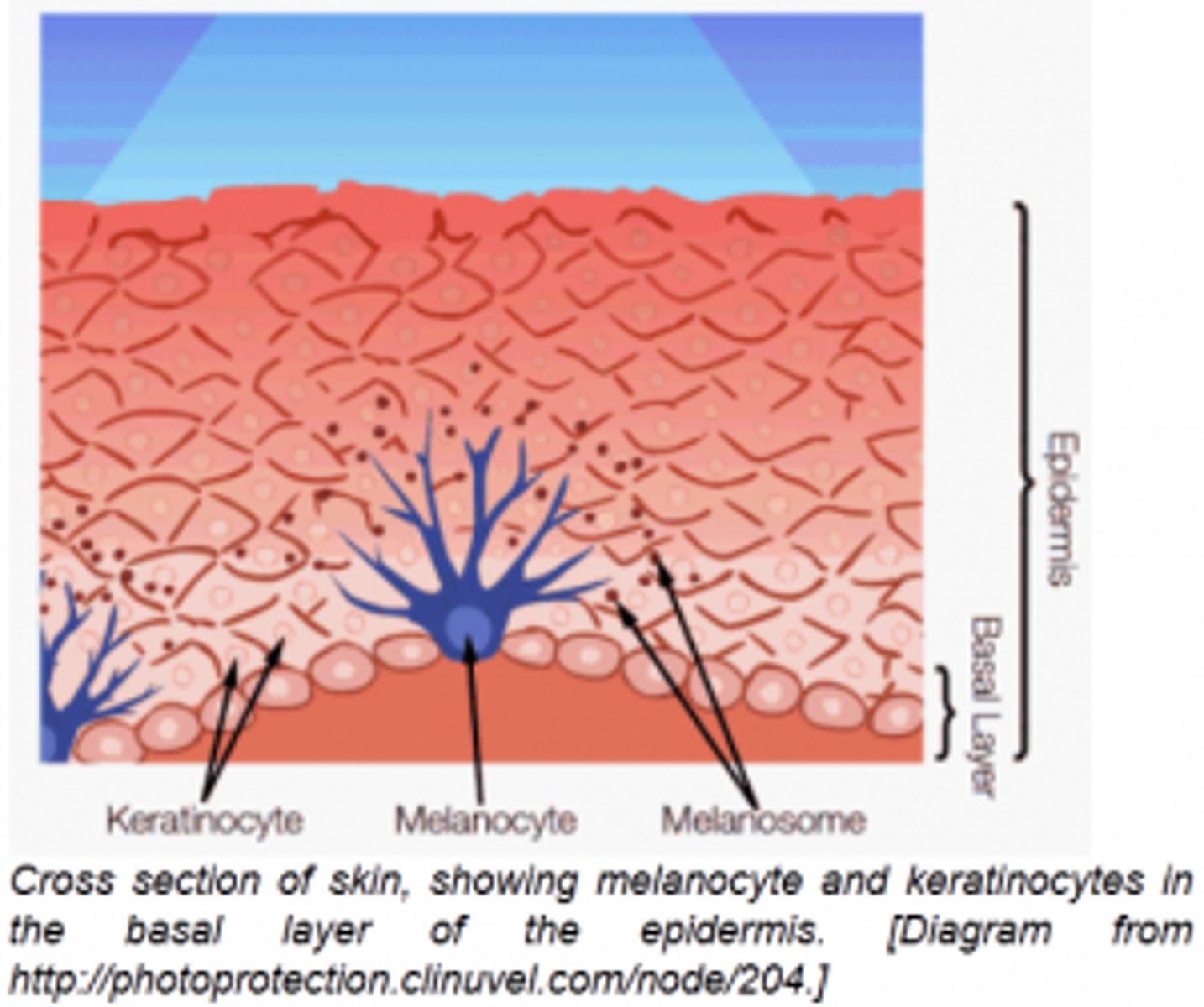
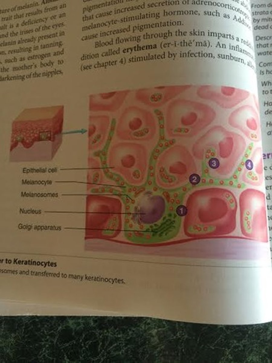
-Melanocytes produce Melanin inside melanosomes (vesicles that move into the cell processes of the melanocytes) and then transfer the melanin to keratinocytes
-golgi apparatus of melanocytes packages melanin into melanosomes
-size and distribution of melanosomes determines skin color
What three things determine melanin production?
1. genetic factors
-responsible for variations in skin color among different races and among people of the same race
2. exposure to light (tanning, UV)
3. hormones
-during pregnancy, estrogen and melanocyte-stimulating hormones cause the mother to have increased melanin production, which causes darkening of the nipples, genitalia, and areolae
What is melanin produced by?
melanocytes (green)
-irregular shaped cells with long processes that extend between keratinocytes of the stratum basale and stratum spinosum
-although all keratinocytes can contain melanin, only melanocytes produce it

What is Albinism and its result?
have melanocytes (everyone) but are unable to produce melanin
-lacks tyrosinase that catalyzes production of melanin
-results in absence of pigment in the skin, hair and irises of the eyes
Extra notes about Melanin transfer to keratinocytes
melanocytes make melanin, which is packaged into melanosomes and transferred to many keratinocytes
1. Melanosomes (little balls) are produced by the Golgi apparatus of the melanocyte (irregular shaped cells, green)
2. Melanosomes move into melanocyte cell processes
3. Epithelial cells (keratinocytes) phagocytize the tips of the melanocyte cell processes (where the melanosomes are)
4. the melanosomes, which were produced inside the melanocytes, have been transferred to the epithelial cells (keratinocytes) and are now inside them
-although all keratinocytes can contain melanin, only melanocytes produce it
What does an increased and decreased blood flow do to skin color?
1. Increased- produces a red skin color
-called erythema
-it is an inflammatory response
-can be stimulated by infection, sunburn, allergic reactions, insect bites, or other causes
-can be exposure to the cold or flushing when angry or hot
2. Decreased- results in pale skin
-occurs in shock
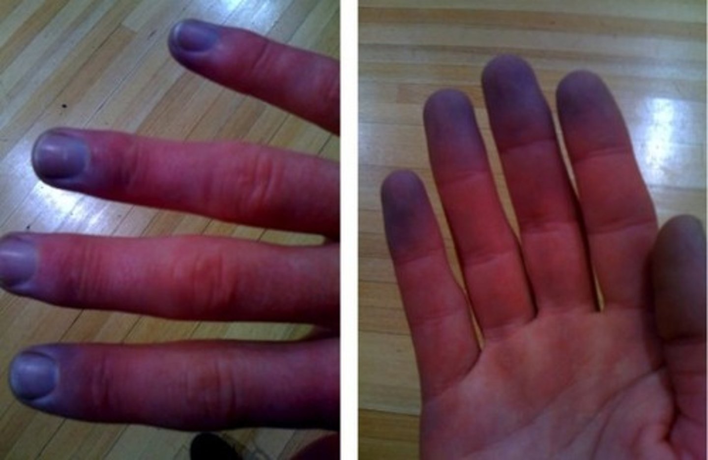
-bluish skin color can result with a decrease in blood oxygen content called cyanosis
Increased blood flow-red
Decreased blow flow- white
Decreased blood oxygen content- blue
What is cyanosis?
a bluish skin color that results from decreased blood oxygen content
What is Carotene?
an ingested plant pigment, can cause the skin to appear yellowish
-carrot diet can turn skin yellow
-source of Vitamin A
-when large amounts of carotene are consumed, the excess accumulates in the stratum corneum and adipocytes (because it is lipid soluble) of the dermis and hypodermis
What are the two layers of the dermis and what are they composed of?
1. Papillary layer- loose connective tissue (upper)
2. Reticular layer- dense irregular connective tissue (much thicker) (main)
Hypodermis- loose connective tissue (beneath the dermis) (not a part of the skin)
What is the papillary layer of the dermis composed of?
has projections called dermal papillae and is composed of loose connective tissue that is well supplied with capillaries
-blood vessels supply the overlying epidermis with oxygen and nutrients, remove waste products, and aid in regulating body temp.
-responsible for finger prints
-upper layer
What is the reticular layer of the dermis composed of? What happens when the skin is overstretched?
consists mostly of collagen
-much thicker than the papillary layer, is dense irregular connective tissue
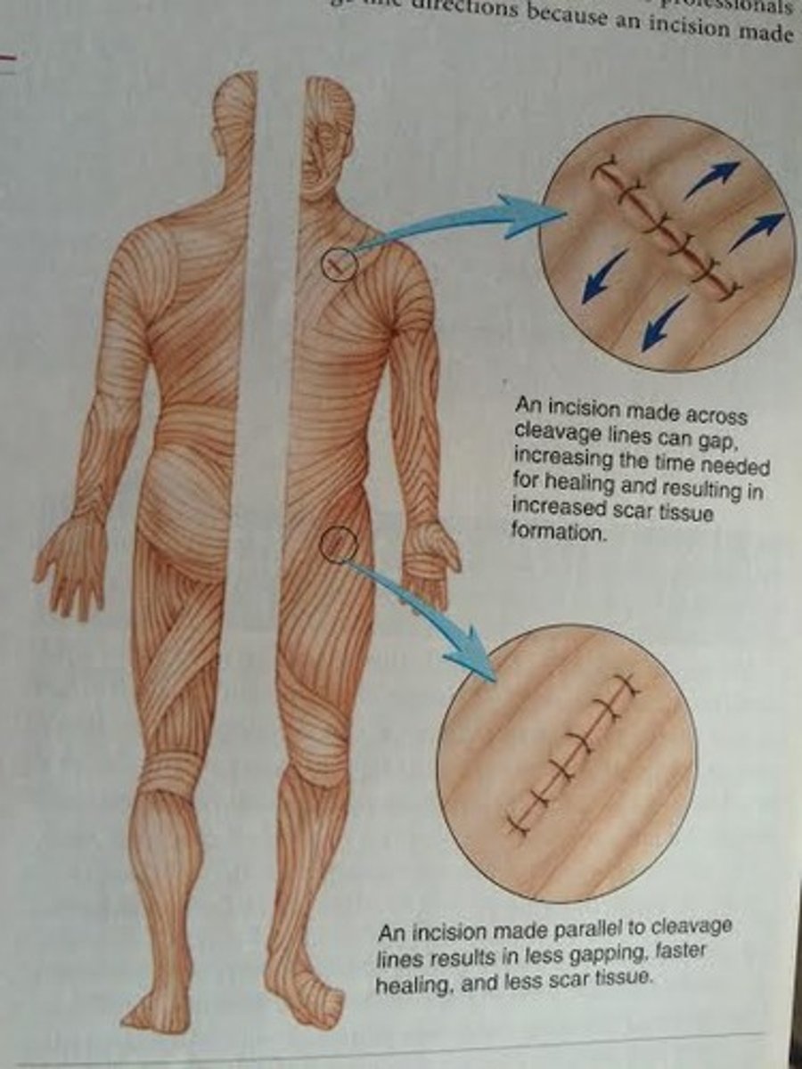
-parallel collagen fibers form cleavage lines (like on wrist when clench)
-responsible for cleavage lines/ tension lines
if skin is overstretched, striae (stretch marks) occur
-amount of elastic fibers (determined by genes) makes stretch marks/ flab
-the dermis ruptures and the lines of scar tissue are stretch marks
-ex. on abdomen and breast during pregnancy or on skin of athletes who have quickly increased muscle size
What is the main connective tissue fiber, but which ones are also present, in the dermis? What do these fibers contribute to?
collagen is the main
-formation of collagen fibers produces cleavage lines in the skin
-collagen is the most abundant protein in the body that holds the body together
-collagen provides strength and structure
elastic and reticular are also present
-amount of elastic fibers makes stretch marks
What is the papillary layer of the dermis responsible for?
finger prints
-the dermal papillae under the thick skin of the palms and soles lie in parallel, curving ridges that shape the overlying epidermis into fingerprints and footprints
-everyone's are unique, even identical twins
What is the reticular layer of the dermis responsible for? Why is it important for health professionals to understand this?
cleavage/tension lines
-formation of collagen fibers produces cleavage lines in the skin
-important for them to know cleavage line directions because an incision made parallel to the cleavage lines is less likely to gap compared to an incision made across them
-development of infections and formation of scar tissue is reduced in wounds where the edges are close together/ have parallel cleavage lines
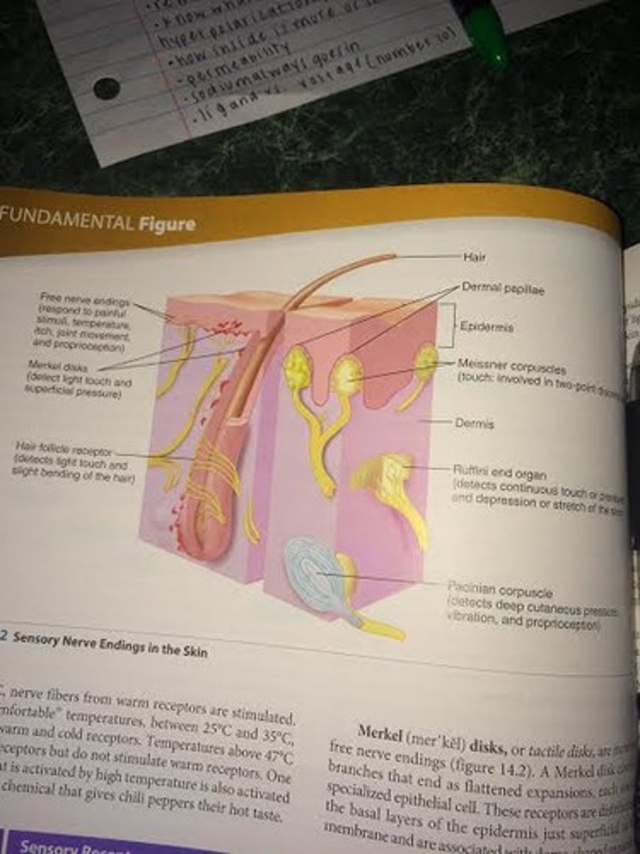
In the dermis, what are the functions of the nerve endings?
-free nerve endings for pain, itch, tickle, and temperature sensations
-hair follicle receptors for light touch
-Pacinian corpuscles for deep pressure
-Meissner corpuscles for detecting simultaneous stimulation at two points on the skin
-Ruffini end organs for sensing continuous touch or pressure
What does the subcutaneous tissue, located beneath the dermis, consist of? Is this a part of the skin?
known as hypodermis, is loose connective tissue that contains collagen and elastic fibers
-what the skin rests on
*****NOT a part of the skin or integumentary system
What does the hypodermis attach the skin to? What is it a site of?
attaches the skin to underlying structures (bone and muscle)
site of fat storage
-half of the body's stored lipids are in this tissue, where they function in insulation, padding, and a source of energy
What are the functions of the adipose tissue within the subcutaneous layer?
insulation, padding, and a source of energy
What can the subcutaneous tissue (hypodermis) estimate?
total body fat by pinching the skin at selected locations and measuring the thickness of the skin fold and tissue
-thicker the fold, greater amount of total body fat
-amount of adipose tissue varies with age, sex, and diet
- most babies have a chubby appearance because they have more adipose tissue, proportionally, than adults
-women have more adipose tissue than men (breasts, thighs, etc.)
What are the two types of hair and when do they occur, what do they replace?
1. Terminal
-long, coarse, and pigmented
-replace lanugo (fetal hair ) at the time of birth (scalp, eyelids, eyebrows)
-replace vellus hair at puberty especially in pubic and axillary (arm pit) regions
-axillary and pubic hair are a sign of sexual maturity
-chest, legs, and arms is 90% terminal for male and 35% for female
-males replace vellus hair for terminal hair on face
2. Vellus
-short, fine, and usually unpigmented
-replace lanugo on the rest of the body at the time of birth
lanugo- delicate, unpigmented hair that covers the fetus
What are the three parts of hair?
1. shaft- protrudes above the surface of the skin
2. root- located below the surface of the skin
3. hair bulb- an expanded knob at the base of the root
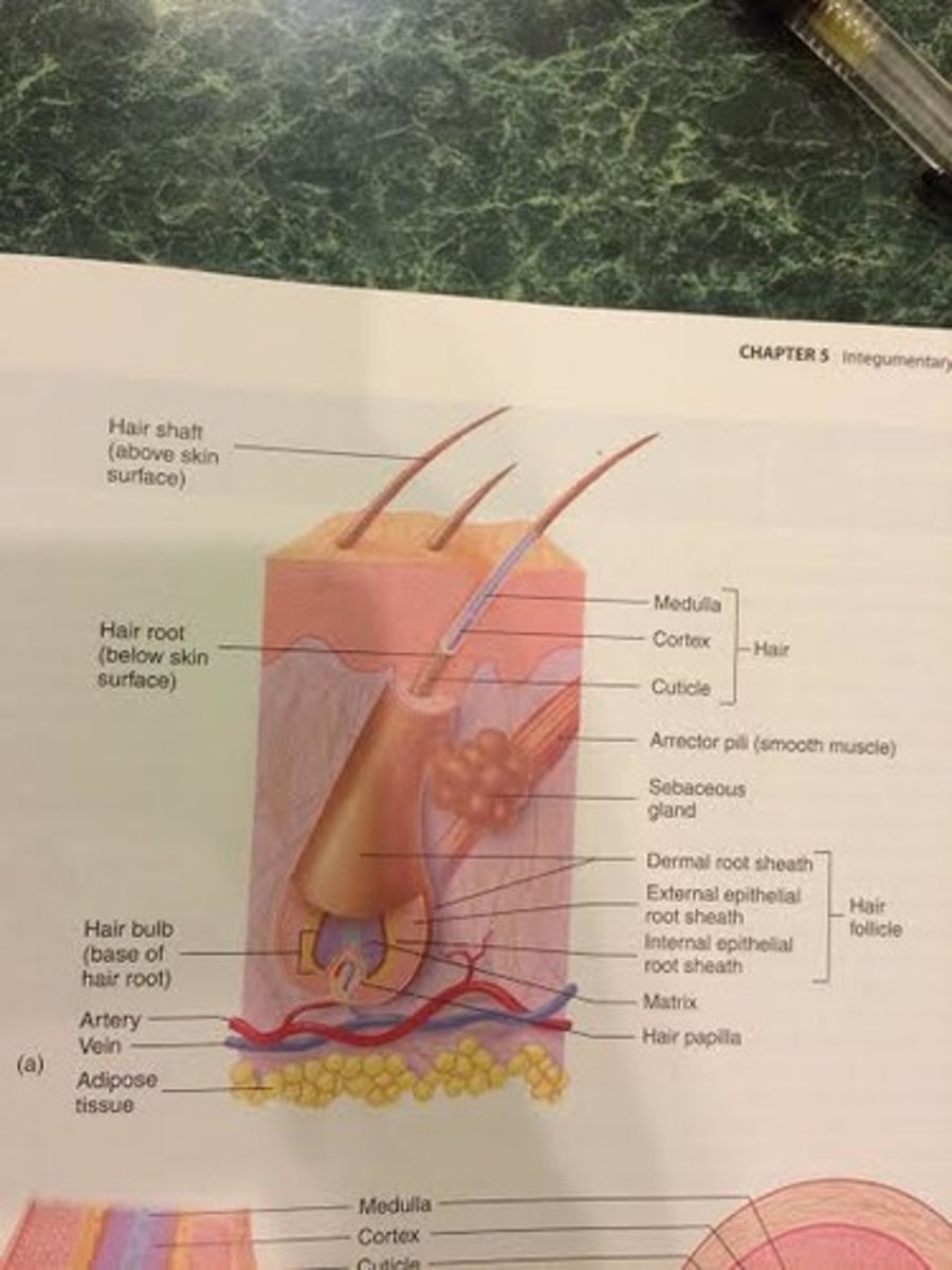
What is the tubelike invagination of the epidermis that extends into the dermis from which hair develops?
hair follicle
-consists of dermal root sheath (portion of dermis that surrounds the epithelial root sheath) and an epithelial root sheath (external and internal)
external epithelial root sheath- where strata is found in thin skin
sheath= close-fitting cover

Where is all the strata found in thin skin? What happens to the number of cells when you go deeper in the hair follicle?
At the opening of the follicle, the external epithelial root sheath has all the strata found in thin skin
-deeper in the hair follicle, the number of cells decreases until at the hair bulb there is only the stratum basale
-if the epidermis and superficial part of the dermis are damaged, the undamaged hair follicle that lies deep in the dermis can be a source of new epithelium
sheath= close-fitting cover

What are the three concentric layers of the root and shaft? What types of cells are they composed of; what types of keratin do they contain?
composed of columns of dead, keratinized epithelial cells
1. medulla-central axis of the hair that consists of two to three layers of cells containing soft keratin
-most interior part of the hair shaft
-medulla=middle
2. cortex- the bulk of the hair that consists of hard keratin
-middle part of the hair shaft
-where melanin is found
3. cuticle- covers the cortex- single layer of cells that contain hard keratin
-outermost part of the hair shaft
-gives protective barrier and mechanical strength
-edges of cuticle cells overlap like shingles on a roof
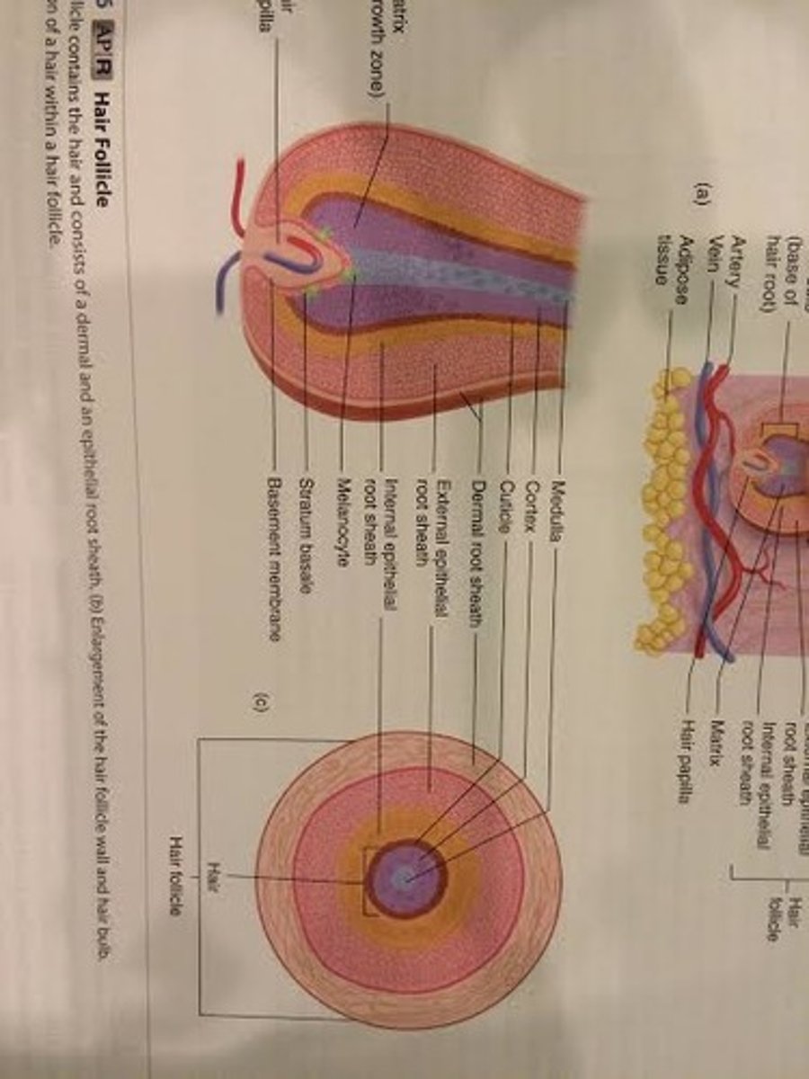
What is the growth zone of the hair? What is the projection of the dermis that goes into the hair bulb?
growth zone: matrix- a mass of undifferentiated epithelial cells
-matrix is in hair follicle
-matrix produces the hair and the epithelial root sheath
hair papilla projects into the hair bulb from the dermis
-contains blood vessels and provides nourishment to the cells of the matrix

What are the two cycles that the hair bulb produces the hair?
1. Growth Stage
-hair is formed in matrix cells that differentiate, become keratinized, and die
-hair grows longer when more matrix cells are added at the base of the hair root (hair bulb)
2. Resting Stage
-hair growth stops, the hair follicle shortens and holds the hair in place
-follows after the growth stage when a new cycle begins and a new hair replaces the old hair, which falls out of the hair follicle
-the stages change with age
-length of each stage depends on the hair (eyelash vs. scalp)
What does losing a hair normally mean?
the hair is being replaced
-loss of 100 scalp hairs per day is normal
-"pattern baldness" is most common kind of permanent hair loss- hair follicles shrink and revert to producing vellus hair
-eventually all hair production in these smaller follicles may completely cease
What determines hair color?
amount and kind of melanin present
-melanocytes within the hair bulb matrix produce melanin and pass it to keratinocytes in the hair cortex and medulla
-blonde hair has little black-brown melanin where black hair has the most, intermediate amounts of melanin account for different shades of brown
-with age, the amount of melanin in hair decreases, causing it to fade or become white (no melanin)
-grey hair= dying melanocytes- air bubbles form so it is not all one grey shade- grey hair is a mixture of unfaded, faded, and white hair
-hair color is controlled by several genes- dark hair is not necessarily dominant over light
What are three structures that are associated with hair?
1. sebaceous gland- empty sebum into hair follicle (so does apocrine sweat gland)
2. arrector pili muscle- smooth muscle cells
3. nerve endings near root
What does the arrector pili muscle do? What was the original reason for this?
causes hair to "stand on end" and produce "goosebumps"
-contraction of the arrector pili muscles, are smooth muscles***** do not have control/ involuntary
-associated with each hair follicle
-extend from the dermal root sheath of the hair follicle to the papillary layer of the dermis
originally used as a protective precaution (makes you appear bigger) but does not have much function now-- like a mad cat

Why does it hurt when hairs are pulled as opposed to getting a hair cut?
When your hair is pulled, you feel it in your hair follicle that is connected to nerve cells and embedded in your skin. Your hair doesn't have any nerve cells. So when it gets cut, you don't feel the scissors cutting your hair, but you probably feel your hair being touched or moved. That's because the strands of hair are moving within the follicles and relaying that to your nerve cells.
-internal root sheath (part of the hair follicle) is usually pulled out as well, plainly visible as white tissue around the root of the hair
What are the two major glands of the skin and where do they excrete into?
1. sebaceous gland
-empty into a hair follicle
2. sweat gland (sudoriferous)
-eccrine sweat gland- open directly onto the surface of the skin through sweat pores
-apocrine sweat gland- empty into a hair follicle
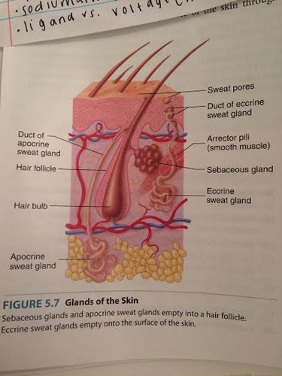
What do sebaceous glands produce and where is it secreted?
sebum (an oily, white substance rich in lipids)
-oils the hair and the surface of the skin
-glands connected by a duct to the upper part of the hair follicle, from which the sebum oils the hair and the skin surface
-not all sebaceous glands are associated with hairs but open directly onto the skin surface (lips, eyelids, genitalia)
-gland very active at puberty
sebaceous-sebum
What is the most common type of sweat gland and what are the secretions?
Eccrine (merocrine) sweat gland
-produces sweat, which cools the body
-secretions are primarily water
-open directly onto the surface of the skin through sweat pores (not hair follicle)
-most numerous in palms and soles (where there is thick skin)
-can be used in lie detector (polygraph) tests because sweat gland activity increases when a person tells a lie-detects sweat because the salt solution conducts electricity
What happens when a duct in a gland gets clogged or blocked?
acne
-sebaceous gland very active at puberty
Which sweat gland helps regulate body temperature?
eccrine sweat gland
-produces sweat, which cools the body
What do apocrine sweat glands produce? What regions of the body is this gland found?
produce an organic secretion that can be broken down by bacteria to cause body odor
-empty into hair follicle
-become active at puberty
-found in genital and axillary regions (stinky armpits)
What do ceruminous glands make? What gland is it a modification of?
cerumen (earwax)
-modified eccrine sweat gland located in the ear canal
-protect entry of dirt and insects
-too much can block the ear canal and make hearing more difficult
What do mammary glands produce? What gland is it a modification of?
milk
-modified apocrine sweat gland located in the breasts
-apocrine gland- becomes active at puberty
What epidermal layer of cells does a nail consist of?
layers of dead stratum corneum cells that contain very hard keratin
-located at the distal ends of digits (fingers and toes)
The nail consists of a proximal nail root and distal nail body. Which is covered by skin and which is the visible part of the nail?
1. nail root- covered by skin
-extends distally from the nail matrix
-nail matrix produces nearly half the nail
2. nail body- visible part of the nail
-nail body consists of stratum corneum (superficial epidermal layer), several layers of cells containing hard keratin
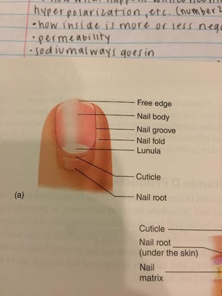
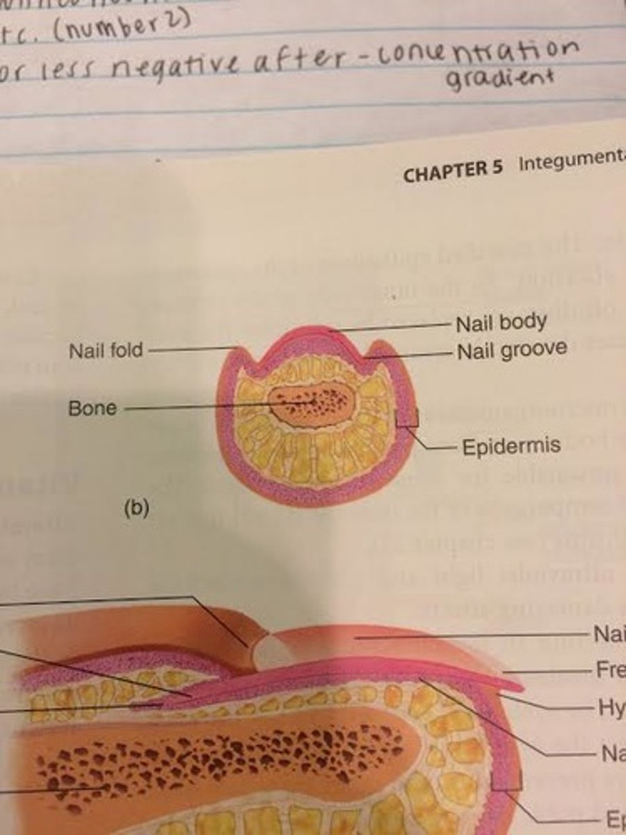
The lateral and proximal edges of the nail are covered by skin called what?
nail fold
-skin near bottom towards lunula

The edges of the nail are held in place by what?
nail groove
-sides of nail
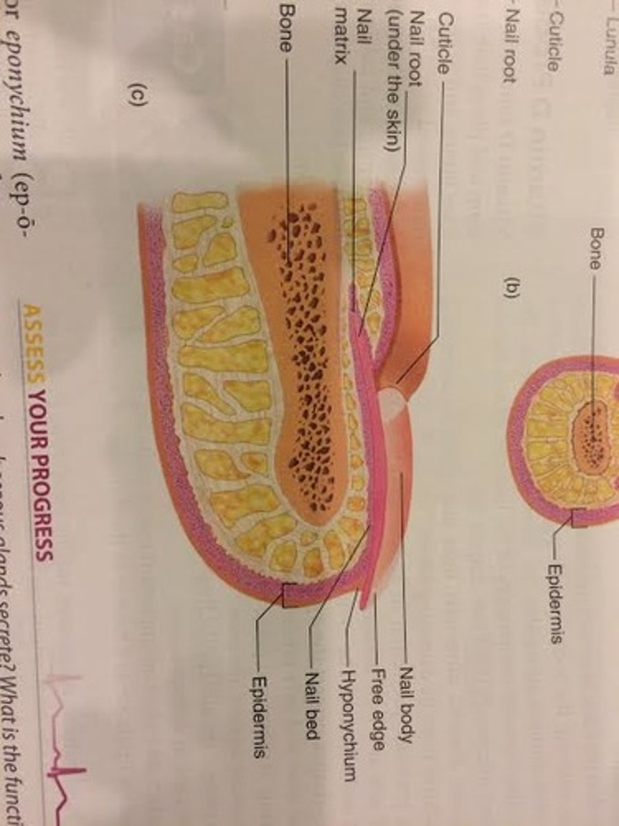
The stratum corneum of the nail fold grows onto the nail body as the cuticle. What is another name for cuticle?
eponychium
-superficial to nail body

What is the stratum corneum beneath the free edge of the nail body?
hyponychium
-thickened region of stratum corneum

What is the area called where the nail attaches between the nail matrix and the hyponychium (cuticle)? Why does it appear pink?
nail bed
-nail matrix and bed are composed of epithelial tissue, with a stratum basale that gives rise to the cells that form the nail
-nail bed is visible through the clear nail and appears pink because the blood vessels in the dermis
-nail matrix is thicker than nail bed
-nail matrix produces nearly all the nail
What is the pale crescent area at the base of the nail called? Why does it appear white?
lunula
-formed by matrix
-seen through the nail body
-appears white because the blood vessels do not show through the thicker nail matrix
-color to nails=blood supply
How is the growth of nails different than that of hair? How many mm/day do fingernails grow?
grows continuously- does not have a resting stage
-fingernails grow .5-1.2mm per day; faster than toenails
-as nail forms in the nail matrix and bed, it slides over the nail bed toward the distal end of the digit
The major function of the integumentary system is protection. What do the skin, hair, and nails protect against?
1. Skin- protects against abrasion (strength from dermis) and UV lights (melanin absorbs it and protects underlying structures from its effects), prevents the entry of microorganisms (secretions create environment unsuitable for some microorganisms and skin contains components of the immune system that act against microorganisms), helps regulate body temp., and prevents water loss (lipids act as barrier to the diffusion of water)
2. Hair- protects against abrasion and UV light and is a heat insulator
3. Nails protect the ends of the digits
What contains sensory receptors, and for what?
skin
-for pain, temperature (hot and cold), and pressure that allow proper response to the environment
How does the skin control heat loss from the body?
through dilation and constriction of blood vessels
What lowers body temperature?
sweat glands (only eccrine, not apocrine)
-produce sweat, which evaporates and lowers body temp.
What does skin exposed to UV light produce?
cholecalciferol
-modified in the liver and then in the kidneys to form active vitamin D
-all vitamin D required in body can be produced by this process if enough UV light is available
vitamin D- calcium- cholecalciferol
In what three ways does vitamin D increase blood calcium levels?
1. promoting calcium uptake from the small intestine
2. calcium release from bone
3. the reduction of calcium loss from the kidneys
cholecalciferol eventually turns into vitamin D
Are skin glands important in excretion?
skin glands remove small amounts of waste products (urea, uric acid, and ammonia) but are NOT important in excretion
-even when large amounts of sweat are lost, the quantity of waste products is insignificant
-urinary system excretes most of the body's waste products
A burn is injury to a tissue caused by heat, cold, friction, chemicals, electricity, or radiation. How are burns classified?
according to the extent of surface area involved and the depth of the burn
How deep do first-degree burns go? What is the result?
damages only the epidermis
-partial-thickness burn
-results in redness, pain, and slight edema (swelling)
-can be caused by sunburn, etc.
-healed in around a week without scarring
How deep do second-degree burns go? What is the result?
damages the epidermis and dermis
-partial-thickness burn
-minimal dermal damage can cause redness, pain, edema, and blisters- healed in around 2 weeks without scarring
-if the burn goes deep into the dermis, the wound appears red, tan, or white- takes several months to heal and might scar
How deep do third-degree burns go? What is the result and what might be necessary?
epidermis and dermis are completely destroyed, and deeper tissue may be involved
-full-thickness burn
-often surrounded by first and second-degree burns
-usually painless because the sensory receptors have been destroyed
-appear white, tan, brown, black, or bright red and skin can only regenerate from the edges
-skin grafts are often necessary
As the body ages, why is the skin more easily damaged?
epidermis thins and the amount of collagen in the dermis decreases
-skin infections are more likely
-skin repair occurs more slowly
-decrease in number of elastic fibers in the dermis and a loss of adipose tissue from the hypodermis cause the skin to sag and wrinkle
What are the four components of the skeletal system? (Ch.6)
bones, cartilage, tendons, and ligaments
What are the five major functions of the skeletal system?
1. Support
-rigid, strong bones can bear weight and is the major supporting tissue of the body
-cartilage provides firm but flexible support
-ligaments are strong bands of fibrous connective tissue that attach to bones and hold them together
2. Protection
-bone is hard and protects the organs it surrounds (ex. skull encloses and protects the brain)
3. Movement
-tendons are strong bands of connective tissue that attach skeletal muscles
-joints allow movement between bones
-smooth cartilage on the ends of bones within some joints allows bones to move freely
-ligaments allow some, but not excessive, movement between bones
4. Storage
-some minerals (mostly calcium and phosphorus) in the blood are taken into bone and stored
-adipose tissue is stored within bone cavities
5. Blood Cell Production
-many bones contain cavities that are filled with red bone marrow, which gives rise to blood cells and platelets
What are the three types of cartilage? Which type is most associated with bone?
1. Hyaline Cartilage
-precursor for most bones in the body
-bone lengthening and repair often involve producing hyaline cartilage first, then replacing it with bone tissue
2. Fibrocartilage
3. Elastic Cartilage
What specialized cells does hyaline cartilage consist of?
chondroblasts
-produce the hyaline cartilage matrix surrounding themselves
What does the hyaline cartilage matrix consist of?
collagen (provides strength) and proteoglycans (makes cartilage resilient by trapping water (can recover quickly from bending, stretching or compression)
-hyaluronic acid is a nonfibrous molecule that also is part of the matrix
-hyaluronic acid and proteoglycans make up the ground substance of the matrix, which is the background which the collagen fibers are seen through in the microscope
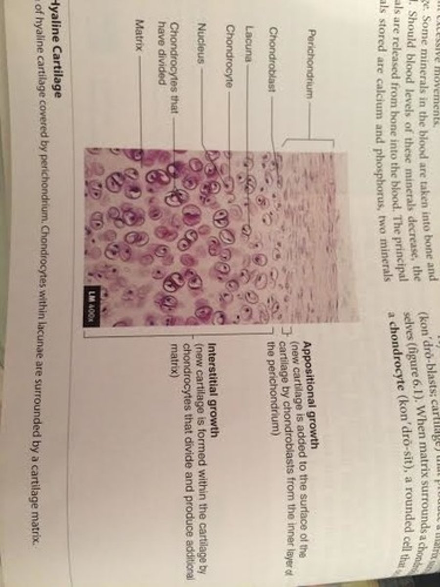
What does the chondroblast turn into when it is surrounded in matrix?
chondrocyte
-a rounded cell that occupies the lacuna
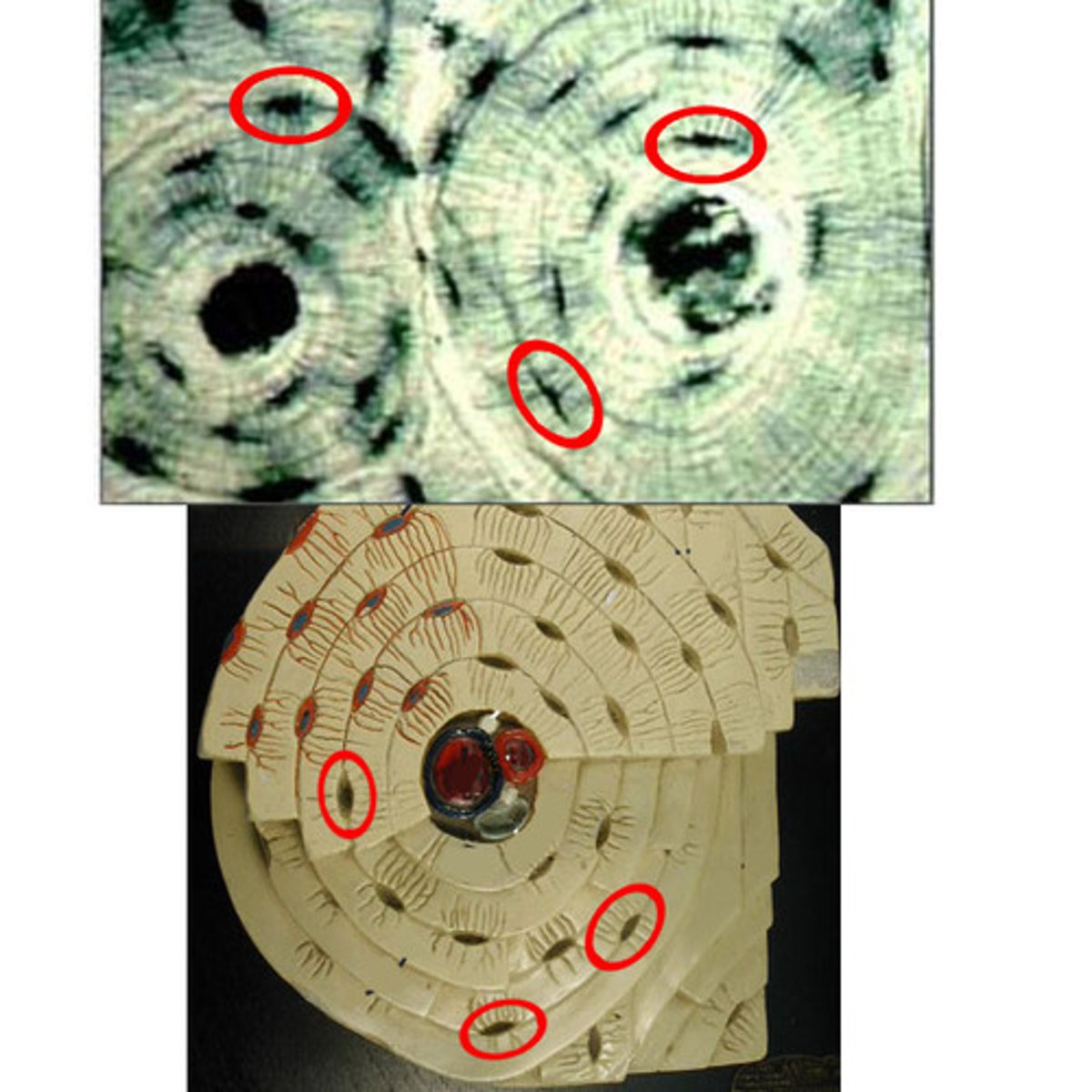
What are lacunae?
the spaces occupied by the osteocyte cell bodies
What is the double-layered sheath that covers most cartilage?
perichondrium
-outer layer is dense irregular connective tissue containing fibroblasts
-inner layer is more delicate and has few fibers and contains chondroblasts
-does not exist in articular cartilage
peri=around
chondrium=cartilage
Blood vessels and nerves penetrate the outer layer of the perichondrium but do not enter the cartilage matrix. How does nutrients reach the chondrocytes?
nutrients must diffuse through the cartilage matrix to reach the chondrocytes
What is the type of hyaline cartilage that covers the ends of bones where they come together to form joints? What does it not contain that other hyaline cartilages do?
articular cartilage
-has no perichondrium, blood vessels, or nerves
What are the two ways in which cartilage grows?
1. Appositional growth
-involves addition of new cartilage to the outside of existing cartilage tissue
-chondroblasts from the inner layer of the perichondrium lay down new matrix and add new chondrocytes to the outside of the tissue
-ossification of osteoblasts for the formation of bone
2. Interstitial growth
-adds new cartilage to spaces within existing cartilage
-chondrocytes within the tissue divide and add more matrix between existing cells
*****bone is incapable of interstitial growth

Muscles are attached to bone via ___________.
tendons
-tendons attach to bone through the periosteum
Bones are attaches to other bones via ____________.
ligaments
-ligaments attach to bones through the periosteum
Tendons and ligaments are both composed of dense regular (collagenous) connective tissue, containing collagen fibers produced by what cell type?
fibroblast
-a cell in connective tissue that produces collagen fibers
Characteristics of bone are primarily due to composition of its what?
matrix
Bone consists of extracellular bone matrix and bone cells. The bone cells produce the bone matrix, become entrapped within it, and break it down so that new matrix can replace the old matrix. The composition of the bone matrix is responsible for the characteristics of bone.
By weight, mature bone matrix is around 35% organic and 65% inorganic material. What are the two organic components?
1. collagen- provides strength
2. proteoglycans- makes resilient
-Vitamin C deficiency inhibits collagen synthesis by osteoblasts so organic components of the bone matrix cannot be properly maintained
What mineral composes the bone matrix's inorganic material? What is it made of?
hydroxyapatite
-crystalized calcium phosphate
-gives bone matrix weight-bearing strength
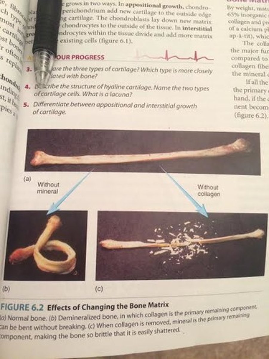
Why are both the organic and the inorganic components of bone matrix necessary?
If all the mineral (hydroxyapatite) is removed from a long bone, collagen is the primary constituent and it becomes too flexible. On the other hand, if collagen is removed from the bone, the mineral component becomes the primary constituent and the bone is very brittle.
without minerals (hydroxyapatite)= can be bent without breaking
without collagen= so brittle that it is easily shattered