Comprehensive CNS and Brain Imaging Techniques in Nuclear Medicine
 Call Kai
 Learn
Learn Practice Test
 Spaced Repetition
 Match
Match Flashcards
 Knowt Play
1/110

There's no tags or description
Looks like no tags are added yet.
Name | Mastery | Learn | Test | Matching | Spaced | Call with Kai | Chat |
|---|
No analytics yet
Send a link to your students to track their progress
111 Terms
What does the Central Nervous System (CNS) consist of?
The brain and spinal cord.

What are the major functions of the CNS?
Generates thoughts and emotions, processes and stores memories, interprets sensory information, and communicates with the body through the peripheral nervous system.
How many neurons are approximately in the adult brain?
About 86 billion neurons.
What is the weight of an adult brain?
Approximately 3 pounds.
What are the four main parts of the brain?
Cerebrum, Cerebellum, Diencephalon, and Brain Stem.

What are the two types of neurons in the CNS?
Motor (efferent) neurons and Sensory (afferent) neurons.
What is the role of motor (efferent) neurons?
They carry impulses away from the CNS to muscles and glands.
What is the role of sensory (afferent) neurons?
They carry impulses toward the CNS from sensory receptors.
What is the largest part of the brain responsible for higher-level functions?
The Cerebrum.

What are the four lobes of the cerebrum?
Frontal, Occipital, Parietal, and Temporal lobes.
What is the function of the Cerebellum?
Controls movement, coordination, and balance.

What does the Diencephalon consist of?
Primarily the thalamus and hypothalamus.

What is the primary function of the thalamus?
Relays sensory impulses to other parts of the brain.
What is the function of the hypothalamus?
Regulates autonomic functions such as body temperature, water balance, and emotions.
What are the components of the brain stem?
Medulla, Pons, and Midbrain.

What is the function of the medulla?
Contains tracts that connect the spinal cord to the brain and is involved in motor function and vital centers.
What is the role of the spinal cord?
Conveys sensory impulses from the periphery and motor impulses from the brain to the periphery.
What is the structure of the vertebral column?
Contains 25 stacked bony vertebrae, separated by discs.

What is the function of Cerebrospinal Fluid (CSF)?
Protects the brain, delivers nutrients, and removes waste.
How many ventricles are in the brain?
There are four ventricles: two lateral ventricles, the 3rd ventricle, and the 4th ventricle.

What is the Blood Brain Barrier (BBB)?
A selective barrier that protects the brain from harmful substances and pathogens.

What is a Cerebrovascular Accident (CVA)?
A stroke that occurs when blood supply to part of the brain is interrupted or reduced.
What are the two major types of strokes?
Ischemic Stroke and Hemorrhagic Stroke.
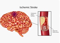
What causes an Ischemic Stroke?
Decreased or blocked cerebral blood flow, often due to thrombosis, embolism, or atherosclerosis.
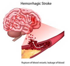
What occurs during a Hemorrhagic Stroke?
Rupture of a cerebral blood vessel leading to bleeding in or around brain tissue.
What is the average total volume of CSF in an adult?
Approximately 125 to 150 mL.
What is the primary source of energy for the brain?
Glucose.
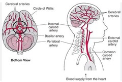
What is the Circle of Willis?
An arterial network at the base of the brain that supplies blood to the brain.
What is a thrombus?
A blood clot that forms in a blood vessel and remains there.
What is an embolus?
A blood clot or other debris that travels through the bloodstream and lodges in a vessel.
What are common signs and symptoms of a stroke?
Facial drooping, weakness or numbness, difficulty speaking, loss of coordination, dizziness, and sudden severe headache.
What are some risk factors for stroke?
Hypertension, diabetes, hyperlipidemia, smoking, obesity, physical inactivity, atrial fibrillation, and family history.
What is the initial management for a stroke?
Rapid neurologic assessment, blood glucose testing, laboratory studies, and non-contrast head CT.
What is the purpose of IV thrombolytic therapy in ischemic stroke?
To restore blood flow by dissolving the clot.

What is a transient ischemic attack (TIA)?
A temporary episode of neurologic dysfunction caused by a brief reduction of blood flow to the brain.

What is the duration of symptoms in a TIA?
Usually less than 24 hours.
What are the characteristic pathologic changes in Alzheimer's Disease?
Beta-amyloid plaques and neurofibrillary tangles.

What is the most common cause of dementia?
Alzheimer's Disease.
What are early signs of Alzheimer's Disease?
Short-term memory loss, confusion, and difficulty communicating.
What is cerebral palsy (CP)?
A permanent disorder affecting movement and muscle tone caused by abnormal brain development or injury.
What is the main characteristic of epilepsy?
A predisposition to recurrent unprovoked seizures.
What causes seizures in epilepsy?
Abnormal, excessive, synchronized electrical activity of neurons in the brain.
What is the treatment focus for cerebral palsy?
Maximizing function and independence through therapy and assistive devices.
What differentiates Alzheimer's Disease from general dementia?
Alzheimer's is a specific disease, while dementia is a syndrome describing significant cognitive decline.
What is the cause of Alzheimer's Disease?
The cause is unknown, but genetics and age are significant risk factors.
What is the treatment outlook for Alzheimer's Disease?
There is no cure; treatments aim to slow progression and manage symptoms.
What is the significance of a TIA being referred to as a 'Warning Stroke'?
One-third of patients with a TIA will experience a CVA within 5 years.
What is the role of mechanical thrombectomy in stroke treatment?
To physically remove a clot from a large cerebral artery.
What are the treatment options for hemorrhagic stroke?
Control bleeding, manage blood pressure, stop anticoagulants, and possibly perform surgery.
What are the symptoms of a TIA?
Symptoms similar to stroke, including facial drooping and difficulty speaking.
What is a seizure?
A single episode of abnormal electrical activity within the brain.
What distinguishes epilepsy from a seizure?
Epilepsy is characterized by an enduring tendency to have unprovoked seizures.
How is epilepsy diagnosed?
After two or more unprovoked seizures.
What are common symptoms of epilepsy?
Staring/unresponsiveness, loss of awareness, muscle stiffening, rhythmic jerking movements, sudden loss of muscle tone, repetitive automatic movements, confusion following the seizure.
What are the treatment options for epilepsy?
Medication, special diets, and surgery.
What is Parkinson's Disease (PD)?
A progressive neurodegenerative disorder that primarily affects movement.
What causes Parkinson's Disease?
Degeneration of dopamine-producing neurons in the substantia nigra of the midbrain.
What are the characteristic symptoms of Parkinson's Disease?
Tremor, rigidity, akinesia/bradykinesia, and postural instability.
What does the acronym TRAP stand for in Parkinson's Disease symptoms?
Tremor, Rigidity, Akinesia/Bradykinesia, Postural instability.
What is hydrocephalus?
An abnormal accumulation of cerebrospinal fluid (CSF) within the ventricular system of the brain.
What are the three types of hydrocephalus?
Noncommunicating (obstructive), communicating, and normal pressure hydrocephalus (NPH).
What is noncommunicating hydrocephalus?
Physical obstruction prevents CSF from moving normally through the ventricular system.
What is communicating hydrocephalus?
CSF can move through the ventricular system, but there is impaired circulation or absorption into the venous system.
What is normal pressure hydrocephalus (NPH)?
Characterized by enlarged ventricles and a classic clinical triad: urinary incontinence, gait disturbance, and cognitive impairment.
What is the treatment for hydrocephalus?
Placement of a ventriculoperitoneal (VP) shunt to drain excess CSF.
What are the two groups of brain imaging?
Planar brain imaging and SPECT brain imaging.
What is the purpose of nuclear medicine brain imaging?
To assess function and perfusion of cerebral abnormalities and CSF imaging.
What radiopharmaceutical is used in planar brain imaging?
99mTc Pertechnetate, which accumulates in the choroid plexus of the brain.
What is the significance of 99mTc-DTPA in brain imaging?
It rapidly clears the blood and localizes in the brain, providing a higher target-to-non-target ratio.
What are the indications for planar brain imaging?
Evaluate brain perfusion, brain death, strokes, localization of radiopharmaceuticals, metastatic brain tumors, cerebral infections, and inflammation.
What does a positive brain death scan indicate?
Tracer seen in carotids but no cerebral arteries visible.
What does a primary brain tumor present as in imaging?
A single focus of activity that may or may not be seen on flow.
What does cerebrovascular disease show in imaging?
Asymmetrical flow with a wedge-shaped defect seen on delayed images.
What is the 'doughnut sign' in brain imaging?
A focal area of increased uptake with an area of decreased activity in the center, indicating inflammation or infection.
What is the patient preparation for planar brain imaging?
No patient prep; focus on history and keep the patient calm and still for at least 20 minutes pre-injection.
What is the Oldendorf technique in imaging?
A method used for rapid bolus IV injection of the radiopharmaceutical.
What is the imaging procedure for dynamic or rapid sequential imaging?
Acquire images every 2-3 seconds for 60 seconds after IV injection of the radiopharmaceutical.
What does a normal planar brain flow study indicate?
Activity seen in the heart, carotid arteries, and cerebral arteries indicates brain perfusion.
What does SPECT stand for in brain imaging?
Single Photon Emission Computed Tomography
What is the primary purpose of SPECT brain imaging?
To evaluate cerebral blood flow and diagnose neurological conditions.
What are the two main radiopharmaceuticals used in SPECT brain imaging?
99mTc HMPAO and 99mTc ECD.
What is a key characteristic of 99mTc HMPAO?
It is lipophilic and crosses the blood-brain barrier.
What is the typical dose range for 99mTc HMPAO in SPECT imaging?
10-30 mCi IV bolus injection.
What imaging time frame is recommended after injecting 99mTc HMPAO?
SPECT images are obtained 15-20 minutes post injection.
What is a disadvantage of 99mTc HMPAO?
It has limited in-vitro stability and must be injected within 30 minutes after preparation.
What is a key advantage of 99mTc ECD over HMPAO?
It has greater post-preparation stability and a longer prep to injection window.
What is the typical imaging time frame after injecting 99mTc ECD?
SPECT images are obtained 15-20 minutes post injection.
What are the indications for SPECT brain imaging?
Evaluate cerebrovascular disease, dementia, trauma, and localize seizure foci.
What patient preparation is required before SPECT imaging?
No specific patient prep, but the patient should be calm and still for at least 20 minutes pre-injection.
What is the significance of the sagittal plane in SPECT imaging?
It provides a right to left cut of the brain.
What does a normal SPECT brain image show?
Bilaterally symmetrical activity with higher activity in the gray matter.
How does SPECT imaging assist in diagnosing brain death?
It shows no perfusion to the brain, indicating brain death.
What is the typical appearance of SPECT images in Alzheimer's disease?
Decreased perfusion to bilateral posterior temporal and parietal lobes.
What is the imaging agent of choice for epilepsy localization?
PET imaging is preferred for localization of seizure foci.
What does a TIA show on a SPECT scan?
A temporary hypoperfusion defect that is time-dependent.
What is the role of 111In-DTPA in cerebral spinal fluid imaging?
It is used to diagnose normal pressure hydrocephalus and identify CSF leaks.
What is the normal flow pattern of 111In-DTPA after injection?
It ascends the spinal column and should reach the basal cisterns within 2-4 hours.
What indicates normal pressure hydrocephalus on a cisternogram?
Persistent activity in the lateral ventricles at 24-48 hours.
What is the significance of shunt patency imaging?
It evaluates whether a shunt is open and functioning properly.
What is the typical dose for 111In-DTPA in CSF imaging?
0.5-1.0 mCi injected intrathecally.