Protozoa
1/75
There's no tags or description
Looks like no tags are added yet.
Name | Mastery | Learn | Test | Matching | Spaced | Call with Kai |
|---|
No analytics yet
Send a link to your students to track their progress
76 Terms
What are pseudopodia?
Extensions of the cytoplasm used for movement, sensing and engulfing food.
How do protozoa take in nutrients?
By pinocytosis or phagocytosis.
What is a direct/monoxenous/homoxenous lifecycle?
Only one host is used for the entire lifecycle.
What is an indirect/heteroxenous lifecycle?
Multiple hosts are used for different stages of the lifecycle.
What are the 3 methods of reproduction used by protozoa?
Binary fission
Shizogony (multiple fission)
Sexual reproduction (gametogony)
What is the host range of Eimeria spp.?
Very broad, with the exception of humans. Nearly always host-species specific.
What disease does Eimeria spp. cause?
Coccidiosis
What does it mean that Eimeria is self-limiting?
It has a predetermined number of replication cycles which it can carry out within the host.
What is the lifecycle of Eimeria?
A non-infectious oocyst is shed in the faeces
Sporulation occurs. The sporulated oocyst contains 4 sporocysts, each containing 2 sporozoites.
Sporulated oocysts are ingested and sporozoites are released and enter enterocytes.
Sporozoites become trophozoites
Trophozoites become schizonts, which are packed full of merozoites, which are released.
Merozoites become male microgametocytes or female macrogametocytes.
Microgametocytes fertilise macrogametocytes, forming oocysts.
What are the two groups of Eimeria spp.
Malabsorptive and haemorrhagic.
What species are in the malabsorptive group and what pathology do they cause?
E.acervulina
E.maxima
E.mitis
E.praecox
They cause villous atrophy, mucoid enteritis, malabsorption, decreased weight gain, increased food conversion ratio
What species are in the haemorrhagic group and what pathology do they cause?
E.tenella
E.necatrix
E.brunetti
They cause cells to rupture, releasing merozoites which create deep erosions and haemorrhage - leading to melena and death
Which Eiemeria species infect the upper intestine?
E.acervulina, E.praecox
Which Eimeria species infect the middle intestine?
E.maxima, E.necatrix, E.mitis
Which Eimeria species infect the lower intestine?
E.brunetti
Which Eimeria species infect the cloaca?
E.tenella
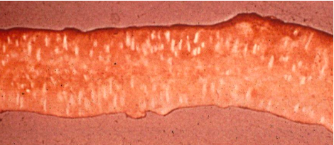
Which Eimeria species creates white ladder regions in the duodenal loop?
Eimeria acervulina
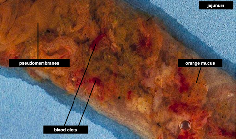
Which Eimeria species creates orange mucus and clotting in the middle intestine?
Eimeria maxima
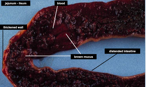
Which Eimeria species creates brown mucus and blood in the middle intestine?
Eimeria necatrix
Which Eimeria species causes the caeca to fill with blood and form a caseous core?
Eimeria tenella
What is the most significant cause of coccidiosis in the UK?
Eimeria tenella
What is the prepatent period and sporulation time of Eimeria tenella?
6 days prepatent period and 2 days sporulation time.
How do ionophores treat Eimeria species?
They create a pore in the sporozoite which allows Na+ influx. The sporozoite fills with water and runs out of energy as it pumps out Na+.
Why are ionophores effective?
The sporozoites still stimulate the immune response as they are dying, allowing chickens to develop immunity so they don’t become ill once the drug is withdrawn.
How do live Eimeria vaccines work?
They contain oocysts and are given as an edible gel. Chickens must ingest the appropriate amount in order to develop immunity and not become sick.
How do attenuated Eimeria vaccines work?
The chickens shed oocysts in their faces which are continually re-ingested to stimulate the immune system.
What Eimeria species cause stable coccidiosis in cattle?
Eimeria bovis and Eimeria zuernii
What is crowding disease?
When non-immune animals are exposed to E.bovis or E.zuernii.
What Eimeria species causes pasture coccidiosis in cattle?
Eimeria alabamensis
What is the most pathogenic Eimeria species that infects birds?
Eimeria zuernii
What is the lifecycle of Cryptosporidium spp.?
Unsporulated oocysts (thick walled) are shed into the environment
They sporulate and contain 4 sporozoites
Sporozoites hatch and move to the brush border, where they form trophozoites
Trophozoites form schizonts which are full of merozoites
Merozoites become microgametocytes and macrogametocytes
Microgametocytes fertilise macrogametocytes which form an unsporulated oocyst.
What does Cryptosporidium cause?
Scours in calves less than a week old
What are thin-walled Cryptosporidium oocysts?
Unsporulated oocysts which are not shed into the environment but remain in the gut, causing autoinfection.
How can cryptosporidium be detected?
Using Ziehl-Neilsen stain - it is acid fast, or immunoassay
Which animals are vaccinated against Cryptosporidium?
Gravid cows
What drug can be given to reduce oocyst excretion?
Halofuginone
What shape is Giardia spp?
Pear shaped, with 8 flagella
How does Giardia reproduce?
Trophozoites attach to the intestinal wall and reproduce by binary fission. They intermittently form cysts which are immediately infective. When consumed by antoher host, they release trophozoites.
What species is commonly affected with Giardia spp. and where?
Dogs in developed countries.
Which Giardia species causes beaver fever?
Giardia duodenalis
How is Giardia spp. diagnosed?
Requires faecal samples taken over 3+ days, as cyst excretion is intermittent. Requires specific high density flotation fluids like ZnSO4.
What drug is used to treat Giardia spp. infection?
Metronidazole, which is a DNA damaging antimicrobial or fenbendazole
What disease does Histomonas cause in turkeys?
Blackhead disease
What pathology does Blackhead disease cause?
Lesions in the liver and caeca which become necrotic, yellow droppings, cyanotic colouring to the head and wattles.
What is the lifecycle of Histomonas spp.?
Hetarakis (nematode) eggs which contain Histomonas are ingested
When Heterakis hatches, Histomonas is released which travels via the hepatic portal system to the liver, where it forms lesions and replicates.
It then returns to the gut and infects other adult Heterakis, and is shed in their eggs.
Which species carries Histomonas asymptomatically do should not be kept with turkeys?
Chickens
What pathology does Tritichomonas foetus cause in cats?
Chronic, large intestine diarrhoea.
What pathology does Tritichomonas foetus cause in cattle?
Infertility and abortion
What species does Toxoplasma gondii infect?
Almost all homeotherms
What is the heteroxenous (indirect) lifecycle of T.gondii?
Unsporulated oocyts are shed in the faeces and sporulate in the environment.
Sporulated oocysts contain 2 sporocysts which each contain 4 sporozoites.
Oocysts are ingested by intermediate hosts.
Sporozoites become tachyzoites
In chronic cases, tachyzoites become bradyzoites, which form cysts in the muscle and nervous tissue.
The intermediate host is eaten by a cat.
Tachyzoites and bradyzoites undergo schizogony to form shizonts
Shizonts release merozoites which become microgametocytes or macrogametocytes. They undergo gametogony to produce unsporulated oocyts.
What is the monoxenous (direct) lifecycle of Toxoplasma gondii?
Cats ingest faecal oocysts from other cats. The prepatent period is 3 weeks, after which T.gondii can be transmitted in utero, to humans if they ingest the meat of intermediate hosts, or to oocysts.
How does Toxoplasma gondii affect immunocompromised cats differently?
They are treated as intermediate hosts, leading to neurological disease.
What does Toxoplasma gondii cause if it infects sheep in the first 1/3 (40 days) of gestation?
Foetal death and resorption
What does Toxoplasma gondii cause if it infectes sheep in the second third (40 -110 days) of gestation?
Foetal death, abortion or development of a mummified foetus
What does Toxoplasma gondii cause if it infects sheep in the last third (110-147 days) of gestation?
Stillbirth or weak lambs
What visible changes does T.gondii cause to the placenta?
Strawberry pip lesions - multifocal white, raised, gritty lesions on the cotyledons where bradyzoites have disrupted blood supply - leading to necrosis.
Why is oocyst detection an ineffective method of T.gondii diagnosis in cats?
Only a small proportion of infected cats excrete oocysts
What is the lifecycle of Neospora caninum?
Dogs shed unsporulated oocyts in the faeces.
Oocysts sporulate. They contain 2 sporocysts which contain 4 sporozoites.
Ruminants are intermediate hosts, within which oocysts hatch.
Sporozoites form tachyzoites.
Tachyzoites form bradyzoites, which are thick-walled cysts.
Once ingested by the dog, tachyzoites and bradyzoites undergo shizogony to form shizonts.
Shizonts release merozoites, which become microgametocytes or macrogametocytes, and fertilise each other to produce oocysts.
What is the epidemiology of canine neosporosis?
Dog acts as intermediate host, infects puppies. In a litter, some puppies will have paralysis, muscle wasting, myocarditis.
What is the epidemiology of bovine neosporosis?
Transplacental infection occurs - leading to an abortion storm or the birth of persistently infect calves.
What is the most common cause of bovine abortion?
Bovine neosporosis
What is exogenous vertical transmission of bovine neosporosis?
Cow becomes infected during pregnancy and tachyzoites cross the placenta
What is endogenous transmission of bovine neosporosis?
PI cow becomes pregnant and tachyzoites cross the placenta.
What disease is Neospora hughesi associated with?
Equine protozoal myeloencephalitis.
What disease does Babesia spp. cause?
Redwater fever
How does Babesia spp. cause pathology?
It replicates inside red blood cells, producing a waste antigen which is released when they rupture. It binds to other RBCs, causing the immune system to destroy them - leading to fever, anaemia, haemoglobinuria, pipe stem faeces (constant urge to defacate)
What is the lifecycle of Babesia?
The definite host (Ixodes tick) feeds on an infected animal
In the tick, Babesia undergoes sexual replication in the intestine and asexual reproduction in other tissues.
Vermicules form, which move to the salivary glands, ovary, eggs
Babesia is passed to offspring (transovarian transmission) and it persists as the larvae develop (transstadial transmission)
Babesia is passed on to another animal when the tick feed
It undergoes asexual reproduction in the red blood cells.
What Babesia species is found in the UK and where?
Babesia divergens, in hilly areas in spring and autumn.
What is the lifecycle of Leishmania spp.
In the vertebrate host, it exists as amastigotes, which replicate within macrophages.
Infected macrophages are ingested by the invertebrate host - the sandfly - while feeding.
In the sandfly gut, the amastigote develops into a promastigote, which migrates to the proboscis, allowing it to pass into another vertebrate host during feeding.
Why is it hard to trace transmission of Leishmania spp.
Incubation period is months to years, and many dogs act as reservoirs without clinical disease.
How does Leishmania spp. cause pathology in dogs?
Skin macrophages are initially infected causing cutaneous leishmaniosis - ulceration of the lip, eyelids, pinna. It then spreads to organs, causing visceral leishmaniosis - chronic wasting, intermittent fever.
What does Sacrocystitis spp. have in common with Toxoplasma and Neospora?
Cyst-forming
What is the lifecycle of Sarcocystitis spp?
Sporulated socysts shed in faeces. Their thin wall easily ruptures, releasing free sporocysts (containing sporozoites).
In the intestine of the intermediate host, sporozoites from shizonts which enter capillaries.
Shizonts from merozoites which move to the cardiac and skeletal muscle to form sarcocysts, which contain bradyzoites.
Sarcocysts are ingested in poorly cooked meat.
Bradyzoites infect the intestinal epithelium and produce shizonts
Shizonts release merozoites, which become microgametocytes or macrogametocytes, and fertilise each other to produce oocysts.
What disease does Sarcocystitis neurona cause?
Equine protozoal myeloencephalitis
What kind of host are horses for Sarcocystitis neurona?
Dead-end hosts - they cannot transmit the protozoan.
What can serological tests for Sarcocystitis spp. cross react for?
Toxoplasma spp.