AnP I Lab Practical
 Call Kai
 Learn
Learn Practice Test
 Spaced Repetition
 Match
Match Flashcards
 Knowt Play
1/107

There's no tags or description
Looks like no tags are added yet.
Name | Mastery | Learn | Test | Matching | Spaced | Call with Kai | Chat |
|---|
No analytics yet
Send a link to your students to track their progress
108 Terms
Anatomical position
Universally accepted position used when describing specific areas of the human body
Directional terms
Describes the relationship of one body part with another
Anterior (ventral)
Refers to front, in humans. Example is kneecaps are on the anterior of the legs
Posterior (dorsal)
Refers to the back. Example is shoulder blades are posterior to chest
Superior (cranial)
Toward the head or upper body; example eyes are superior to mouth
Inferior
Away from the head or towards the tail; example is chin is inferior to the nose
Proximal
Something being close to the point of origin. Example is elbow is proximal to wrist
Distal
Refers to being more distant or further away from the same point of origin. Example is fingers are distal to the wrist
Body’s midline
Imaginary line that runs down the middle of the body
Medial
Refers to a position that is closer to the midline. Example is the nose is medial to the eyes
Lateral
Refers to a position that is further away from the midline. Example is the ears are lateral to the eyes
Superficial
Refers to structures that are closer to the surface of the body. Example is the skin is superficial to the muscle
Deep
Refers to structures that are further away from the surface. Example is bone is deep to the muscle
Regional terms
Body can be divided into two; axial
And appendicular
Axial region
This includes the head, neck, and trunk
Appendicular region
Includes upper and lower limbs or appendages
Planes of section
Provide a means of studying form and function of a body region by dividing body or a body part up for examination
Sagittarius plane
Divides body or body part into right and left sections
Midsagittal plane (median plane)
Divides body or body part into equal left and right sections
Parasagittal plane
Divides body or body part into unequal right and left sections
Skull regional names

Frontal plane (coronal plane)
Divides body or body part into anterior and posterior sections
Transverse plane (horizontal plane)
Divides body or body part into superior and inferior sections or proximal and distal sections when describing structures of appendicular region
Compound light microscope
Uses light and two lenses to produce an image with excellent resolution
Resolution
The capability to distinguish between two separate points
Microscope parts

Microscope total resolution

Bone tissue

Bone tissue cell

Tendons are deep to
dermis/skin
Tissue
a group of structurally and functionally related cells and their
extracellular matrix (ECM) that together perform common functions
Histology
study of normal structures of tissues
Extracellular materials
Composed of substances in a liquid, thick gel, or solid that surround cells of a tissue; consists of ground substance and protein fibers. These include extracellular fluids, cellular secretions, and extracellular matrix
Extracellular fluids
Body fluids including interstitial fluid, blood plasma, and cerebrospinal fluid
Interstitial fluid
cells are submersed (bathed) in this fluid
blood plasma
fluid of the blood
Cerebrospinal fluid
fluid surrounding nervous system organs
Cellular secretions
These are salivia, mucus, gastric fluids
Extracellular matrix
substance that acts as glue to hold cells together
Protein fibers
embedded within ground substance and include collagen fibers, elastic fibers, and reticular fibers.
Epithelial Tissue
these consist of tightly packed cells that are linked together by tight junctions and desmosomes. they are fairly impermeable and resistant to physical stresses and mechanical injury. Classified by layers and shape
Basal lamina
ECM synthesized by epithelial cells; consists of collagen fibers and ground substance
reticular lamina
synthesized by underlying connective tissue; consists of reticular fibers and ground substance
Simple epithelial
consist of a single layer and are adapted for transportation of substances between different tissues
Stratified epithelial
consist of more than one layer; best suited as protective barriers in
locations subjected to high degrees of mechanical stress
Squamous
these are flattened
Cuboidal
short
Columnar
tall and elongated
Simple Squamous epithelium
very thin single layer of cells; adapted for rapid diffusion of substances; found in air sacs of lung, specific segments of kidney tubules, and lining blood vessels

Simple Cuboidal Epithelium
Single layer of cube-shaped cells with a large central nucleus;
found in segments of renal tubules, respiratory passages, ducts of many glands, and thyroid gland. They are for secretion and absorption

Simple columnar epithelium
single layer of rectangular-shaped cells with nuclei located in the basal portion of the cell, found in the small intestine. They are for absorption and secretion, with added protection and more surface area.

Pseudostratified Columnar Epithelium
Appears to be layered because nuclei are found at various heights; found in the trachea and other segments of the respiratory tract and nasal cavity; ciliated. They secrete mucus and move it through the respiratory tract using cilia. The mucus is secreted through goblet cells and they trap dust, pathogens, and debris.

Nonkeratinized stratified squamous epithelium tissue
apical cellular layers retain nuclei; still alive; found in regions subjected to mechanical stress where surface must remain moist but still protected; i.e. esophagus.

Keratinized stratified squamous epithelium
Apical cellular layers are dead; lack nuclei; are filled with protein keratin; make tissue tough and resistant to friction; are well adapted for outer layers of skin. They also protect from pathogen entry and helps prevent dehydration

Transitional epithelium
only found in urinary system; lines interior of kidney, ureters, urinary bladder, and urethra; ability of apical cells to flatten contributes to ability of urinary tissues to stretch. The stretch readily which permits urine to distend urinary organ. They are stratified and surface cells are either domeshaped or squamous like depending on stretch. The basal cells are cuboidal or columnar.

The main components of connective tissue are
ground substance, fibers, and cells
Collagen fiber
the strongest and most abundant type and is tough while providing high tensile strength
Elastic Fiber
they form a network while being long and thin and allow for stretch and recoil
Reticular fibers
they are short, fine, and are highly branched collagenous to maintain structure and integrity
Resident cells
permanently inhabit the tissue in which they are found
Migrant cells
migrate into different areas of body depending on situation
Fibroblast
these are the most common resident cell and make protein fibers (especially collagen) and ground substances
Adipocytes
these are fat cells found in many different connective tissues. They store the energy as fat
Mast cells
these are immune sentinel cells that detect threats and release chemical mediators to coordinate immune responses, allergic reactions, and tissue repair
Phagocytes
immune cells that engulf foreign entities and objects to destroy them.
Connective Tissue Proper
Divided into loose and dense
Loose Connective Tissue
characterized by loosely arranged collagen, elastic, and reticular fibers within a gel-like ground substance that function in support, cushioning, diffusion, and immune defense. It includes areolar, adipose, and reticular tissue
Dense Connective Tissue
characterized by tightly packed collagen fibers that provide strength and resistance to mechanical stress. It includes regular, irregular, and elastic
Areolar Connective Tissue
contains a matrix with all three fiber types as well as all cells. They wrap and cushion organs while its macrophages phagocytizes bacteria. They play an important role in inflammation and hold tissue fluids. They are widely distributed under epithelia of the body while forming a lamina propria of mucous membranes packaging organs and surrounding capillaries

Adipose Connective Tissue
These consist of fat-storing adipocytes and surrounding fibroblasts and ECM. They function as a fat storage reserving food fuel, insulating against heat loss, and supporting and protects organs. They are found under the skin in subcutaneous tissue, around kidneys and eyeballs, within the abdomen, and in breasts.

Reticular Connective Tissue
composed mostly of reticular fibers that form fine networks supporting small structures like blood and lymphatic vessels. The fibers form a soft internal skeleton that support other cell types including wbc, mast cells, and macrophages. They are also found in lymph nodes and spleen while forming weblike nets that trap old and foreign cells. They form part of basement membrane that support all epithelia and internal structure of liver and bone marrow.

Dense Regular Connective Tissue
primarily parallel collagen fibers with a few elastic fibers and fibroblast as the major cell type. They attach muscles to bones or to muscles, and bones to bones. They withstand great stress when pulling force is applied in one direction (resist tension in one plane). They are found in tendons, most ligaments, and aponeuroses.

Dense Irregular Connective Tissue
irregular arranged collagen fibers. They withstand tension in all three planes of movement while providing structural strength. They are found in fibrous capsules of organs and of joints, dermis of the skin and surrounding organs and joints, and submucosa of digestive tract.

Dense Regular Elastic Connective Tissue
They are parallel-oriented elastic fibers with randomly oriented collagen fibers. They allow tissue to recoil after stretching, maintain pulsatile flow of blood through arteries, and aid in passive recoil of the lungs following inspiration. They are found in the walls of large arteries, within ligaments associated with the vertebral column, and within the walls of the bronchial tubes.

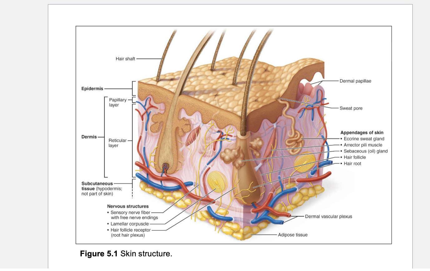
Epidermis
the outermost layer of the skin, made of stratified squamous epithelial cells that form a protective barrier against environmental damge.
Dermis
consists of strong, flexible connective tissue, fibroblasts, macrophages, and occasionally mast cells and white blood cells. The fibers bind body together. It contains nerves, blood vessels, lymphatic vessels, epidermal hair follicles, oil glands, and sweat glands. Consist of papillary layer (areolar connective tissue) and reticular layer (dense irregular connective tissue).
Papillary Dermis
the thin, superficial layer of areolar connective tissue consisting of loose, interlacing collagen and elastic fibers and blood vessels. The loose fibers allow phagocytes to patrol for microorganisms.
Dermal papillae
superficial region of dermis that sends fingerlike projections up into epidermis. it contains capillary loops, free nerve endings (pain receptors), and touch receptors (tactile corpuscles, also called meissner's corpuscles)
Reticular dermis
consists of dense irregular connective tissue, elastic fibers that provide stretch-recoil properties, and collagen fibers that provide strength and resiliency that bind water, keeping skin hydrated. It contains eccrine glands, hair, sebaceous glands, and lamellated (Pacinian) corpuscles that sense deep pressure
Sebaceous glands
associated with hair follicles, these produce sebum that empties into follicle and coats shaft
Skin Structure
Specialized Connective Tissues include
Cartilage, Bone, and blood
Cartilage
ough, flexible tissue; absorbs shock and resists tension, compression, and shearing forces; oxygen and nutrients must diffuse from blood vessels in perichondrium through ECM which limits the thickness of living ones.
Chondroblasts
immature cells that produce the cartilage ECM and undergoes mitosis.
Chondrocytes
specialized cells that maintain the cartilage ECM; reside within lacunae (small cavities).
Perichondrium
a dense irregular connective tissue that serves as the source for new chondrocytes
Hyaline Cartilage
the most abundant cartilage; it is found on ends of bones in joints, linking sternum to ribs, framing sections of respiratory tract (slide in lab was trachea), and in nose; most of the fetal skeleton contains it and it is replaced with bone during development; contains pairs of chondrocytes in lacuna and has chondroblasts making fibers; the main function is to support and reinforce while serving as a resilient cushion to resist compressive stress

Fibrocartilage
filled with bundles of collagen fibers with little room for ground substance in ECM; the tissue has great tensile strength with some degree of elasticity to absorb compressive shock, it is found between bones of fibrous joints; forms articular discs that improve fit of bones in joints

Elastic Cartilage
mostly elastic fibers in its ECM; this allows the tissue to vibrate and maintain its shape with great flexibility; it is found in external ear and larynx

Compact Bone
tissue is subjected to great stress; contains tightly packed osteons (Haversian Systems) and lamellaes; it is the dense, hard outer layer of bones that provides strength, protection, and structural support, making up about 80% of the human skeleton

Osteons
the primary structural and functional unit of compact bone, consisting of concentric layers of bone tissue surrounding a central Haversian canal.
Lamellaes
thin layers of mineralized bone matrix—3–7 µm thick—made of collagen and hydroxyapatite, arranged in concentric, circumferential, or interstitial patterns to give compact and spongy bone its strength.
Concentric Lamellaes
circular layers of bone matrix that form the structural rings of an osteon, providing strength and resistance to twisting forces in compact bone.
Circumferential Lamellaes
layers of bone matrix that encircle the entire bone, providing structural strength and helping bones resist bending and torsional forces.
Interstitial Lamellaes
fragments of older bone tissue located between osteons, providing structural support and filling gaps in compact bone.
Osteoblasts
“Bone-builders” found on outer surface of bones; carry out process of bone deposition; synthesize and secrete organic ECM
Osteocytes
osteoblasts that have surrounded themselves with ECM in lacunae; mature cells
Osteoclasts
large, multinucleated bone destroyers; carry out process of bone resorption
Perforating canals
microscopic channels in compact bone that connect the periosteum to the inner bone structures, allowing blood vessels, nerves, and nutrients to reach osteons. It also connects the osteons together through the central canals
Anatomy of Compact Bone
