General Pathology
 Call Kai
 Learn
Learn Practice Test
 Spaced Repetition
 Match
Match Flashcards
 Knowt Play
1/108

There's no tags or description
Looks like no tags are added yet.
Name | Mastery | Learn | Test | Matching | Spaced | Call with Kai | Chat |
|---|
No analytics yet
Send a link to your students to track their progress
109 Terms
Gross lesions
Structural abnormalities that are visible to the naked eye (without a microscope).
Pathognomonic lesions:
Structural abnormalities that can only be seen under a microscope.
Pseudo-lesions
Artefacts produced after death due to the normal process of decomposition rather than real disease processes.
Aetiology
Refers to the cause of a disease
Pathogenesis
Refers to the stepwise mechanism by which a disease develops.
In other words the sequence of events, that leads from the cause (aetiology) of a disease to the development of lesions or clinical signs.
List and describe the tools available for the recognition of diseases.
Tissue samples
- surgical/biopsies or postmortem tissue samples used for histological examination
Cytology
- Use a microscope to examine samples of collections of cells that are smeared or blotted onto a microscope slide before examination.
Clinical and laboratory techniques
- Can be used to to detect structural or functional changes in tissues.
- For example, clinical biochemistry which analyzes bodily fluids such as blood and urine.
Algor mortis
The body cools after death until it reaches the same temperature as the environment.
How does a body in a colder environment affect Algor Mortis?
Colder environments increase the temperature gradient, which drives a faster cooling curve
How does a body in a windy environment affect Algor Mortis?
Greater wind blows away warm air and replaces it with cold air, increasing the rate of heat loss.
How does a insulation affect Algor Mortis?
The greater the insulation the slower the heat loss.
E.g. A well-insulated wooly carcass will retain heat much longer than a lean, short-haired animal
How does the initial temperature of the body affect Algor Mortis?
The rate of cooling is directly proportional to the temperature gap between the body and its surroundings.
The larger the difference, the faster the heat loss.
What is the cooling of the body after death called?
Algor Mortis
Rigor Mortis
Post-mortem muscle stiffening due to gradual ATP depletion.
Why does rigor mortis cause stiffening of the body?
ATP depletion results in a failure of muscle relaxation due, as ATP is needed to break actin-myosin cross-bridges in muscle fibres.
Thus the muscle becomes fixed in contraction→ Rigor Mortis
What is the stiffening of the body after death called
Rigor Mortis
Does Rigor Mortis occur immediately post mortem? And why?
Rigor Mortis does not occur immediately→ ATP persists for 1-9 hours via limited residual ATP stores and anaerobic glycogen metabolism (glycogen stores can be used temporarily to produce ATP)
What effects the onset of rigor mortis?
Glycogen stores
- Develops later in well-nourished individuals with larger glycogen stores (ATP takes longer to deplete)
Environmental temperature
- ATP is used up more quickly at higher temperatures.
What is the predictable pattern rigor mortis is said to follow?
Beginning in the head and neck, progressing to the heart and diaphragm, and finally affecting the limbs.
Head/neck → trunk → limbs
What causes the end of rigor mortis?
Muscle fibres undergo decomposition (due to autolysis + putrefaction) and cross-bridges physically degrade.
Hypostatic congestion/Lividity/ Liver Mortis
Blood settles in dependent areas (lower portion of the organ/body) due to gravity causing red/blue discolouration.
This becomes fixed after clotting
And will remain in place even when the body is moved.
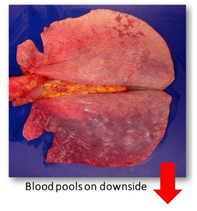
What is this post mortem change called?
Livor Mortis, or Hypostatic Congestion
What are the differences between post-mortem and ante-mortem clots?
Post- Mortum
Smooth, glistening
Fills vessel
Takes vessel shape
Yellow serum layer + Red cells below
Ante-Mortem (thrombus)
Rough, firm
Attached to vessel wall
No clear layering
Forms in living animal
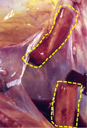
Is this a post-mortem or ante-mortem clot?
Post-mortem
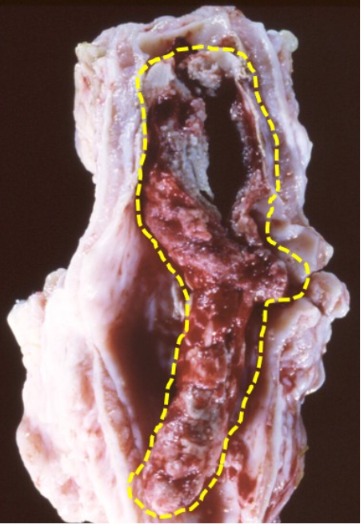
Is this a post-mortem or ante-mortem clot?
Ante-Mortem
What is autolysis?
Breakdown of cells and tissues by the body’s own cellular enzymes.
What is putrefaction?
Breakdown of cells and tissues by bacteria
That invade from the environment or from within the body (e.g., gut flora).
What two factor contribute to the break down of the bodies tissues?
Autolysis and putrefaction
What changes occur to tissue due to autolysis and putrefaction?
Putrefaction results in the breakdown of cells and connective tissue.
Resulting in pale to greenish, soft, and fragile (friable) tissues
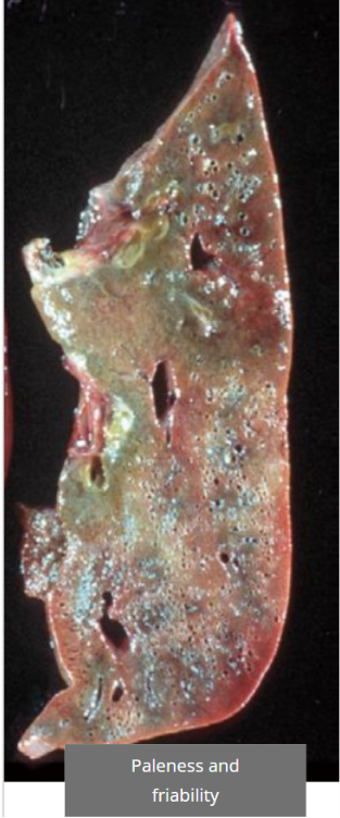
What is this post mortem change called?
Putrefaction
results in the breakdown of cells and connective tissue.
Resulting in pale to greenish, soft, and fragile (friable) tissues
What is does Pseudonecrosis look like?
Patchy discoloration + softening
Mimics the the appearance of groups of dead (necrotic) cells.
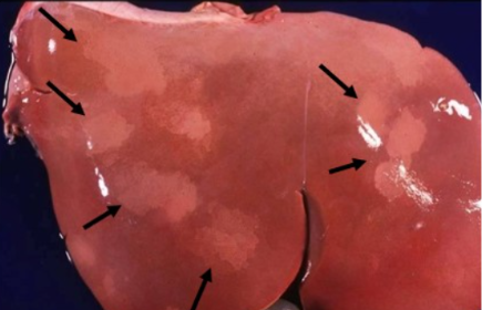
What is this post mortem change called?
Pseudonecrosis
Patchy discoloration + softening
Mimics the the appearance of groups of dead (necrotic) cells.
What is does Pseudomelanosis look like?
Green-black staining
Due to iron (from red blood cells) + hydrogen sulphide (produced by the anaerobic bacteria → reacting to from green-black iron sulphide
What causes Pseudomelanosis green black staining?
Iron (from red blood cells) + hydrogen sulphide (produced by the anaerobic bacteria → reacting to from green-black iron sulphide
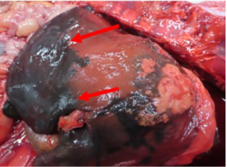
What is this post mortem change called?
Pseudomelanosis
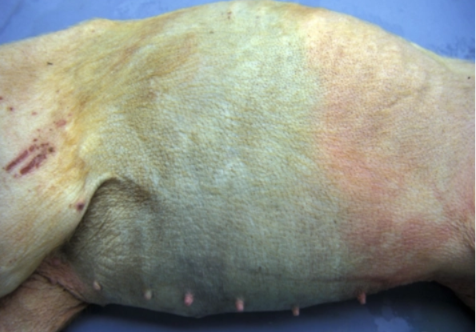
What is this post mortem change called?
Pseudomelanosis
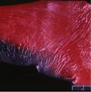
What is this post mortem change called?
Pseudomelanosis
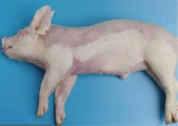
What is this post mortem change called?
Hypostatic Congestion
What is haemoglobin imbibition?
Bright red to pink staining from lysed Red Blood Cells diffusing hemoglobin.
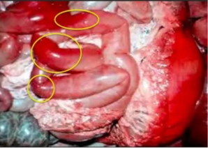
What is this post mortem change called?
Haemoglobin Imbibition
What is bile imbibition?
Orange-green staining near the gall bladder or bile ducts.
Due to post-mortem leakage of bile.
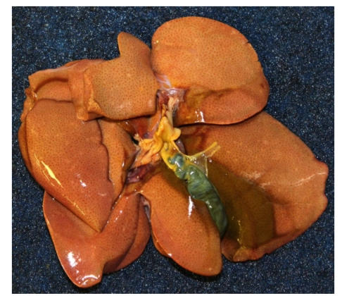
What is this post mortem change called?
bile imbibition
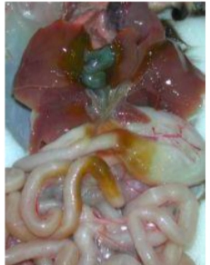
What is this post mortem change called?
bile imbibition
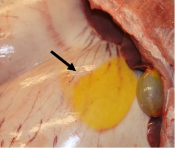
What is this post mortem change called?
bile imbibition
What causes gas production (post-mortem bloat)?
Gas is produced by putrefactive anaerobic bacteria
Where can gas produced by putrefactive bacteria accumulate post mortem?
Body cavities
Mimicking bloat (abdominal distension)
Tissues (emphysema)
What causes post-mortem organ displacements and/or rupture?
gas production and thus distention of the gastrointestinal tract
It can also caused by moving the carcass.
What causes post-mortem mucosal sloughing in the rumen?
Autolysis and Putrefaction.
The ruminal mucosa (the innermost lining of the rumen) begins to deteriorate rapidly after death.
What causes the lens of the eye to become cloudy/opaque post-mortem?
Chilling or partial freezing of the carcass.
Extra Info for fun:
Cooling or partial freezing of the carcass alters the arrangement of lens proteins and water.
Light is scattered instead of passing cleanly through the lens, making it appear cloudy.
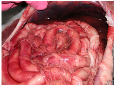
What is this post mortem change called?
Haemoglobin Imbibition
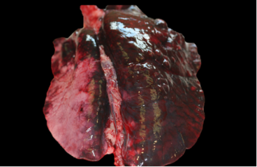
What is this post mortem change called?
Hypostatic Congestion
How can you tell the difference between haemoglobin imbibition and hypostatic congestion?
Haemoglobin imbibition (caused by diffusion of haemoglobin from lysed red blood cells)
Appears as bright red to pink tissue staining.
Hypostatic congestion (caused by blood pooling in the bodies lower/dependent parts)
Appears a dark red to purple.
Additionally, in paired organs hypostatic congestion is typically more affected and hence darker than the other.
The three key cell structures that can be affected by injury are:
1. - The cell membrane
2. - Enzymes (interferes with ATP synthesis and/or protein synthesis).
3. - Direct damage to DNA.
The three types of response to injury by cells are? and Which ones are reversible?
Cell degeneration (reversible)
Cellular adaption (reversible)
Cell death (irreversible)
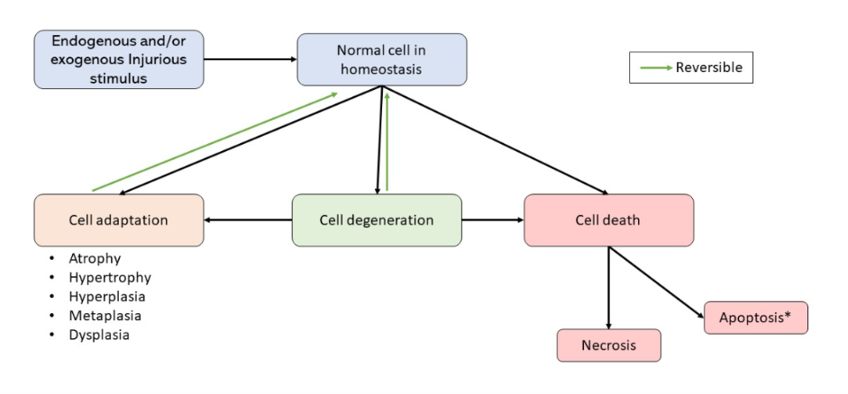
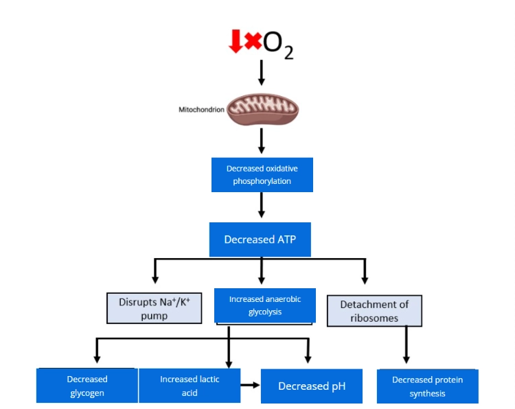
How does inadequate oxygen cause cell injury?
Without oxygen:
Oxidative phosphorylation stops.
ATP production falls dramatically.
Anaerobic Glycolysis does not require oxygen but produces very little ATP (only 2 ATP per glucose molecule)
And thus without aerobic glycolysis cannot meet the cell's energy demands for long
Energy-dependent cellular processes begin to fail.
E.g. the sodium potassium pump- which results In cell swelling ect.
Cell injury develops.
When does cellular degeneration occur?
The injury is reversible as long as the damage is mild and/or of short duration.
What is hydropic degeneration?
Hydropic degeneration is the abnormal accumulation of water inside cells, causing them to swell.
What causes hydropic degeneration?
Cell injury (e.g. Inadequate Oxygenation-Anoxia/hypoxia, toxins, trauma) decreases ATP production.
Without enough ATP, the Na⁺/K⁺-ATPase pump fails.
The normal ion gradients can no longer be maintained.
The Na⁺/K⁺ pump maintains the normal electrolyte and water balance of cells.
For every 1 ATP used, it transports:
3 Na⁺ out of the cell
2 K⁺ into the cell
Due to Na⁺/K⁺ pump Failure
K⁺ and Mg²⁺ move OUT of the cell
Na⁺ and Ca²⁺ move INTO the cell (down their concentration gradients)
Water follows sodium by osmosis, producing cell swelling (Hydropic degeneration).
Anoxia vs hypoxia definition:
Hypoxia = partial reduction in oxygen supply to tissues or cells.
Anoxia = complete absence of oxygen.
What cellular structures can be effected by inadequate oxygen?
Mitochondria (lack of oxygen effects ATP creation)
Cell membrane (decrease in ATP creation effects the ATP dependent sodium potassium pump)
The Endoplasmic Reticulum (Cell swelling causes the Rough endoplasmic reticulum to swell and may become severe enough to detach ribosomes from the endoplasmic reticulum)
What is the gross appearance of organs that are experiencing cellular degeneration?
Increased weight and size. Pallor (paleness).
Cell swelling will hence increase the volume and weight of the affected organ and will also give it a pale colour (pallor).
Swollen cells contain excess water, diluting the normal tissue colour and compressing blood-filled capillaries (less blood flow also contributes to pale color).
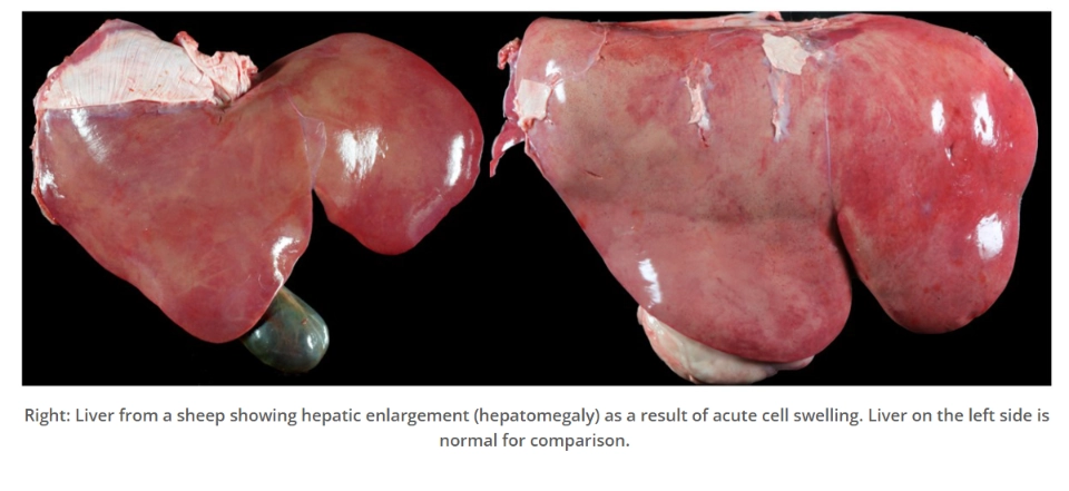
Is this the gross appearance of a tissue that had cellular degeneration, cell death or cell adaption?
Cellular degeneration
Increased weight and size. Pallor (paleness).
Cell swelling will hence increase the volume and weight of the affected organ and will also give it a pale colour (pallor).
Swollen cells contain excess water, diluting the normal tissue colour and compressing blood-filled capillaries (less blood flow also contributes to pale color).
What is the microscopic appearance of cells that are experiencing cellular degeneration?
Under the microscope, affected cells appear:
Enlarged
Enlarged and cloudy
Have a variable number of vacuoles.
Filled with extracellular fluid
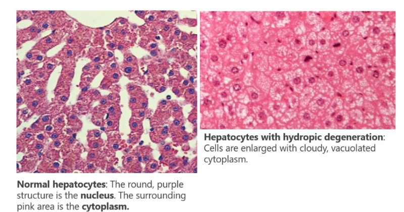
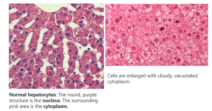
Is this the microscopic appearance of cells that have undergone cellular degeneration, cell death or cell adaption?
Cellular degeneration
Under the microscope, affected cells appear:
Enlarged
Enlarged and cloudy
Have a variable number of vacuoles.
Filled with extracellular fluid
What is Irreversible Cell Injury? and when does it occur?
It is the local death of tissues within a living individual
Irreversible cell injury occurs when cellular damage becomes so severe that the cell can no longer recover, even if the original cause of injury is removed.
What are the two major forms of cell death?
Apoptosis (programmed cell death)
Necrosis (uncontrolled cell death)
What occurs during apoptosis?
The cell and its organelles shrinks
Cell membrane forms blebs
Cell fragments into apoptotic bodies
Apoptotic bodies are phagocytosed by neighbouring cells or macrophages
No leakage of cell contents
No inflammation
What is necrosis?
Necrosis is uncontrolled cell death caused by severe, irreversible injury.
The damage overwhelms the cell's repair mechanisms, resulting in membrane rupture and leakage of intracellular contents.
Thus Necrosis almost always stimulates inflammation.
Does apoptosis or necrosis stimulate inflammation and why?
Necrosis stimulates inflammation as the cell membrane ruptures resulting in leakage of intracellular contents. In contrast apoptosis is controlled cell death in which the cell blebs into membrane bound apoptoic bodies which are phagocytosed by macrophages.
In Cell Death the cell injury is irreversible. This is primarily due to membrane damage. What mechanisms cause this membrane damage.
Influx of calcium across damaged membranes
The Ca²⁺ activates destructive enzymes (endogenous phospholipases) which lyse membrane phospholipids.
Production of lipid breakdown products due to phospholipase activity.
Which has a detergent effect on cell membranes
Cytoskeletal damage caused by cell swelling.
Including detachment of the cell membrane from the cytoskeleton
Production of free radicals
These are unstable molecules unpaired electrons, that in their search for stability "steal" electrons from nearby molecules. This damages lipids, proteins and DNA
Promoting membrane rupture
What is the main mechanism of cell death (irreversible cell injury)?
Severe damage → causes membrane breakdown → results in calcium influx → causing destructive enzyme activation → widespread cell destruction → cell death (necrosis) +leakage of cell contents (causing inflammation)
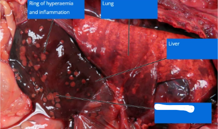
Is this the gross appearance of a tissue that had cellular degeneration, cell death or cell adaption?
Cell Death
What is the gross appearance of organs that are experiencing cell death?
The tissue shows loss of normal structure
If only one part of the organs is affected (which is most common), the necrotic area becomes more obvious.
It is clearly demarcated from the surrounding healthy tissue.
Necrotic cells release their breakdown products into the extracellular space.
These substances irritate neighboring healthy tissue and trigger inflammation.
Producing a red line of demarcation (also called the margin of hyperaemia)between the healthy and necrotic tissue.
Inflammation is a sign of true necrosis.
If the lesion occurs post-mortem inflammation does not occur.
Tissue may become soft and friable (crumbly).
True or false: Necrosis may resemble reversible injury in the early stage.
True:
Because irreversible cell damage can develop from reversible injury, necrosis may resemble reversible injury in the early stage.
I.e. The organ may look enlarged and pale (pallor).
What is the microscopic appearance of the cytoplasm of cells that are experiencing cell death?
The cytoplasm appears pinker.
When a cell is damaged, RNA breaks down, so their is less blue staining.
RNA is basophilic, meaning it binds to haematoxylin (a blue dye).
Additionally, as the cell dies, proteins in the cytoplasm become denatured (they lose their normal structure).
Eosin (an acidic pink dye) binds more strongly to these altered proteins.
This makes the cytoplasm appear pinker.
Cells often become smaller after death
Rather than remaining swollen.
No cell membrane.
This appears as a lack of distinct borders between adjacent cells.
These cells thus seem to blend together (appear uniformly pink)
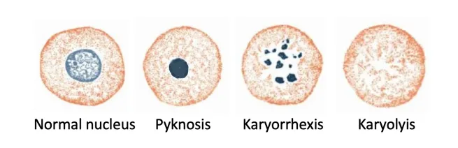
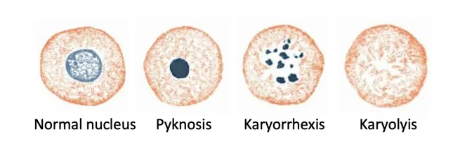
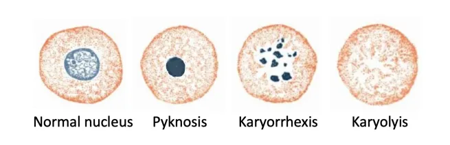
Nuclear changes
Pyknosis (a shrunken, dark-staining nucleus)
Karyorrhexis (fragmentation of the nucleus)
Karyolysis (‘fading’ of the nucleus as it loses affinity for haematoxylin (blue staining dye))
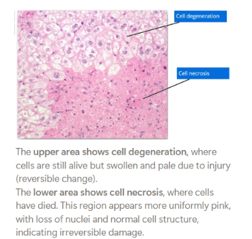
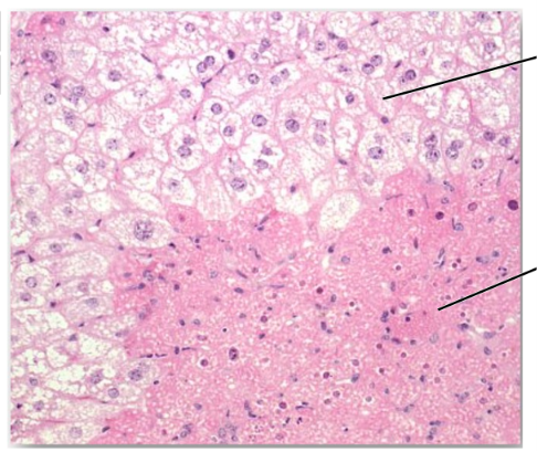
Is this the microscopic appearance of cells that have undergone cellular degeneration, cell death or cell adaption? The bottom and top half have different answers.
The upper area shows cell degeneration, where cells are still alive but swollen and pale due to injury (reversible change).
The lower area shows cell necrosis, where cells have died. This region appears more uniformly pink, with loss of nuclei and normal cell structure, indicating irreversible damage.
What nuclear change caused by necrosis is Pyknosis?
A shrunken, dark-staining nucleus
What nuclear change caused by necrosis is Karyorrhexis.?
Fragmentation of the nucleus
Karyorrhexis= bursting of the nucleus: karyon=nucleus (i.e karyograph) + rrhexis=bursting/tearing
What nuclear change caused by necrosis is Karyolysis?
‘fading’ of the nucleus as it loses affinity for haematoxylin (blue staining dye)
Karyolysis= fading of the nucleus: karyon=nucleus (i.e karyograph) +-ysis= loosen/dissolve
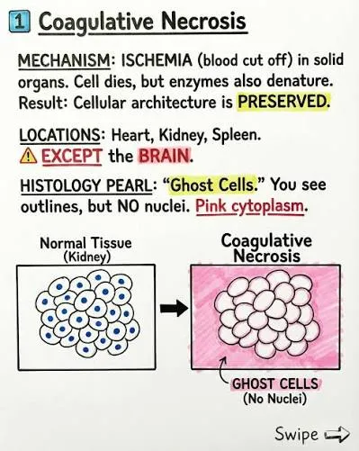
What is Coagulative Necrosis?
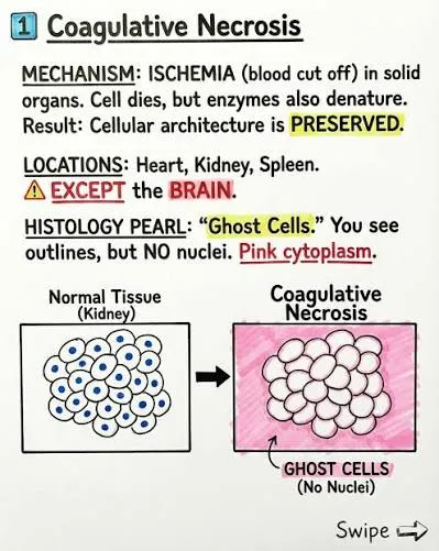
Coagulative necrosis is localised tissue death in which cellular detail is lost, but the overall tissue architecture is preserved.
Think: Coagulative Necrosis= "Ghost tissue."
a.k.a. You can still recognise the organ, but the individual cells are dead.
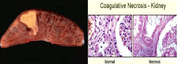
What type of necrosis is this?
Coagulative
Localised tissue death in which cellular detail is lost, but the overall tissue architecture is preserved.
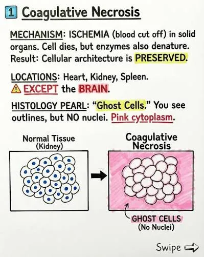
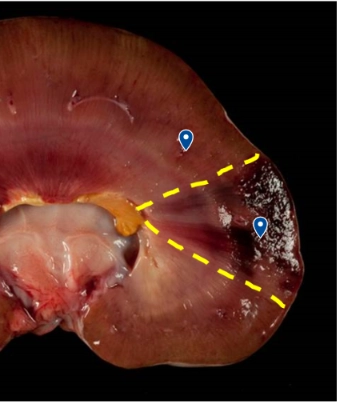
What type of necrosis is this?
Coagulative
Localised tissue death in which cellular detail is lost, but the overall tissue architecture is preserved.
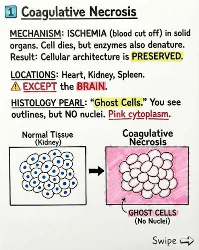
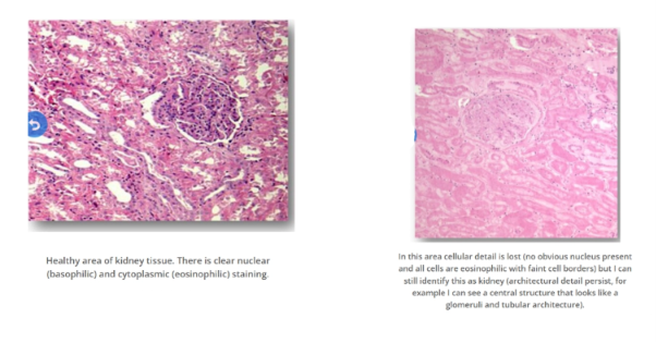
What type of necrosis is this?
Coagulative
Localised tissue death in which cellular detail is lost, but the overall tissue architecture is preserved.
What is Caseous Necrosis?
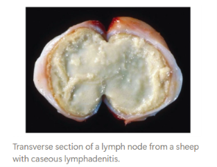
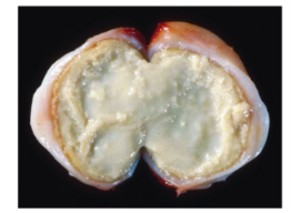
Caseous necrosis is localised tissue death in which both cellular detail and tissue architecture are completely destroyed.
The dead tissue becomes pale granular material resembling crumbly cheese.
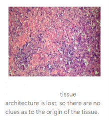
What type of necrosis is this?
Caseous necrosis is localised tissue death in which both cellular detail and tissue architecture are completely destroyed.
The dead tissue becomes pale granular material resembling crumbly cheese.
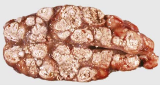
What type of necrosis is this?
Caseous necrosis is localised tissue death in which both cellular detail and tissue architecture are completely destroyed.
The dead tissue becomes pale granular material resembling crumbly cheese.
What type of necrosis is this?
Caseous necrosis is localised tissue death in which both cellular detail and tissue architecture are completely destroyed.
The dead tissue becomes pale granular material resembling crumbly cheese.
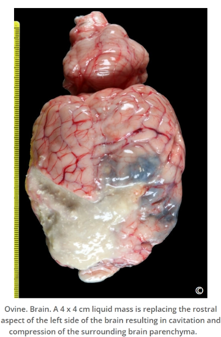
What is Liquefactive (suppurative) Necrosis?
Liquefactive necrosis is complete enzymatic digestion of dead tissue into a liquid mass.
Both tissue architecture and cellular detail are lost.
What is Gangrenous Necrosis?
Gangrene is necrosis affecting a body region.
E.g. distal extremities (distal limbs, teat of udder, pinnae, tip of the tail) and dependent (lower) part of organs (mammary gland and lung lobes).
It is not a separate microscopic type of necrosis, but a clinical pattern based on location and the presence or absence of infection.
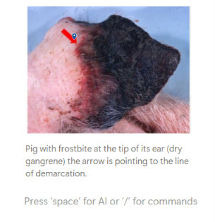
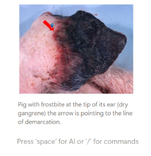
What is dry gangrene?
Necrosis caused only by ischaemia (reduced blood supply).
I.e. without infection or additional factors.
The result is dry, shrivelled tissue.
An example is frostbite, where extreme vasoconstriction cuts off circulation
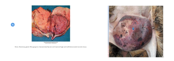
What is wet (moist) gangrene?
When already necrotic tissue becomes infected with bacteria.
The affected area becomes dark, foul-smelling, and wet.
What is gas gangrene?
A form of wet gangrene caused by Clostridium bacteria.
The bacteria produce toxins that destroy nearby cells and produce gas, creating bubbles in the tissue (and a strong, foul smell).
What are the three different types of gangrene necrosis?
Dry = Dead only (caused only by ischaemia (reduced blood supply) without infection ect.)
Wet = Dead + bacteria
Gas = Wet + Clostridium + gas
What is fat necrosis?
The death of fat cells.
Stored fat breaks down into glycerol and fatty acids, which then react with minerals like calcium, potassium, or sodium to form soaps.
A process called saponification.
Necrotic fat appears opaque, firm, and white.
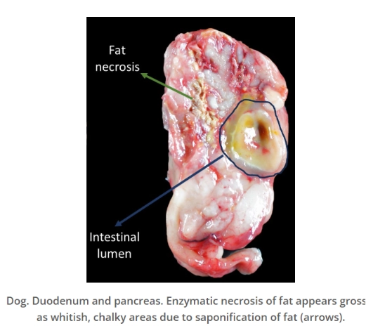
What are the different types of fat necrosis?
Enzymatic necrosis:
Occurs mainly in the adipose tissue surrounding the pancreas.
Due to pancreatitis, damaged pancreatic acinar cells release lipase, which digests surrounding fat.
Traumatic fat necrosis:
Occurs after trauma, or due to chronic pressure, of the subcutaneous adipose tissue.
E.g. a sick cow that has been in recumbency for a long time.
Nutritional:
Occurs in patients with to diets high in polyunsaturated fatty acids and low in antioxidants.
Oxidative damage causes fat cell death.
Idiopathic (unknown cause):
What are the different sequelae (outcome) of necrosis:
Liquefaction and Removal
Occurs in small areas of necrotic tissue that contain large amounts of enzymes and fluid.
The enzymes break down the dead tissue, causing it to liquefy.
The resulting liquid (made of cell debris and small molecules) is then absorbed into the blood or lymphatic system.
And gradually removed by the body.
Encapsulation Without Liquefaction (Sequestration)
The dead tissue causes an inflammatory response, which leads to the growth of fibrous tissue around the tissue, overtime forming a fibrous capsule.
The isolated piece of dead tissue with the surrounding fibrous tissue is called a sequestrum.
The centre becomes drier and may slowly be replaced by fibrous tissue.
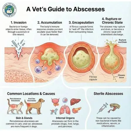
Abscessation
An abscess is a localized collection of pus within tissue (that form when pus builds up in a tissue)
Abscesses can heal in two ways:
By forming a thick fibrous capsule around the pus (like in sequestration)
Or by bursting
Where the pus breaks through surrounding tissue and drains to the surface or into a nearby body cavity.
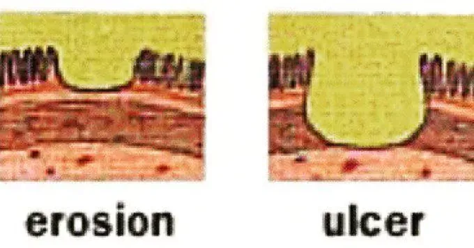
Erosion and Ulceration
These occur when necrotic tissue is located on epithelial surfaces.
Dead cells may lose their attachment to the underlying living tissue and desquamate (or slough) into the surrounding space or lumen causing either ulceration or erosion.
Erosion implies loss of epithelial cells only.
While ulceration occurs when the basement membrane is involved and the underlying connective tissue is exposed.
Regeneration and Repair
Injury to cells and tissues triggers a series of events aimed at limiting damage and promoting healing.
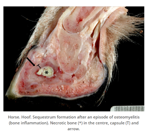
What is a sequestration?
An sequelae (outcome of necrosis): Encapsulation Without Liquefaction (Sequestration)
Sequestrum = a separated piece of dead tissue
The dead tissue causes an inflammatory response, which leads to the growth of fibrous tissue around the tissue, overtime forming a fibrous capsule.
The isolated piece of dead tissue with the surrounding fibrous tissue is called a sequestrum.
The centre becomes drier and may slowly be replaced by fibrous tissue.
What is Abscessation ? and what are the two outcomes of it?
An sequelae (outcome of necrosis): An abscess is a localized collection of pus within tissue (that form when pus builds up in a tissue)
Abscesses can heal in two ways:
By forming a thick fibrous capsule around the pus (like in sequestration)
Or by bursting
Where the pus breaks through surrounding tissue and drains to the surface or into a nearby body cavity.
What is erosion and ulceration?
An sequelae (outcome of necrosis):
Erosion and Ulceration
These occur when necrotic tissue is located on epithelial surfaces.
The dead cells may lose their attachment to the underlying living tissue and desquamate (or slough) into the surrounding space or lumen, causing either ulceration or erosion.
Erosion implies loss of epithelial cells only.
While ulceration occurs when the basement membrane is involved and the underlying connective tissue is exposed.
When does cell adaption occur over cell death and cellular degeneration?
When injury is repetitive/chronic but not severe (sublethal), cells may be able to adapt.
Rather than undergo degeneration or death.
What is the cellular adaption atrophy? And what are some causes?
Atrophy = decreased cell size and/or number → smaller organ.
Causes include:
Disuse, like when a limb isn’t used for a long time (disuse atrophy)
Loss of nerve supply, especially in muscles, after a spinal injury
Lack of nutrition
Lack of hormonal signals