Cell lineage
 Call Kai
 Learn
Learn Practice Test
 Spaced Repetition
 Match
Match Flashcards
 Knowt Play
1/46

There's no tags or description
Looks like no tags are added yet.
Name | Mastery | Learn | Test | Matching | Spaced | Call with Kai | Chat |
|---|
No analytics yet
Send a link to your students to track their progress
47 Terms
How does early embryonic development become controlled and patterned?
Cleavage, gastrulation and organogenesis transform one cell into a patterned embryo.
During cleavage, maternal determinants are distributed among cells, influencing their future body regions.
As development continues, zygotic gene expression gradually replaces maternal control. Maternal products initially dominant and later in development zygotic gene expression takes over
What are the general principles of nervous-system development?
DRCPMMFN
Determination: establishes neural progenitors and restricts their possible fates.
Regionalisation: assigns positional identities along body axes, such as anterior–posterior and dorsal–ventral.
Cell-type specification: directs cells towards particular neuronal or glial identities.
Proliferation and systems matching: produces enough cells, followed by removal of excess or mismatched cells.
Migration: moves developing cells to the correct place at the correct time.
Morphological differentiation: develops cell-specific structures, including axons and dendrites.
Functional differentiation: establishes neurotransmitter, receptor and electrical properties.
Network integration and maintenance: forms and stabilises appropriate synaptic connections.
What defines a stem cell, what is potency, and how does it change during development?
What is the potency trend?
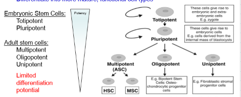
A stem cell can self-renew and differentiate into mature, specialised cells. Its potency is the range of cell types it can produce, which progressively decreases during lineage restriction.
Totipotent → pluripotent → multipotent → oligopotent → unipotent → terminally differentiated.
What are totipotent cells with examples and where they are found?
Cells that can form all embryonic and extra-embryonic tissues, allowing them to produce an entire organism. They are found in the zygote and very early embryo.
What are pluripotent cells with examples and where they are found??
Cells that can form nearly any embryonic cell type but cannot independently form all extra-embryonic tissues or an entire organism. Examples include inner-cell-mass cells, embryonic stem cells and induced pluripotent stem cells.
What are multipotent cells?
Cells that can form several related cell types within a particular tissue ineage. Examples include neural, haematopoietic and mesenchymal stem cells.
What are oligopotent cells?
Restricted progenitor cells that can form only a small number of closely related cell types within a lineage.
What are unipotent cells?
Highly restricted cells that can produce only one mature cell type while retaining the capacity for self-renewal.
What does pluripotent stem-cell technology allow?
It allows fully differentiated cells to be reprogrammed back into a pluripotent state, producing induced pluripotent stem cells (iPSCs). Reprogramming can occur in vitro in tissue culture or in vivo within living organisms.
What are the three patterns of stem-cell division?
Symmetric self-renewal: produces two stem/progenitor cells, expanding or maintaining the precursor pool.
Symmetric differentiation: produces two more-restricted daughter cells, rapidly increasing the committed population.
Asymmetric division: produces one self-renewing cell and one daughter with reduced potency, maintaining the stem-cell pool while generating lineage diversity.
What are neuroblasts and glioblasts?
A neuroblast is an immature neural cell committed to producing neurons, whereas a glioblast is an immature cell committed to producing glia.
How do unipotential and multipotential neuroblasts and glioblasts differ?
Unipotential progenitors produce one specific cell type, such as motor neurons or astrocytes. Multipotential progenitors produce several related cell types within their lineage—for example, different neuronal types or different types of glia.
What are mixed neuronal–glial progenitors?
They are progenitors not yet committed to a neuronal or glial lineage. They can produce a neuroblast → neuron or a glioblast → glial cell, depending on developmental signals
What determines a neural progenitor’s lineage? (4) IEST
What is the progression of cell lineage?
Intrinsic factors: genes and transcription factors within the cell.
Extrinsic factors: growth factors, signalling molecules, neighbouring cells and the extracellular matrix.
Spatial factors: the cell’s location—for example, forebrain versus spinal-cord progenitors.
Temporal factors: developmental timing—for example, a progenitor may produce neurons early and astrocytes later.
Neural progenitor→ Neuroblast/Glioblast→ mature neuron and glial cell
How are neuronal and glial development coordinated from embryogenesis to adulthood?
Neurons form first, followed by glia, which coordinate circuit formation, pruning, stabilisation and myelination through bidirectional neuron-glia and glia-glia interactions.
How does BMP signalling promote epidermal fate?
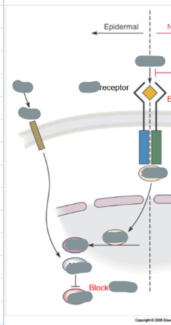
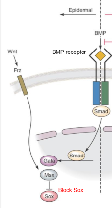
BMP binds its receptor and activates SMAD signalling pathway, while Wnt binds to its receptor Frizzled (Frz) and their signalling cooperates with BMP pathway. GATA & MSX are TF that activate epidermal specific genes reinforcing skin fate. SMAD, GATA and MSX activate epidermal genes and inhibit neural SOX (TF needed for neural development) activity, directing ectoderm towards epidermis
BMP + Wnt → BMP receptor + Frizzled → SMAD activation → GATA & MSX activate epidermal genes + SOX inhibited → Epidermis (skin)
How does BMP inhibition promote neural fate?
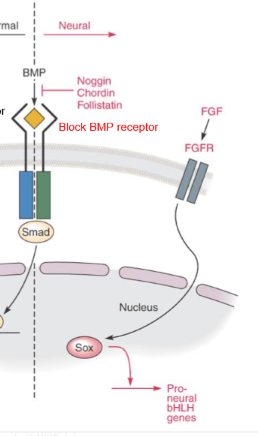
Noggin, chordin and follistatin (all BMP antagonists) block BMP signalling by binding to BMP and preventing from activating the BMP receptor, preventing SMAD activation so GATA and MSX not activated so epidermal genes not expressed. FGF binds to FGFR activating SOX which activate proneural genes, directing ectoderm towards neural tissue.
Noggin/Chordin/Follistatin block BMP → No SMAD → No GATA/MSX activation → FGF → FGFR → SOX activated → Proneural genes ON → Neural tissue
What does deleting noggin and chordin demonstrate?
Noggin and chordin have redundant BMP-antagonist functions. Deleting either alone causes mild defects, but deleting both causes severe head and nervous-system abnormalities, showing that BMP inhibition is essential for neural development.
How is the neural tube formed?
Once BMP is inhibited by Noggin, Chordin, and Follistatin, ectodermal cells commit to a neural fate. These cells form the neural plate, which invaginates (folds inward) and fuses to form the neural tube, the precursor of the brain and spinal cord.
How do BMP and SHH pattern the neural tube?
What do their opposing gradients mean?
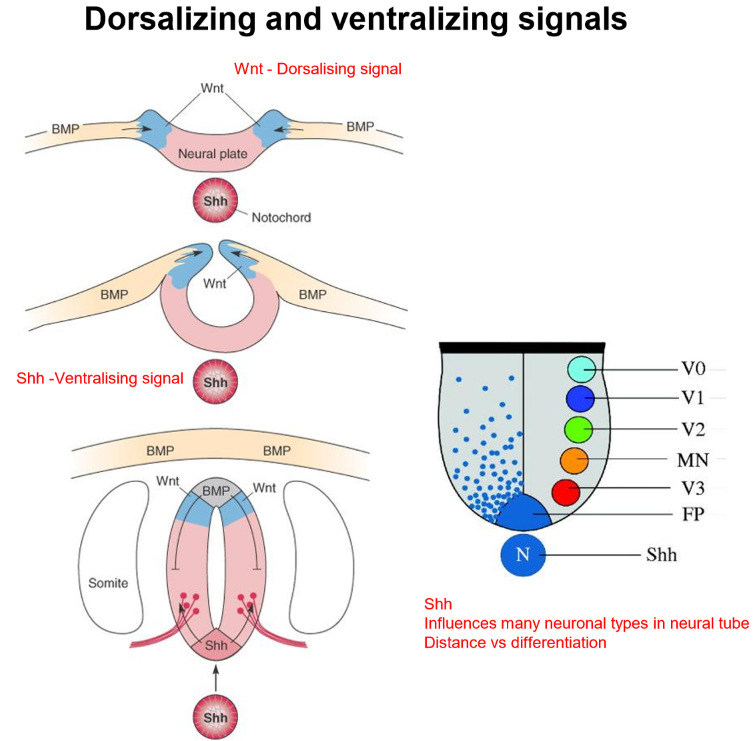
Opposing morphogen gradients regionalise the neural tube:
SHH from the notochord (beneath neural tube) and floor plate specifics ventral (bottom) neural-tube identities, including motor-neuron types. Cells closest to notochord have highest SHH concentration while further away get less. SHH has centralizing gradient.
BMP from the roof plate (dorsal) specifies Dorsal neural-tube identity and neural-crest cells. BMP from dorsal side of has an opposing derailing gradient. Cells closest to roof plate have highest BMP concentration. BMP has dorsalizing gradient.
Together, these opposing gradients provide each cell with positional information, allowing it to determine where it is within the neural tube.
How do morphogen gradients determine neural-tube cell fate?
Cells respond to their position and local concentrations of SHH and BMP, activating different transcription factors:
High SHH ventrally → motor neurons
Intermediate SHH/BMP → interneurons
High BMP dorsally → sensory interneurons
Different concentration thresholds and exposure durations activate different transcription-factor programmes so the morphogens can generate several cell types
What happens to neural crest cells?
Neural crest cells detach from the neural-tube border through EMT, (epithelial-to-mesenchymal transition) migrate throughout the embryo and form structures including PNS neurons, Schwann cells, melanocytes, the adrenal medulla and craniofacial tissues.
What are Hox genes?
Hox genes are mammalian homeotic selector genes that encode homeobox transcription factors with a highly conserved DNA-binding homeodomain. They regulate gene expression in a particular way to help set up body access (anterior and posterior access). As TFs they regulate downstream genes that control development
What is the function of Hox genes and positional memory?
Hox genes establish and maintain the body’s anterior–posterior (head-to-tail) axis by telling cells where they are and what body structures they should form, such as the head, neck, thorax or lumbar region. They also provide positional memory, allowing cells to remember and maintain their regional identity even after many rounds of cell division.
How is Hox gene expression controlled?
Hox genes are regulated in space and time. Spatial regulation controls where different Hox genes are expressed in the embryo, while temporal regulation controls when and in what sequence they are activated. Together, this establishes and maintains the correct body plan.
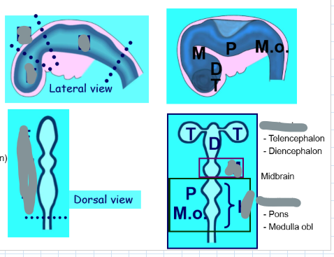
What does the forebrain or prosencephalon form?
What does the mesencephalon form?
What does the rhombencephalon form?
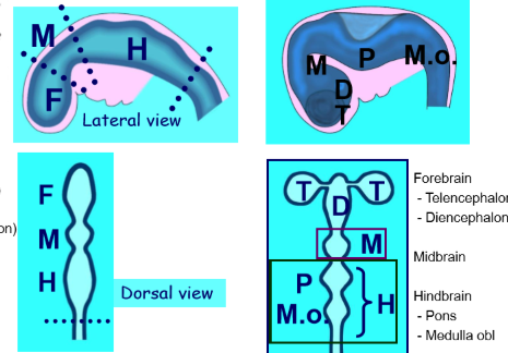
The telencephalon and diencephalon.
Midbrain structures.
The pons and medulla oblongata.
What is an organising boundary?
Give an example of an organising boundary.
A signalling boundary between developmental compartments that helps establish distinct regional identities.
The isthmus between the midbrain and hindbrain.
Which cells occupy the ventricular region during cortical development?
What do ventricular-region progenitors generate?
Neuroepithelial cells, radial glia, and neural progenitors.
Cortical neurons and glia.
Where are neurons and glia produced in the developing brain?
They are produced by neuroepithelial cells and radial glia near the inner ventricular surface. Neuroepithelial cells are the earliest multipotent neural stem cells and later develop into radial glia.
How do neural progenitors divide?
Symmetric division produces two progenitors to expand the progenitor pool. Asymmetric division produces one progenitor and one differentiating daughter, allowing new neurons or glia to form while keeping the progenitor pool.
What does multipotency mean in radial glia?
Radial glia are not all committed at the same time and can produce both neuronal and glial cells. They mainly produce neurons early in development and more glia later, depending on genes, environmental signals and developmental timing.
What are the two main roles of radial glia?
What happens if radial glia or their adhesion molecules are disrupted?
Are all radial glia committed at the same time?
Radial glia act as both “parents and ladders”:
Parents: they produce neurons and glia.
Ladders: their long fibres guide new neurons from the ventricular surface to the outer cortex. (provide scaffold for radial migration)
If radial glia or their adhesion molecules are disrupted, neurons lose their scaffold, causing faulty migration and abnormal cortical layers.
Neuronal migration becomes faulty because cells lose their scaffold.
No. They can retain neuronal/glial multipotency for different periods.
How did birth-dating experiments show when neurons are born?
Scientists used ³H-thymidine(tritiated thymidine) which is added to DNA when cells divide. By finding labelled neurons later, they could see when each neuron was born and where it ended up in the cortex. The label becomes weaker if the cell continues dividing.
What is inside-out cortical development?
Neurons are born at different times:
Earlier-born neurons stop first and form the deep cortical layers.
Later-born neurons migrate past them and form the outer, superficial layers.
So, the cortex develops from the inside out.
Which cells primarily undergo radial migration?
Most excitatory cortical neurons.
They are born at the ventricular zone.
They migrate vertically (radially) toward the brain surface.
Radial glial cells provide the scaffold that guides this migration.
What is the difference between radial and tangential migration?
Radial migration: excitatory neurons climb “up the ladder” of radial glial fibres from the ventricular zone to the cortex.
Tangential migration: GABAergic interneurons are born in the ganglionic eminence and travel sideways (laterally) into the cortex.
Both routes help neurons reach the correct place and form normal cortical circuits.
How do intrinsic changes and extrinsic cues support the switch to gliogenesis?
Transcriptional and epigenetic changes reduce neurogenic competence and increase astrocytic or oligodendroglial competence.
Signals such as Wnt, Notch, growth factors, and receptor activation interact with the progenitor’s changing intrinsic state.
What is epigenetics and what do the changes mean?
Why is epigenetic regulation important for lineage?
Regulation of gene accessibility or expression without changing the DNA nucleotide sequence.
It can stabilise, reverse, or experimentally reprogramme developmental gene programmes.
How do DNA methylation and histone modifications control gene expression?
DNA methylation (adding methyl groups to DNA) usually reduces gene expression by making DNA less accessible.
Histone modifications change how tightly DNA is wrapped around histones.
Open chromatin allows transcription factors to bind as DNA is loosely packed and switch genes on.
Closed chromatin blocks transcription-factor access as DNA tightly packed and switches genes off.
Other epigenetic mechanisms include non-coding RNAs, phosphorylation, ubiquitylation, SUMOylation and nucleosome positioning.
How do HATs histone acetyltransferases and HDACs histone deacetylases affect chromatin?
HATs add acetyl groups → chromatin loosens → transcription usually increases.
HDACs remove acetyl groups → chromatin tightens → transcription usually decreases.
Think: HATs open; HDACs close.
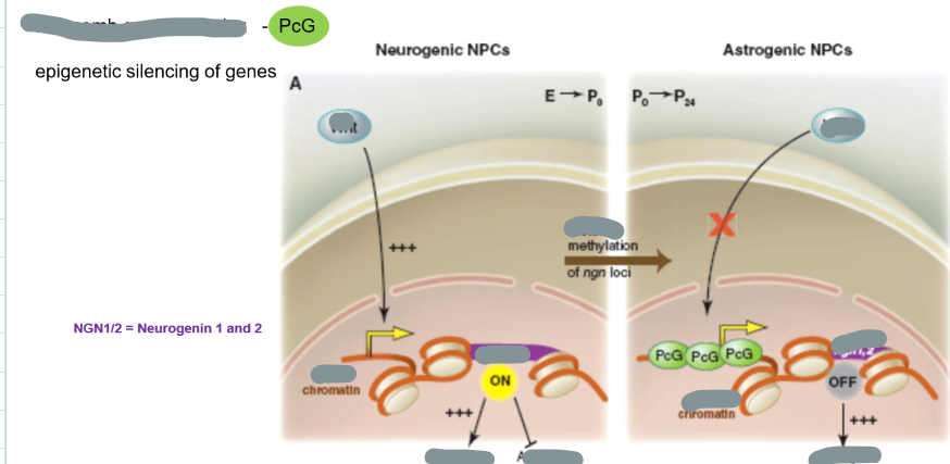
How can epigenetics determine whether a neural precursor becomes a neuron or an astrocyte?
Wnt binds to its receptor on NPC, chromatin opens, Neurogenin-1 and Neurogenin-2 (neurogenins are pro-neuronal TF) activated which switch on neuronal genes and block the astrocyte pathway, producing a neuron.
The Polycomb group complex (PcG is epigenetic gene silencing complex) promotes histone methylation, closing chromatin and repressing gene transcription, silencing Neurogenins and shifting the cell towards an astrocyte fate as chromatin closed.
What is epigenetic reprogramming?
It means changing a cell’s identity by switching specific genes on or off without changing its DNA sequence. Transcription factors act like cell-identity switches.
Why is reprogramming potentially useful after CNS injury with example of NeuroD1
How could NeuroD1 (pro neuronal transcription factor) support neural repair after CNS injury?
It could convert abundant local reactive cells into replacement neurons or glia at the damaged site.
After injury, activated astrocytes migrate to injury site and form glial scar but while glial scar stabilized damanged tissue, it inhibits axon regenration. Researchers use viral vectors to introduce NeuroD1, a pro-neuronal transcription factor, aiming to reprogramme these astrocytes into neurons and improve repair.
Which molecules are linked to different neural identities?
NeuroD1 → neuronal differentiation
Olig2 → oligodendrocyte lineage
GFAP → astrocyte marker rather than a transcription factor; its promoter can be used to target astrocytes experimentally.
What is a neurogenic niche and why is it important?
A neurogenic niche is the specialised environment that keeps neural stem cells alive and tells them when to stay inactive, divide, self-renew or differentiate. It contains astrocytes, blood vessels, extracellular matrix and signals such as Wnt, BMP, Notch and growth factors.
What are microRNAs?
How do microRNAs reduce gene expression?
How do microRNAs affect neural lineage?
Short non-coding RNAs that bind target mRNAs.
They decrease target-mRNA translation or stability.
Different families help switch transcriptional programmes controlling neuronal and glial fate.