The Skeletal System
1/68
There's no tags or description
Looks like no tags are added yet.
Name | Mastery | Learn | Test | Matching | Spaced |
|---|
No study sessions yet.
69 Terms
The Skeletal System
The system that provides support, framing and movement to the body
- Made up of bones and joints
Functions of the Bones
1. Support
2. Storage
3. Blood Cell Formation
4. Protection
5. Body Movement/Leverage
Functions of the Bones - Support
- Skeleton provides a firm framework to support the weight of the body
- Gives shape to the body
- Provides support eg lower limbs and pelvis hold up/support the upper body
Functions of the Bones - Storage
Bones store and release minerals and fats
- Storage of inorganic salts
• calcium, phosphorus, sodium and potassium are stored within bone
• minerals can be removed from bones when needed and distributed around the body by the circulatory system
- Storage of fats
• fat is stored in adipose tissue in the yellow bone marrow which is found in hollow cavity in bones (particularly long bones)
Functions of the Bones - Blood Cell Formation
- red bone marrow which is found in certain bones contains stem cells
• Stem cells can differentiate into blood cells : red blood cells, white blood cells, and platelets
• in adults red marrow is found in spongy bone of flat bones such as skull and sternum and the ends of long bones such as the femur
Functions of the Bones - Protection
Bones protect delicate organs
- e.g. brain is encased with the skull
- spinal cord is contained within the spinal canal formed by the vertebrae
- the heart and lungs are protected by the rib cage
Functions of the Bones - Body Movement
Bones are able to move (articulate) by the contraction and relaxation of the muscles
- Bones act as levers, simple mechanical devices that can provide a force advantage
Parts of the Bone
- Diaphysis
- Epiphysis
- Articular Cartilage
- Periosteum
- Epiphyseal Plate/Line
Parts of the Bone - Diaphysis
A hollow cylinder (shaft) of compact bone surrounding a cavity (the medullary/yellow bone marrow cavity)
- Cavity is used for fat storage: yellow bone marrow

Parts of the Bone - Epiphysis
Enlarged ends of the bone
- Compact bone on outside
- spongy/cancellous bone on inside
• is more porous - many large spaces filled with marrow (red bone marrow - where blood cell production takes place)
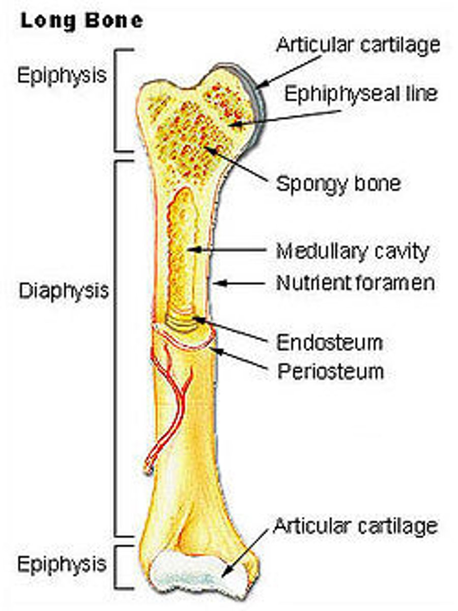
Parts of the Bone - Articular Cartilage
- Covers the end of the epiphysis
• is a layer of cartilage at the ends of the bone where it articulates with another bone

Parts of the Bone - Periosteum
- The dense, white, fibrous, vascularized outer covering of the bone
- It provides for the insertion point of tendons and ligaments
- the inner membrane has bone forming cells use to shape the bone and for repair.
- Not found on joints

Vascularized
provide (a tissue or structure) with vessels, especially blood vessels
Parts of the Bone - Epiphyseal Plate/Line
- A line made up of two cartilage plates
- Located between the diaphysis and epiphysis of long bones

Bone Growth
- Bone elongation occurs at the epiphyseal plates from birth through adolescence by producing new cells and increasing the length of the shaft
- At the end of adolescence the the thickness of the growth plates decreases and ossification occurs (usually finishes by the age of 25)
- Can be used to date the age of a corpse
Two Types of Internal Bone (Parts of Bone)
1. compact
2. spongy/cancellous
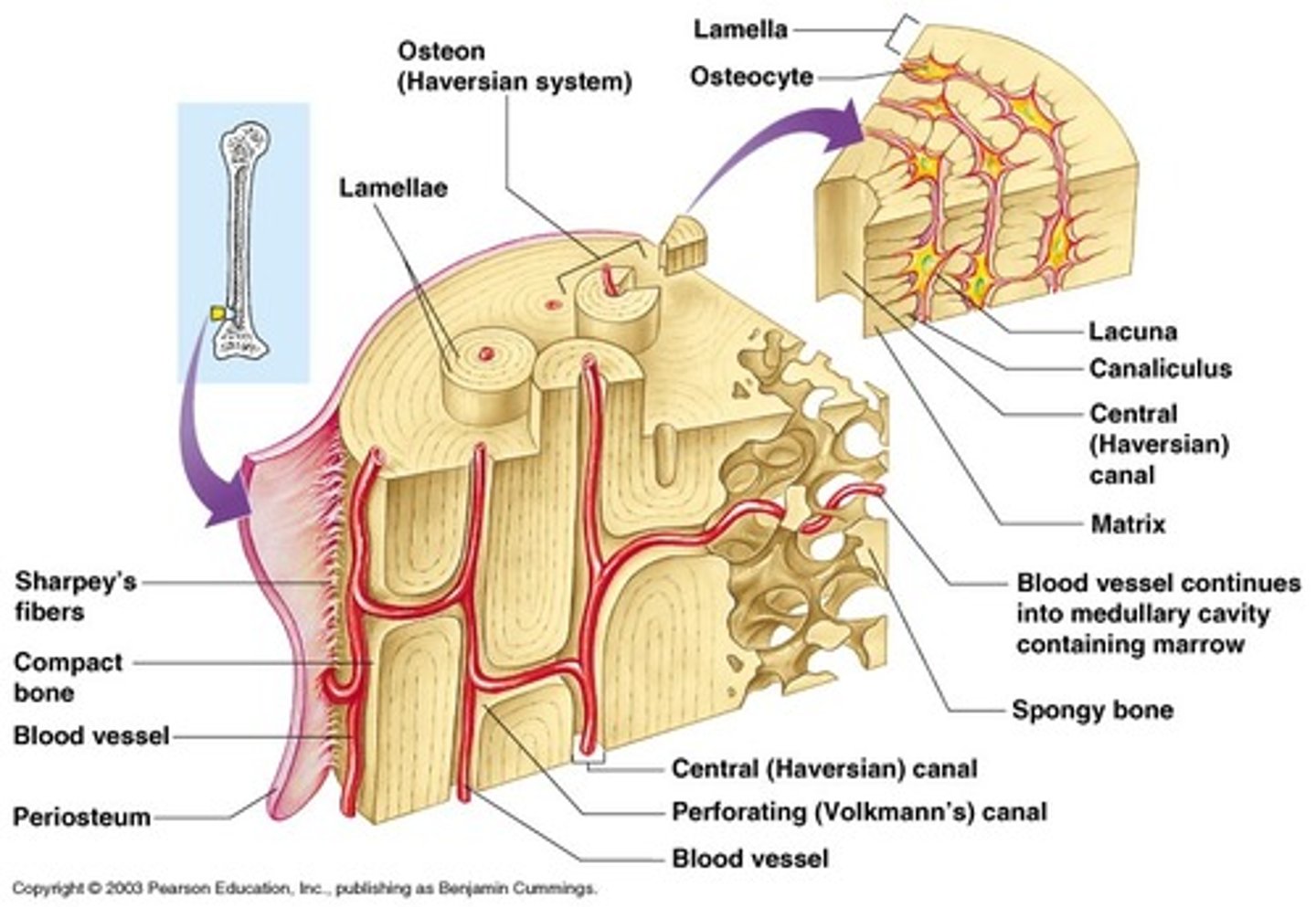
Compact Bone
- Found on the diaphysis of long bones
- Consists of many similar units called osteons (or Haversian systems)
• run parallel to the long axis of the bone for maximum strength
• are cylinder shaped
- gives strength to bones and enables bones to provide strong mechanical levers
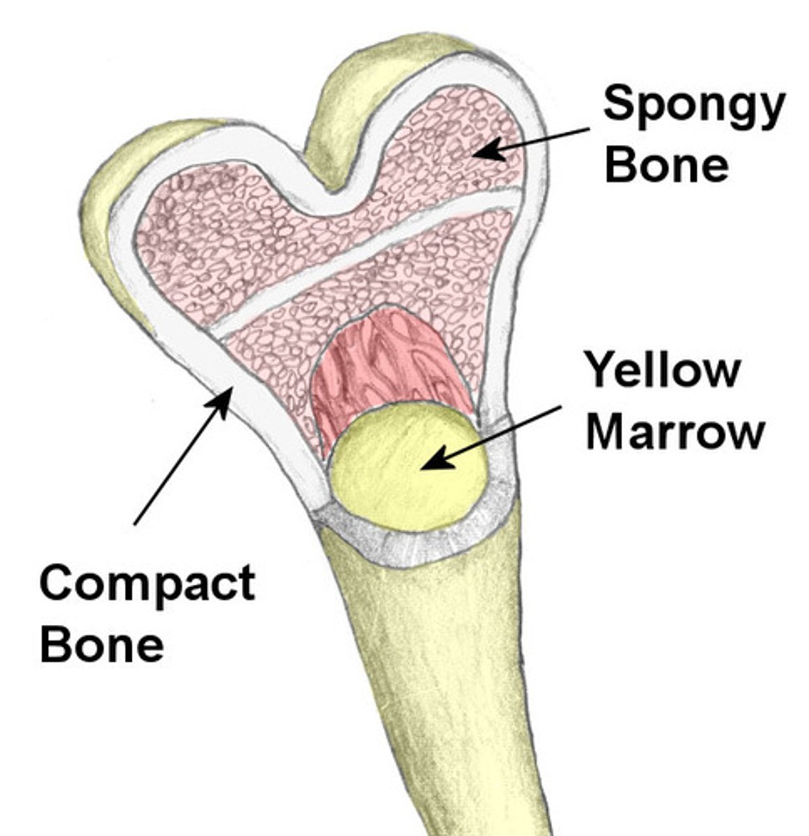
Spongy/Cancellous Bone
- found on the epiphysis of long bones, ribs, skull and pelvis
- consists of an irregular arrangement of thin bone plates (called trabeculae)
- bone cells, nerves and blood vessels are found in the spaces of the trabeculae
- has honeycomb appearance
- reduces density of bone
• allows the ends of long bones to compress as the result of stresses applied to the bone
Parts of an Osteon
1. Central/Haversian canal
- has one blood capillary and may contain nerves and lymph vessels
2. Lamellae
- are concentric layers of bony matrix
3. lacunae
- Small spaces found between the matrix between lamellae
- an osteocyte occupies each lacuna
4. Canaliculi
- Small canals that run between lacunae
Type of Tissue Bone is
Connective Tissue
- has a matrix with inorganic salts of calcium and phosphate
• increases its rigidity
Osteocytes
Bone cells
Cartilage
Is a connective tissue
- Found on the surface of bone joints, trachea, bronchi, nose, larynx and outer ear
Structure of Cartilage
- Contains fibres made from a protein called collagen
• Are embedded in a firm protein-carbohydrate complex called chondrin (is the extracellular matrix in cartilage)
• Firmness allows cartilage to provide structural support and flexibility (from fibres)
- Within chondrin are spaces that contain cartilage cells called chondroblasts
• produce the matrix and are eventually trapped in small spaces called lacunae
• are called chondrocytes after being encased and 'maturing'
Blood Supply to Cartilage
Cartilage does not contain blood vessels
- nutrients & wastes rely on diffusion through the matrix (chondrin)
• is a slow process as chondrocytes have slow metabolism and cell division
- Blood supply to the cartilage is from inner layer of perichondrium
• perichondrium is fibrous membrane of connective tissue
• covers external surface of cartilage
- Injured cartilage takes some time to heal due to less blood supply
Types of Cartilage
1. Hyaline
2. Elastic
3. Fibrous/Fibrocartilage
Types of Cartilage - Hyaline
- Gives strength & flexibility
- has closely packed collagenous fibres through the matrix
• Provides strength to cartilage
- Very fine, not distinguishable under a microscope
Example Locations of Hyaline Cartilage - Knee
- contains cartilage named articular cartilage
- Provides separation and a protective cushion between thigh bone and tibia
• Without cartilage, bones rub together and wear down
• Knees take a lot of weight, creating extreme pain
Example Locations of Hyaline Cartilage - Respiratory Tract
- Nose, larynx, trachea and bronchi
- Tracheal cartilage keeps the trachea open and prevents it's collapse from the negative pressure of the respiratory system
- The septal nasal cartilage separates both right and left nasal cavity which structurally allows air to pass through them
• Provides two cavities
• generates turbulence within the tight space, allowing for air to flow better
Example Locations of Hyaline Cartilage - Epiphyseal Plate
Involved in the growth of long bones
- Cartilage is formed when calcified, degraded and replaced by bone
- Therefore called a growth plate
Types of Cartilage - Elastic
Provides flexible support with elastic recoil
- The most flexible cartilage as it can bounce back to its original shape, even with a strong force.
- Has obvious elastic fibers
• Also has collagenous fibers similar to hyaline cartilage but not as closely packed
• Collagen fibers strengthen the cartilage
Example Locations of Elastic Cartilage - Outer Ear
Auricular cartilage helps maintain the shape of the ear while allowing for flexibility
Example Locations of Elastic Cartilage - Larynx
-Phonation (making sound)
-Cough reflex
- Protection of the lower respiratory tract
- Contains 3 cartilages
•Thyroid cartilage (hyaline) - Supports & protects vocal cords
• Cricoid cartilage (hyaline) - Attachment for muscles, ligaments and cartilage, involved in opening and closing the airway & speech
• Epiglottis (elastic) - Prevents liquids and solids from entering the lower respiratory system
Types of Cartilage - Fibrous
- Regions where weight of the body is supported/withstand heavy pressure
- Has a coarse appearance
• From parallel bundles of thick collagenous fibres
- Not as compact as hyaline
• can be compressed slightly
- Intervertebral discs of spine, articular cartilage of knee, tissue joining two sides of the pelvis
Example Locations of Fibrous Cartilage - Meniscus
is a C-shaped piece of cartilage that acts as a cushion between the shinbone (tibia) and thighbone (femur)
Example Locations of Fibrous Cartilage - Disks between Vertebrae
- Intervertebral discs has a cartilage outer ring called the annulus fibrous
• Resist the stresses to which intervertebral discs are subjected to
Joints
Site at which two or more bones come together
Function of the Joints
- Enable movement
- Allow bone growth
- Allow growth of the brain
- Allow changes in shape during childbirth
- Binds parts of the skeleton
Factors Affecting Joint Stability
- Articular Surfaces - When larger and fit together
• eg. Ball and socket joint is more stable compared to the shallow flatter joint of the knee
- Ligaments - join bones to bones and help to direct bone movement and prevent excessive movement
- Muscle Tone - the tone of the muscles whose tendons cross the joint The greater their strength the more stable the joint
- Bones that fit together tightly have strong joints with little movement
- Bones that are loosely fitted have weaker joints with more mobility but are prone to dislocation
Classifications of Joints
1. Fibrous (Fixed)
2. Cartilaginous (Slightly Moveable)
3. Synovial (Freely Moveable)
Classifications of Joints - Fibrous
- Have no movement
- Bones are held by fibrous connective tissue
- Are difficult to damage
- E.g. Skull sutures
- E.g. teeth
Classifications of Joints - Cartilaginous
- Held in place by cartilage
- Allows slight movement
- E.g. Junction of two pelvic bones, joints between adjacent vertebrae, joints between ribs and sternum
Classifications of Joints - Synovial
- Amount of movement is limited by ligaments, muscles, tendons and adjoining bones
- Occur at shoulder, elbow, wrist, fingers, hip, knee, ankle and toes
- Most commonly injured.
- Can be categorised by type of movement (6 types)
Types of Synovial Joints
1. Ball and Socket
2. Hinge
3. Pivot
4. Saddle
5. Ellipsoid/Condyloid
6. Gliding
Types of Synovial Joints - Pivot
When a rounded, pointed or conical end of a bone articulates with a concaved ring
- ring is lined with a ligament to make movement smooth
- provides for rotation around only one axis
• One bone rotates around another with a concave ring formed in the second bone
- E.g. neck
• Enables neck to rotate left and right
- E.g. forearm
• The ulna & radius of the forearm are connected via a pivot joint
• This allows you to rotate your forearm
Types of Synovial Joints - Hinge
- Allows movement on one plane only
- The convex surface of one bone fits into the concave surface of another
- e.g. knee
Types of Synovial Joints - Ball and Socket
are the most mobile, allowing a wide range of motion
- bones in these joints fit together with a spherical bone sitting inside another bone that has a concave depression
- allows for bending and circular movement as well as rotation of the limb
- E.g. hip joint
• Connects the pelvis and femur
- E.g. shoulder joint
• Connects humerus to shoulder blade
Types of Synovial Joints - Saddle
Allow both side-to-side and back-and-forth movements
- The two bones forming the joint are saddle-shaped (concave in one direction and convex in the other) and bones fit together in such a way to allow movement in two planes
- E.g. only found in carpal bone in thumb
Types of Synovial Joints - Ellipsoid/Condyloid
Allow movement in two directions, such as up-and-down and side-to-side
- Have one surface that is slightly convex that fits into a slightly concave depression in another bone
- E.g. between radius and carpal bone, between metatarsal bones
Types of Synovial Joints - Gliding
Allows movement in either a side-to side or back-and-forth direction
- Only restricted by ligaments or bony processes
- E.g. between carpal bones (in hand), tarsal bones (in feet, between the sternum and clavicle
Movement Around a Joint
- Extension
• Increases angle at a point
- Flexion
• decreases angle at a point
- Abduction
• Movement away from the body
- Adduction
• Movement of a limb towards the body
- Circumduction
• Movement in a rotary action around a joint
- Medial Rotation
• Towards the midline to the body
- Lateral Rotation
• Away from the midline of the body
Movement At the Foot
- Plantar Flexion
• Pointing the toe
- Dorsiflexion
• Pulling toes up (flexing the foot)
- Eversion
• Foot pulled to the side
- Inversion
• Foot pulled to the centre
Parts of the Synovial Joint
1. Joint Capsule
2. Synovial Membrane
3. Articular Cartilage
4. Cruciate Ligament
5. Meniscus
6. Tendon
7. Patella
8. Bursa
9. Synovial Fluid
Parts of the Synovial Joint - Joint Capsule
- Outer layer is tough and fibrous connective tissue (dense fibrous connective tissue)
- Dense CT has a high proportion of tightly packed collagen fibers providing strength
- Connects periosteum of the articulating bone
• Flexibility permits movement and strength prevents dislocation
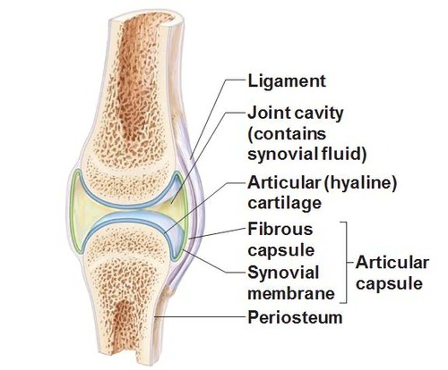
Parts of the Synovial Joint - Synovial Membrane
- inner layer of joint capsule
• Made of loose CT and supplied with blood vessels
• Loose CT provides more movement (loose CT fibers are less densely packed)
- secretes synovial fluid which lubricates the joint to reduce friction between the two bones
Parts of the Synovial Joint - Articular Cartilage
Hyaline cartilage that covers the ends of articulating bones
- Prevents friction by providing a smooth surface for movement
Parts of the Synovial Joint - Cruciate Ligament
- Ligaments are fibrous connective tissue that attaches bones to bones
- A band of strong, fibrous CT
- Joins bone to bone and provides stability
Parts of the Synovial Joint - Meniscus
A wedge of tough, flexible cartilage that divides the synovial cavity in two so that synovial fluid can be directed into the area of greatest friction
- Reduces wear and tear to joint surfaces and increases
- provides stability
Parts of the Synovial Joint - Tendon
are dense connective tissue that attaches muscles to other body parts, usually bone
- Dense fibrous CT made up of primarily collagen fibers
- attaches muscles to bones
Parts of the Synovial Joint - Patella
- A small flat, rounded triangular bone
- improve quadriceps efficiency by acting as a fulcrum
- acts as a bony shield for deeper structures in the knee joint
Parts of the Synovial Joint - Bursa
- Little bags of synovial fluid
- Reduces friction between a bone and a tendon or ligament or a bone and the skin
Parts of the Synovial Joint - Synovial Fluid
Slippery fluid which fills synovial cavity
- Reduces friction
- stops articulating bones from making contact
- nourishes cartilage
- contains phagocytes to get rid of microorganisms and debris
- Only a small amount is normally present but it can increase if the joint is injured which can lead to swelling and pain
Types of Bone
1. Axial
2. Appendicular
Types of Bone - Axial
Bones thats lie around the central axis of the body
- provides main support for erect posture
- protect central nervous system and organs around the thorax
Bones in the Axial Skeleton
- skull
- ribs
- vertebrae
- sternum
Types of Bone - Appendicular
Bones that consist of the upper and lower limbs
- is every other bone to the axial bones
Bones in the Appendicular Skeleton
- pectoral girdle (shoulder)
- upper limbs (arms)
- pelvic girdle (hips)
- lower limbs (legs)
Osteoporosis
a chronic bone disease where the body breaks down broken or older bone quicker than it can replace it
- results in a loss of bone density, minerals (such as calcium) and strength
Osteoarthritis
a degenerative joint disease
- causes the wearing and thinning of cartilage between bones and joints
Bone Shapes
1. Long
2. Short
3. Flat
4. Irregular
5. Sesamoid