General Anatomy upper extremity
1/77
There's no tags or description
Looks like no tags are added yet.
Name | Mastery | Learn | Test | Matching | Spaced |
|---|
No study sessions yet.
78 Terms
how many phalanges are there
14
how many metacarpals are there
5
how many carpal bones are there
8
how many bones are in the hand
27
what do the head of the metacarpal bones articulate with
the base of the phalanges
from radial to ulnar name the proximal carpal bones
scaphoid/navicular
lunate
triquetrum
pisiform
from radial to ulnar side name the distal row of carpal bones
trapezium/greater multangler
trapezoid/ lesser multangler
capitate/ os magnum
hamate/ unciform
what is the largest carpal bone
capitate/ os magnum
what is the most palpable carpal bone
scaphoid/ navicular
what is the senetnce to remember the carpal bones
Sacred Lovers Try Positions That They Cannot Handle
Scaphoid
Lunate
Triquetrum
Pisiform
Trapezium
Trapezoid
Capitate
Hamate
what is the hook like process on the hamate
hamulus
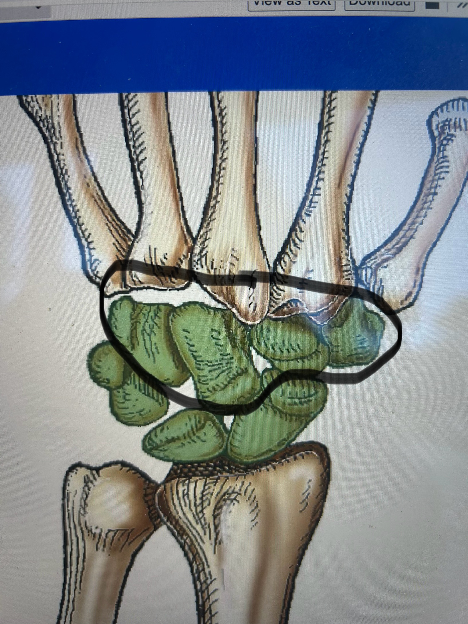
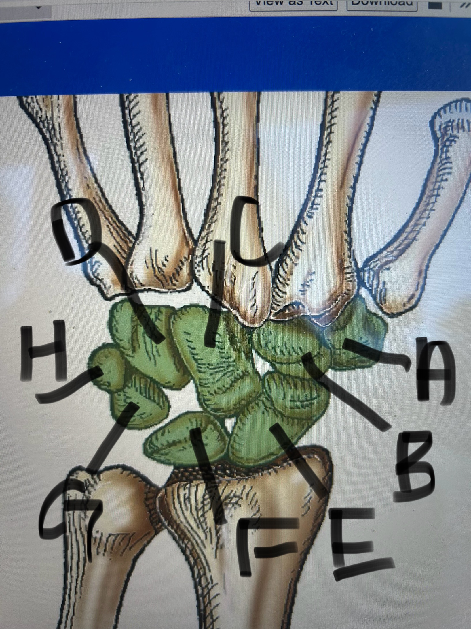
label this image
A) trapezium
B) trapezoid
C) capitate
D) hamate
E) scaphoid
F) lunate
G) triquetrum
H) pisiform
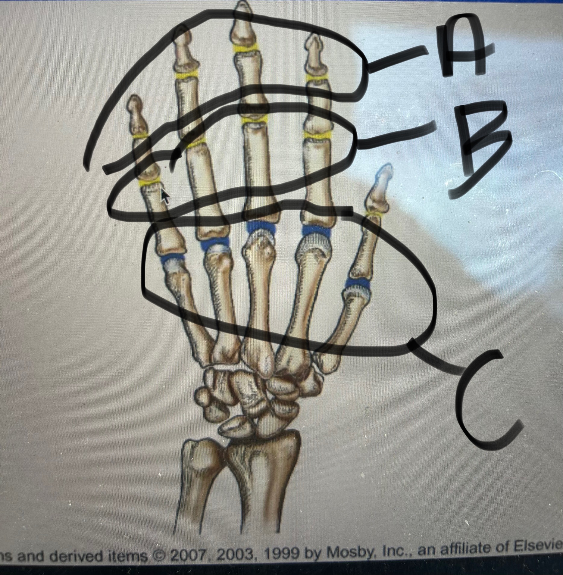
label this image
A) Distal Interphalangeal Joints DIPS
B) Proximal Interphalangeal Joints PIPS
C) metacarpalphalangeal joints MCPJ
what is the interphalangeal joint of the 1st digit
an IP
what are the joints in between metacarpals at their bases
intermetacarpal joints
what are carpometacarpal joints
between bases of metacarpals and distal carpal bones
what kind of joint is the 1st carpometacarpal and between what bones is it
between thumb and trapezium
saddle/ sellar
what are the 2nd-4th carpometacarpal joints classified as
plane/ gliding
what are the joints between carpal bones called
intercarpal
what is the flexor retinaculum (carpal tunnel) attached laterally and medially to
laterally to scaphoid & trapezium
medially to hamulus & pisiform
what are sesamoid bones
small circle bones that develop in tendons
what is the anatomical snuff box
radial fossa
scaphoid and trapezium form floor
where does a bennetts fracture happen
base of first metacarpal
where does a boxers fracture happen
5th metacarpal
what kind of fracture happens at the distal radius, ulnar styloid with posterior displacement
colles
what fracture happens at the distal radius & ulnar styloid with anterior displacement
smiths (reverse colles)
what is a torus/ buckle fracture
bulging of periosteum
what are the 2 radioulnar joints
proximal and distal
what are the 2 beak like process in the proximal ulna
olecrenon process
coranoid process
is the olecrenon process anterior or posterior? Coranoid process?
olecrenon: posterior
coranoid: anterior
what notch is inbetween the olecrenon & coranoid process
trochlear
what is the notch on the ulna that the radial head fits into
radial notch
what are the 2 anatomies on the distal end of the ulna
ulnar head
styloid process
what are the 3 things on the proximal end of the radius
head
neck
tuberosity
what are the parts of the distal radius
styloid process
what is the flat part at the distal end of the radius and what does it articulate with
carpal articular surface
articulates with carpal bones
what is the notch on the distal end of the radius where the ulnar head fits into
ulnar notch
is the styloid process on the radius lateral or medial
lateral
what are the structures that make up the distal humeral condyle
medial epicondyle
coranoid fossa
trochlea
lateral epicondyle
radial fossa
capitulum
is the capitulum medial or lateral on the humerus and what does it articulate with
lateral
radial head
is the trochlea medial or lateral on the humerus
medial
is the coranoid medial or lateral and what does it sit above
medial sits above trochlea
is the radial fossa lateral or medial and what does it sit above
lateral sits above capitulum
where is the ulnar groove
on humerus below medial epicondyle
what is the olecrenon fossa
on humerus posterior groove that olecrenon process goes into
what are the parts of the proximal humerus
head
anatomical neck
greater tubercle
lesser tubercle
surgical neck
intertrabecular groove
body
deltoid tubercle
where does the greater and lesser tubercle sit on the proximal humerus
greater: lateral
Lesser: anterior
where is the surgical neck on the proximal humerus
below tubercles
whats the most common fractures site on the humerus
surgical neck
what is the muscle attachment for the deltoid
deltoid tubercle
what joints makes up the elbow joint proper
proximal radioulnar joint
humerulnar joint
humeroradial joint
what makes the humeroulnar joint
between trochlea& trochlear notch
what makes the humeroradial joint
head of radius and capitulum
what are the 3 fat pads in the elbow and where do they sit
posterior: in olecrenon fossa
anterior: anterior to distal humerus
supinator: anterior to proximal radius
what is the sid for a hand/ digit x ray
40in
what is the patient position for digits 2-5
PA hand
lateral of digit
PA oblique of digit
what is the CR entrance point for PA hand
perpendicular to 3rd MCPJ
what should the collimation be for a PA hand
whole hand and 1-1.5 inches of radius & ulna
what should the hand look like if theres no rotation
see concavities of metacarpal and phalangeal shafts & open joint spaces
what is the CR entrance point for digits 2-5
perpendicular to PIP
what projected is used for the lateral view of digits 2&3
mediolateral
what is the projection used for the lateral view of digits 4&5
lateral to medial
what anatomy should be included in the image of affected digit
distal phalynx to distal portion of metacarpal
what is the CR entrance point for a PA oblique of digits 2-5
perpendicular to PIP
what are the essential projections for the thumb
PA hand
lateral
AP thumb
what is patient position for AP thumb projection
extreme internal rotaion
finger nail in contact with IR
what is the CR entrance point for AP thumb
1st MCPJ perpendicular
what anatomy is included in AP thumb projection
entire thumb to trapezium
whats the other name for the scaphoid
navicular
whats the other name for the trapezium
greater multangler
whats the other name for the trapezoid
lesser multangler
whats the other name for the capitate
os magnum
whats the other name for the hamate
unciform
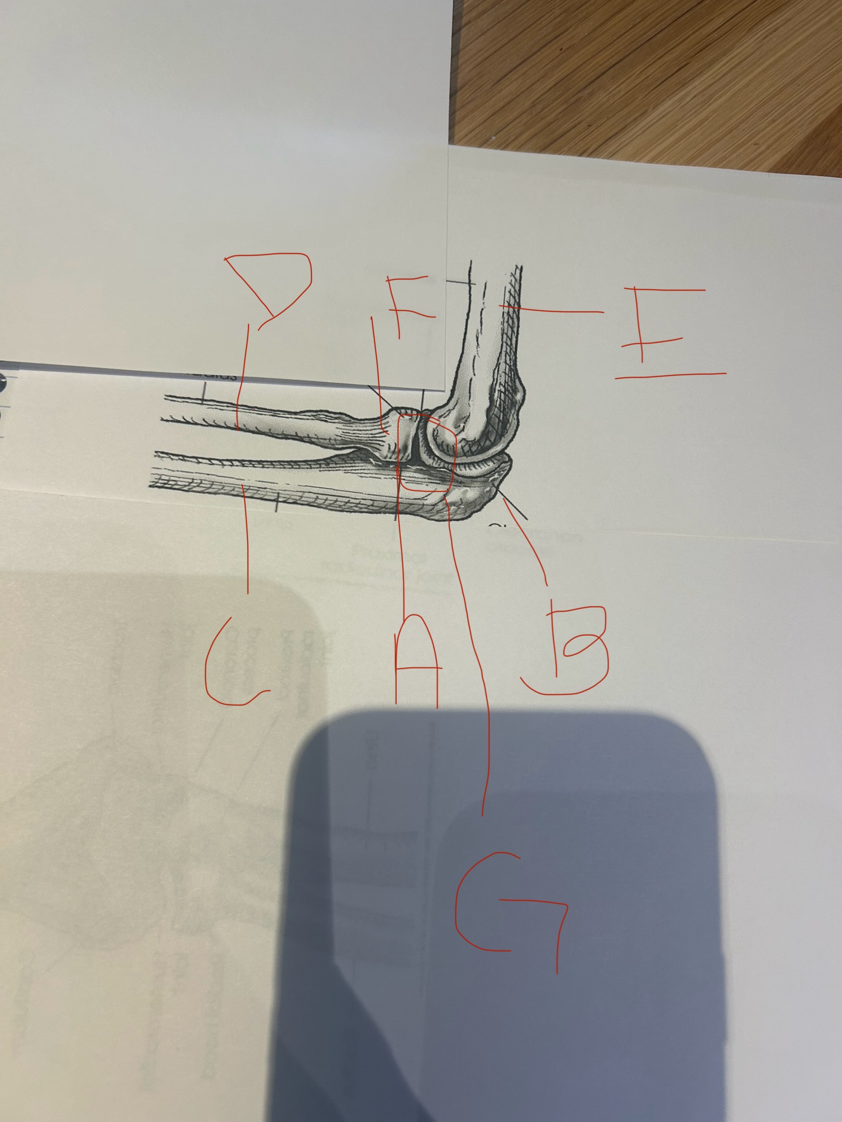
label this image
A) proximal radioulnar joint
B) olecrenon process
C) ulna
D) radius
E) humerus
F) radial head
G) humeroulnar joint
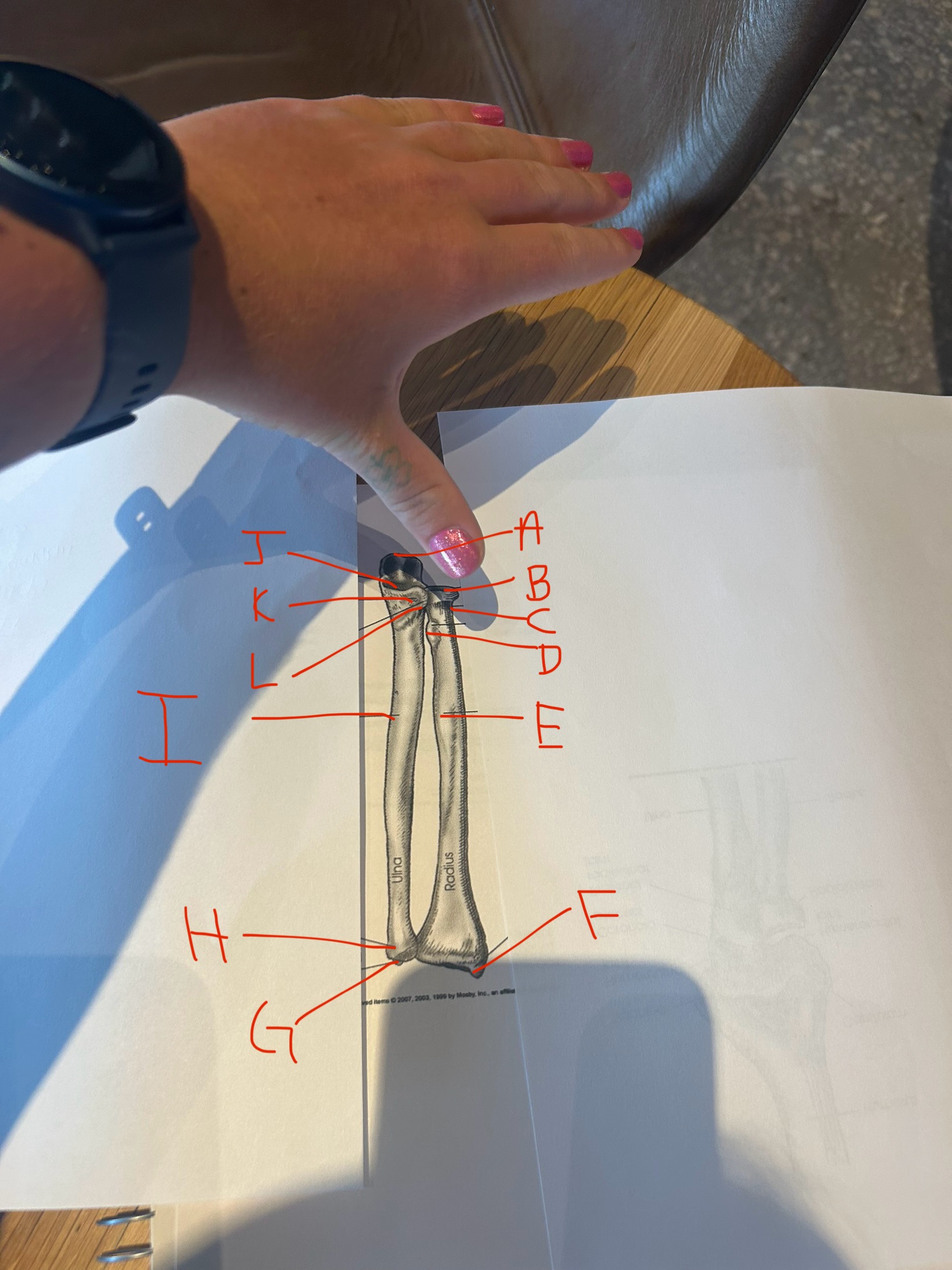
label this image
A) olecrenon process
B)head
C) neck
D) radial tuberosity
E) body
F) radial styloid process
G) ulnar styloid process
H) head
I) body
J) trochlear notch
K) coronoid process
L) radial notch
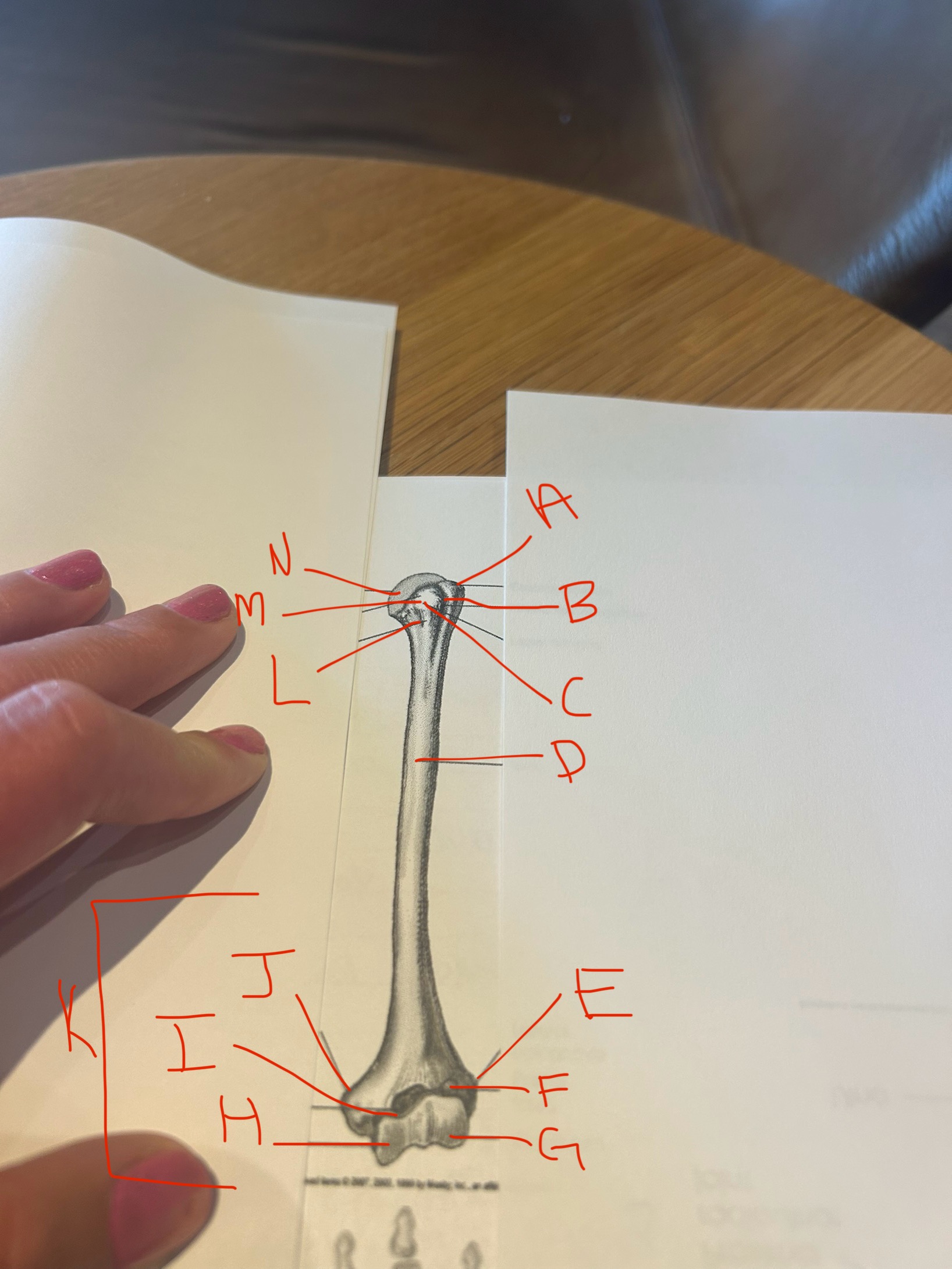
label this image
A) greater tubercle
B) intertubecular groove
C) lesser tubercle
D)body
E) lateral epicondyle
F) radial fossa
G) capitulum
H) trochlea
I) coronoid fossa
J) medial epicondyle
K) humeral condyle
L) surgical neck
M) anatomical neck
N) head
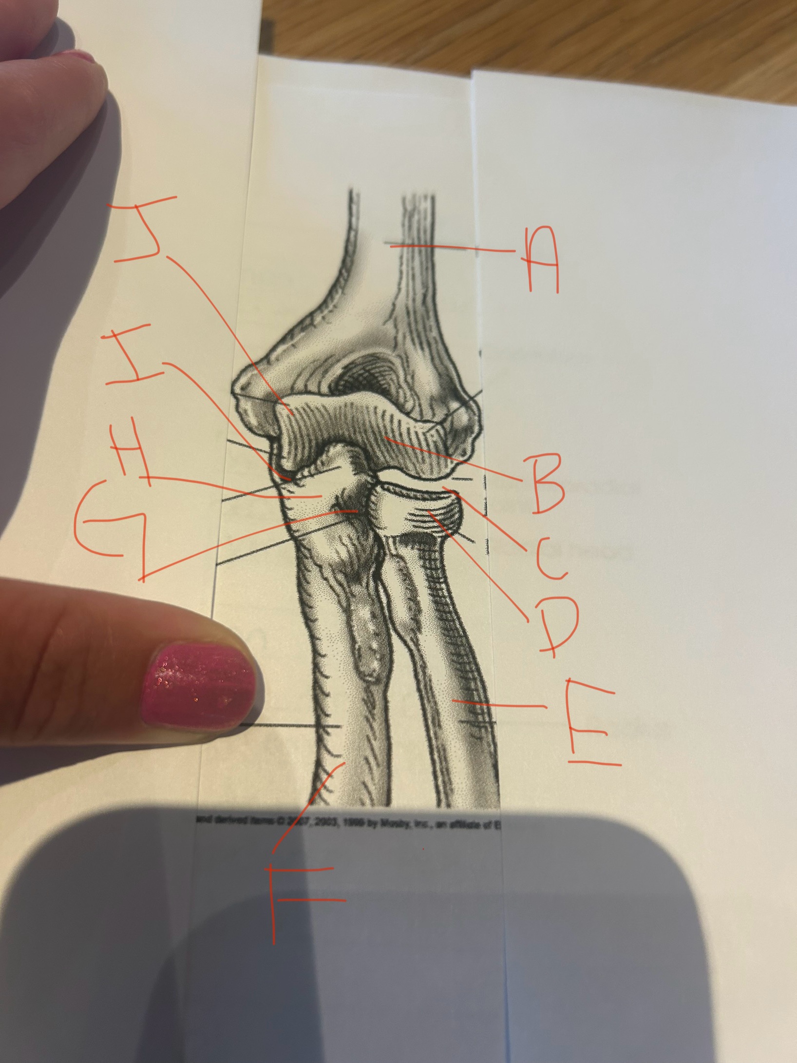
label this image
A) humerus
B) capitulum
C) humeroradial joint
D) radial head
E) radius
F) ulna
G) proximal radioulnar joint
H) coronoid process
I) humeroulnar joint
J) trochlea