Week 4 Lower Limb Anatomy
1/76
There's no tags or description
Looks like no tags are added yet.
Name | Mastery | Learn | Test | Matching | Spaced |
|---|
No study sessions yet.
77 Terms
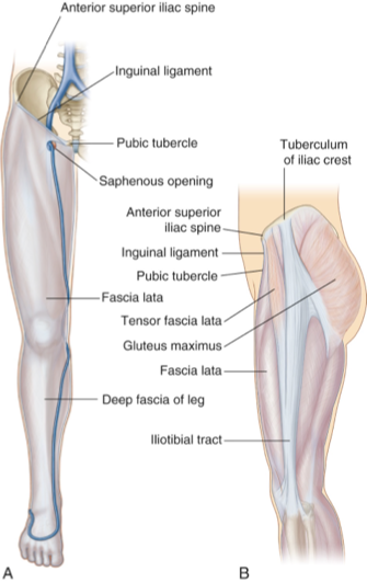
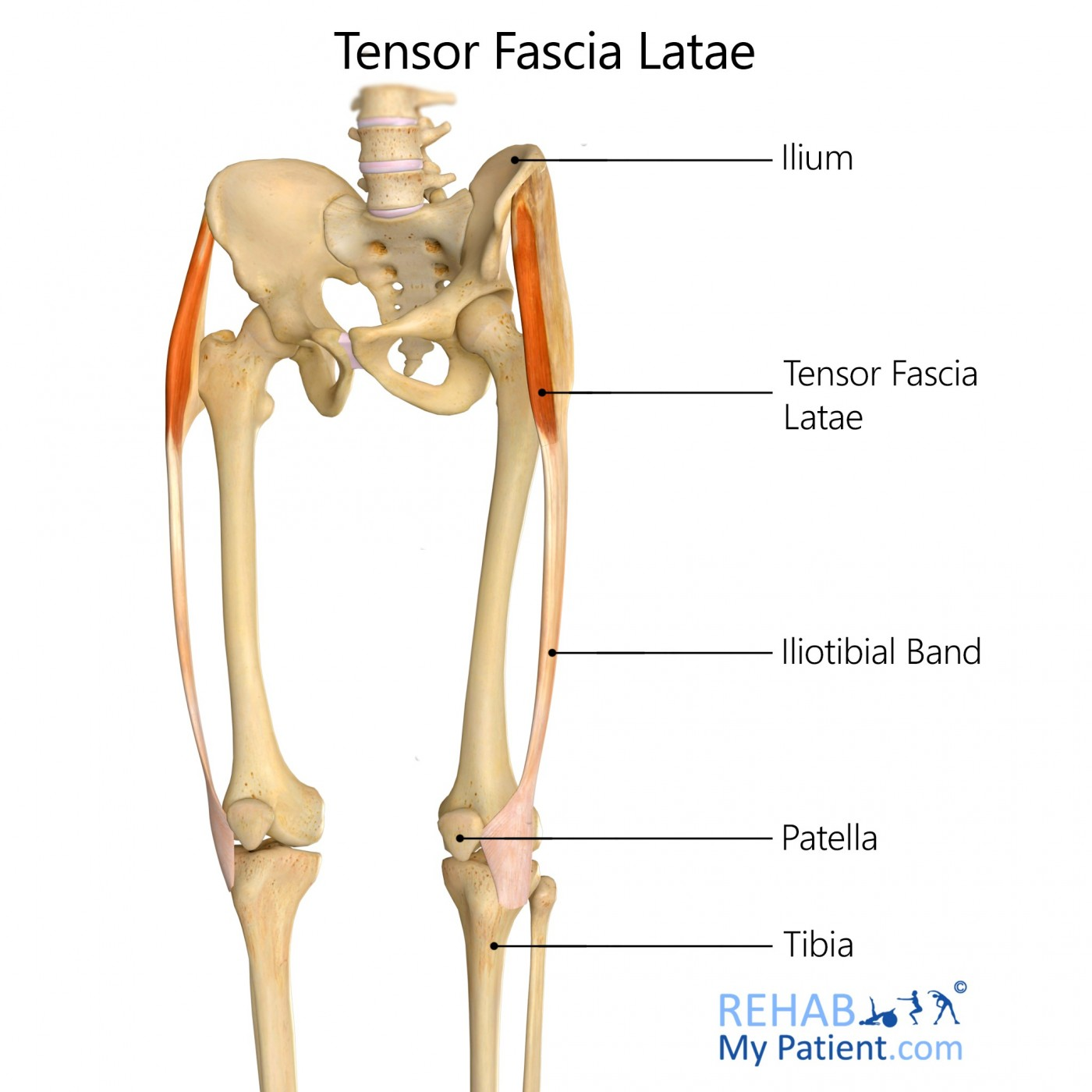
Fascia Lata
“lata” = broad
outer layer of deep fascia that forms a “stocking-like” membrane
from hip to knee
continuous with the Deep fascia of the leg
deep to superficial fascia
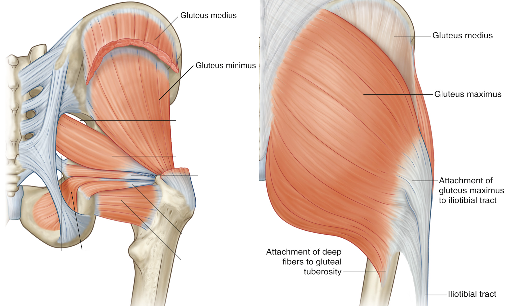
Iliotibial Tract (ITB):
Thickened lateral portion of the fascia lata, from the iliac tubercle to the lateral condyle of the tibia
FASCIAL SEPTA: separates the limbs into compartments.
compartment syndrome
increase in interstitial pressure, resulting in peripheral nerve and muscle ischemia.
Surgical emergency (acute)

thigh compartments
medial
posterior
anterior
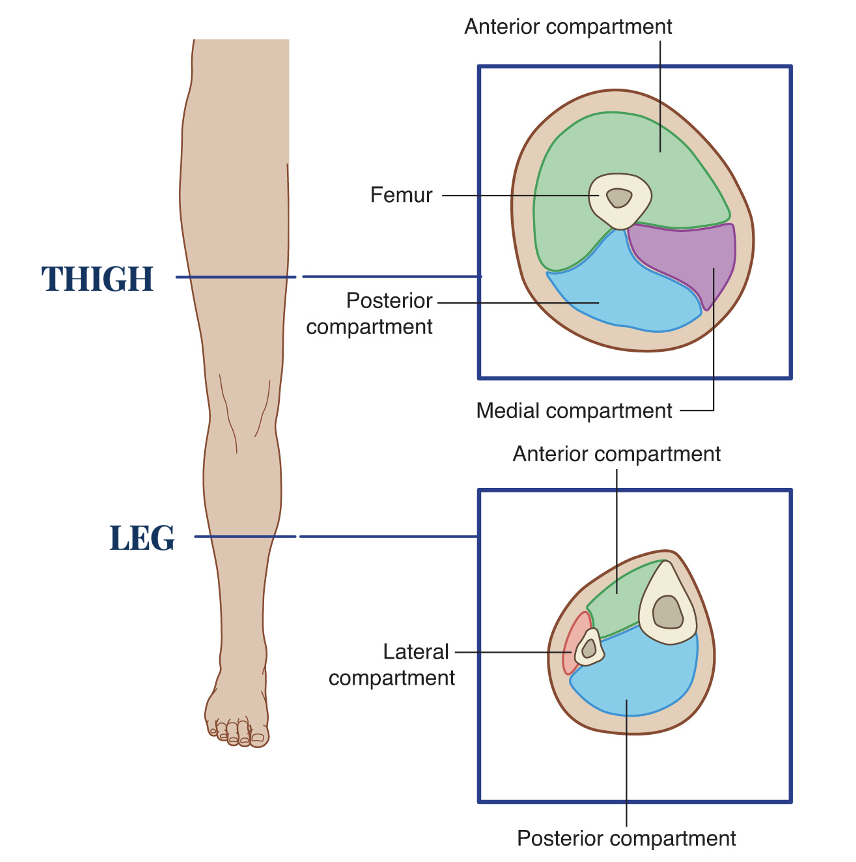
leg compartments
lateral
posterior
anterior

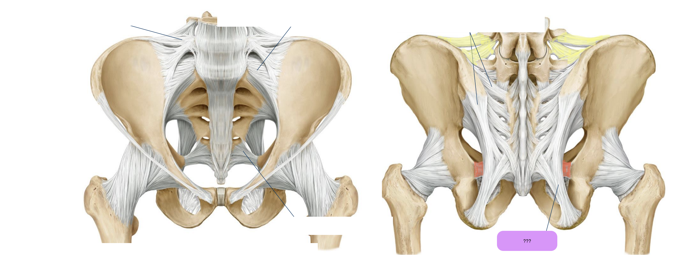
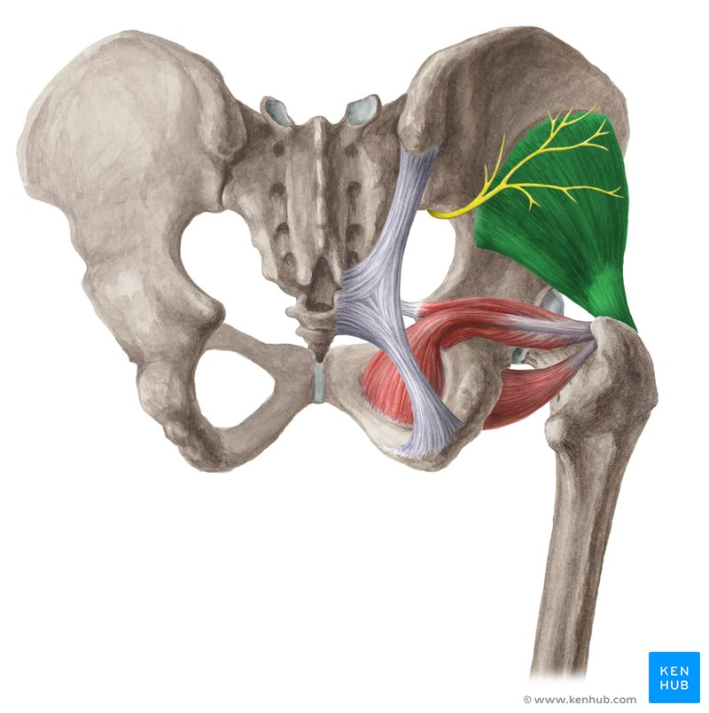
sacrotuberous ligament
connects sacrum to ischial tuberosity
forms greater sciatic foramen
prevents sacrum from collapsing under body weight
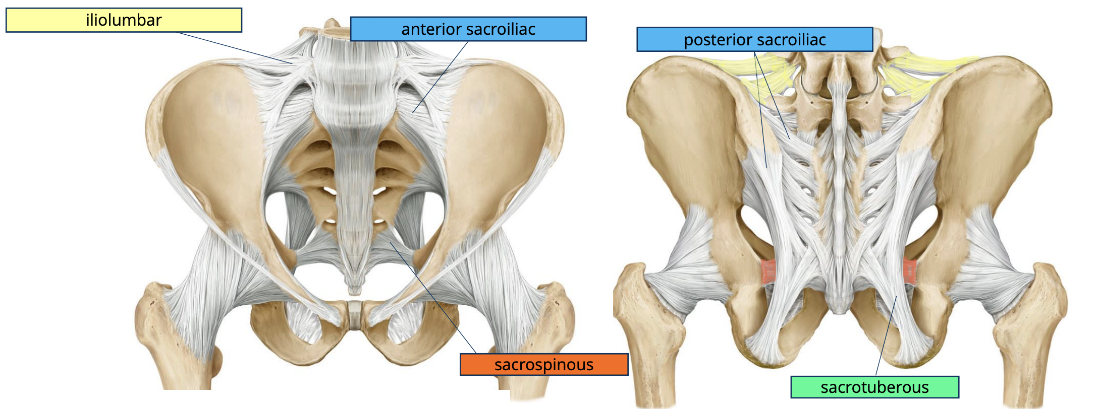
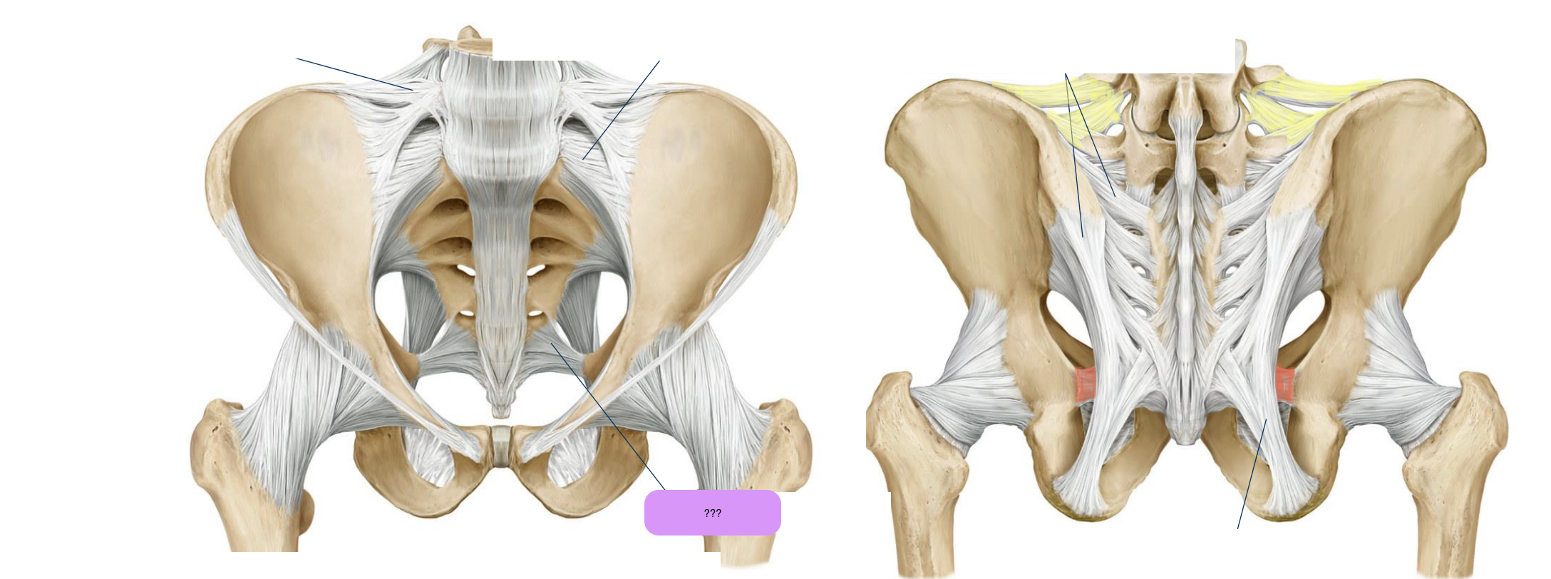
sacrospinous ligament
extends from sacrum to ischial spine
forms the lesser sciatic foramen
prevents sacrum from collapsing under body weight

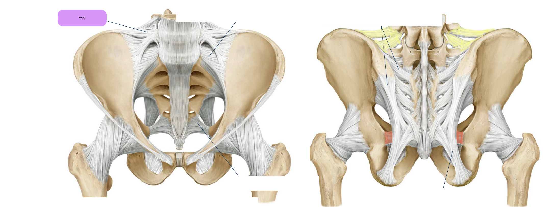
iliolumbar ligament
two bands (anterior and posterior) that extend from L5 to the iliac crest and iliac tuberosity
stabilises lumbosacral spine on the pelvis

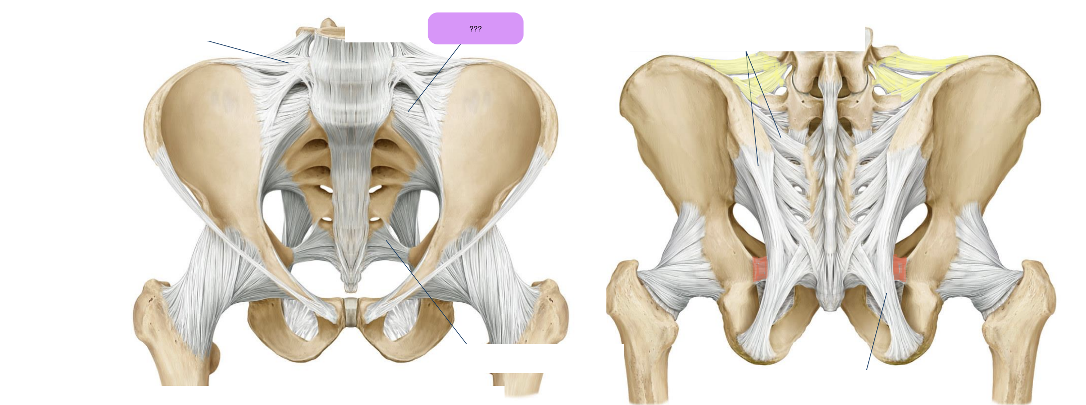
anterior sacroiliac ligament
key for holding pelvic complex together
supports upper body weight

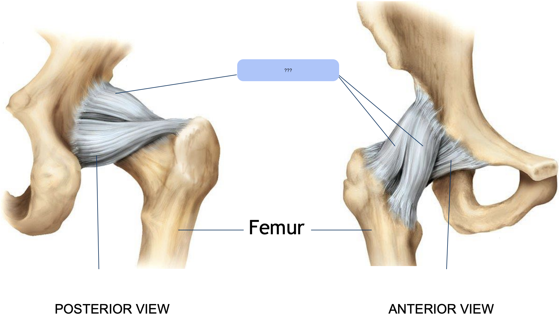
iliofemoral ligament
resists hyperextension

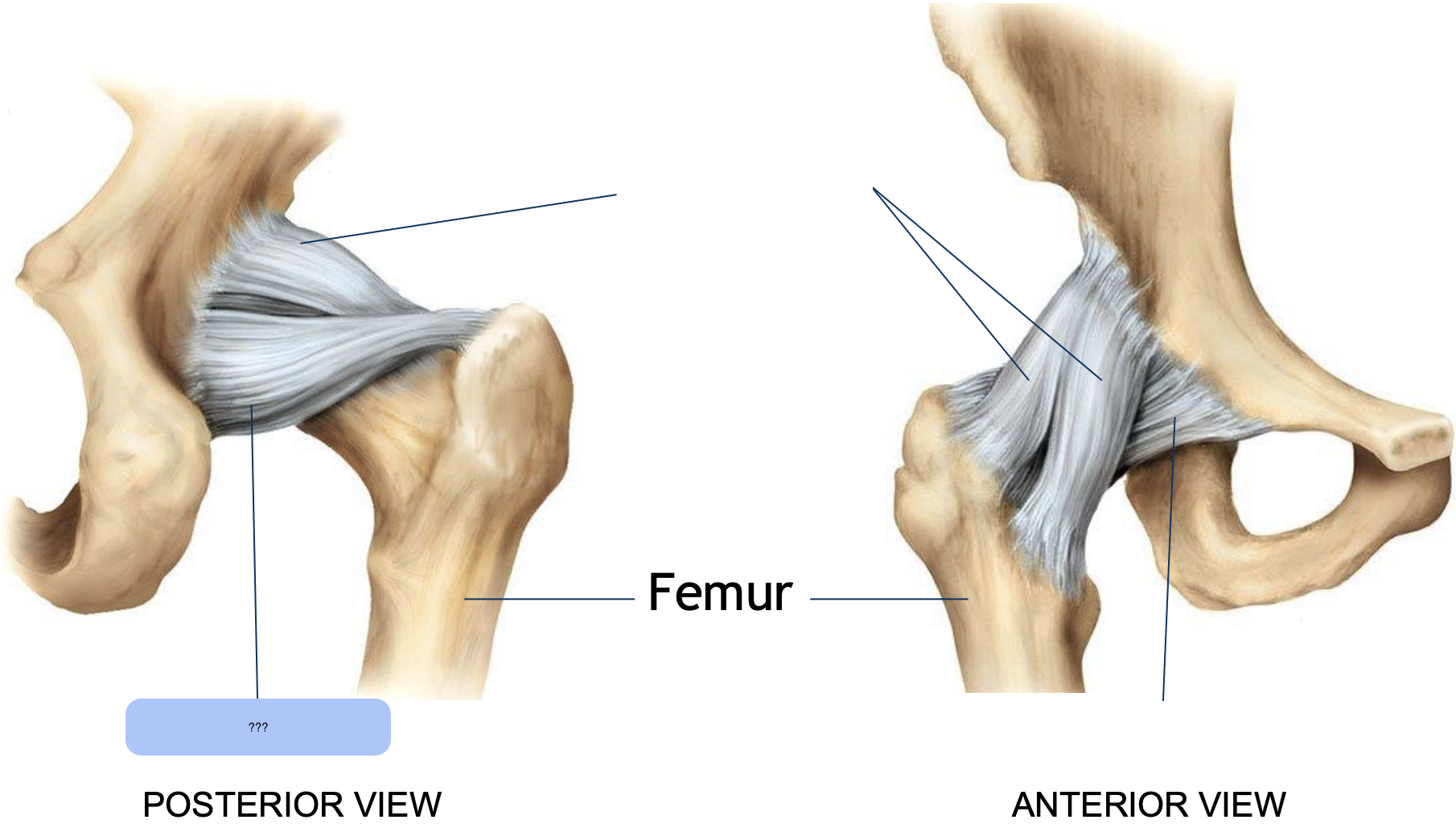
ischiofemoral ligament
resists hyperflexion
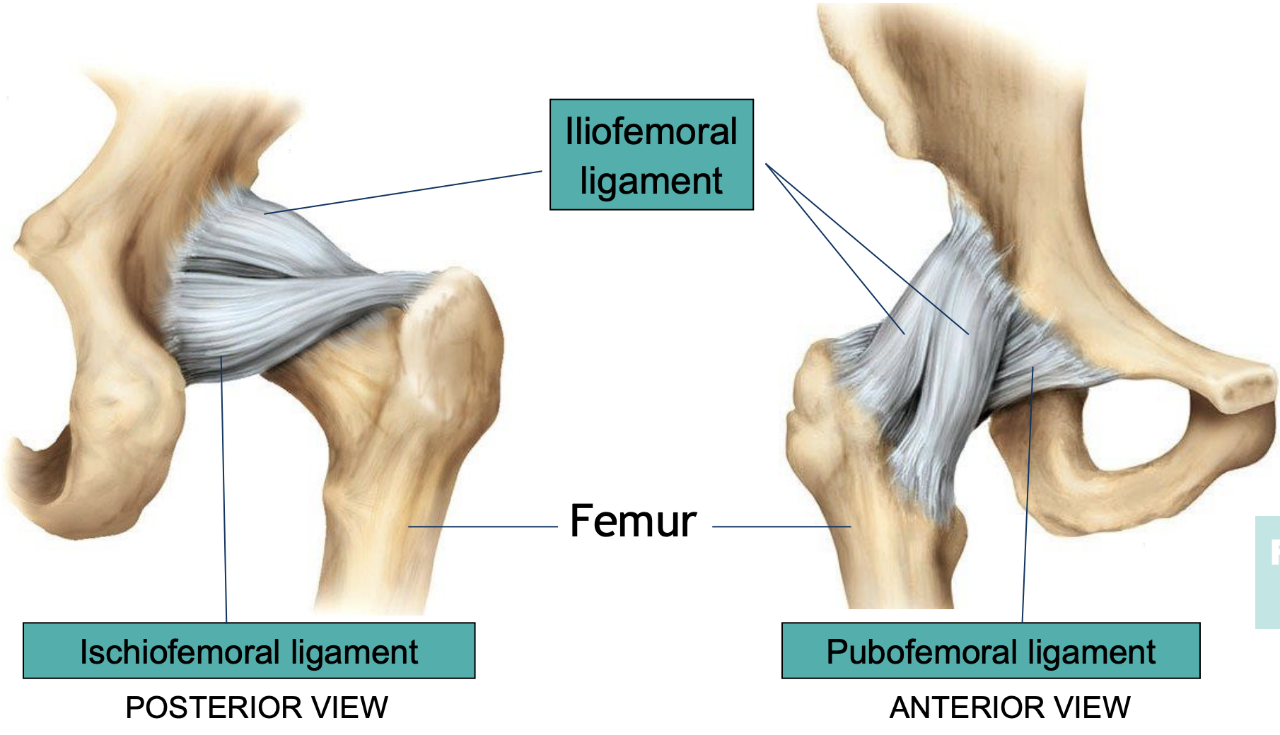
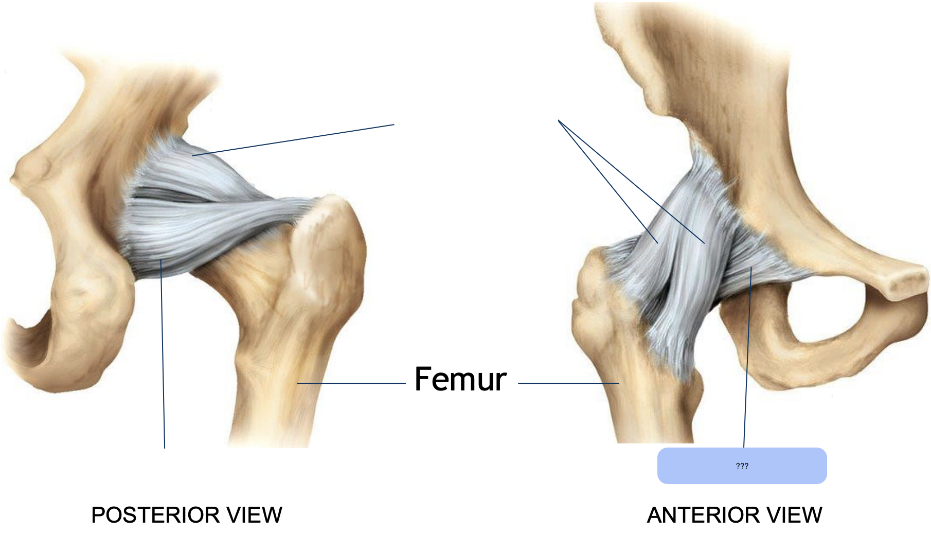
pubofemoral ligament
resists abduction

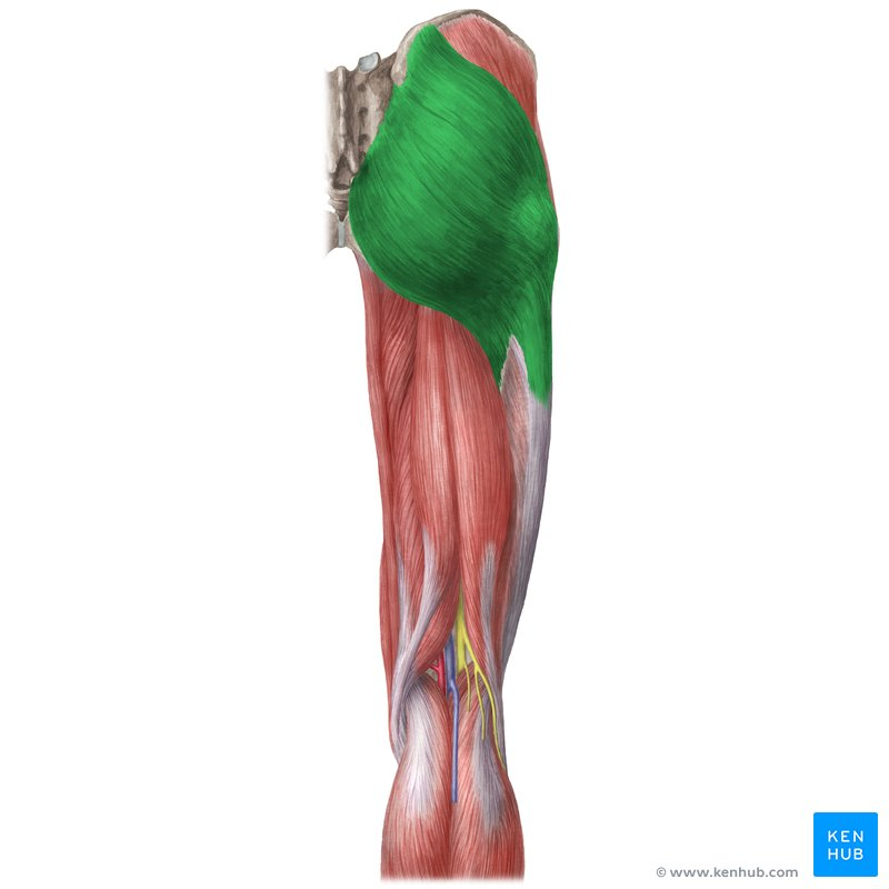
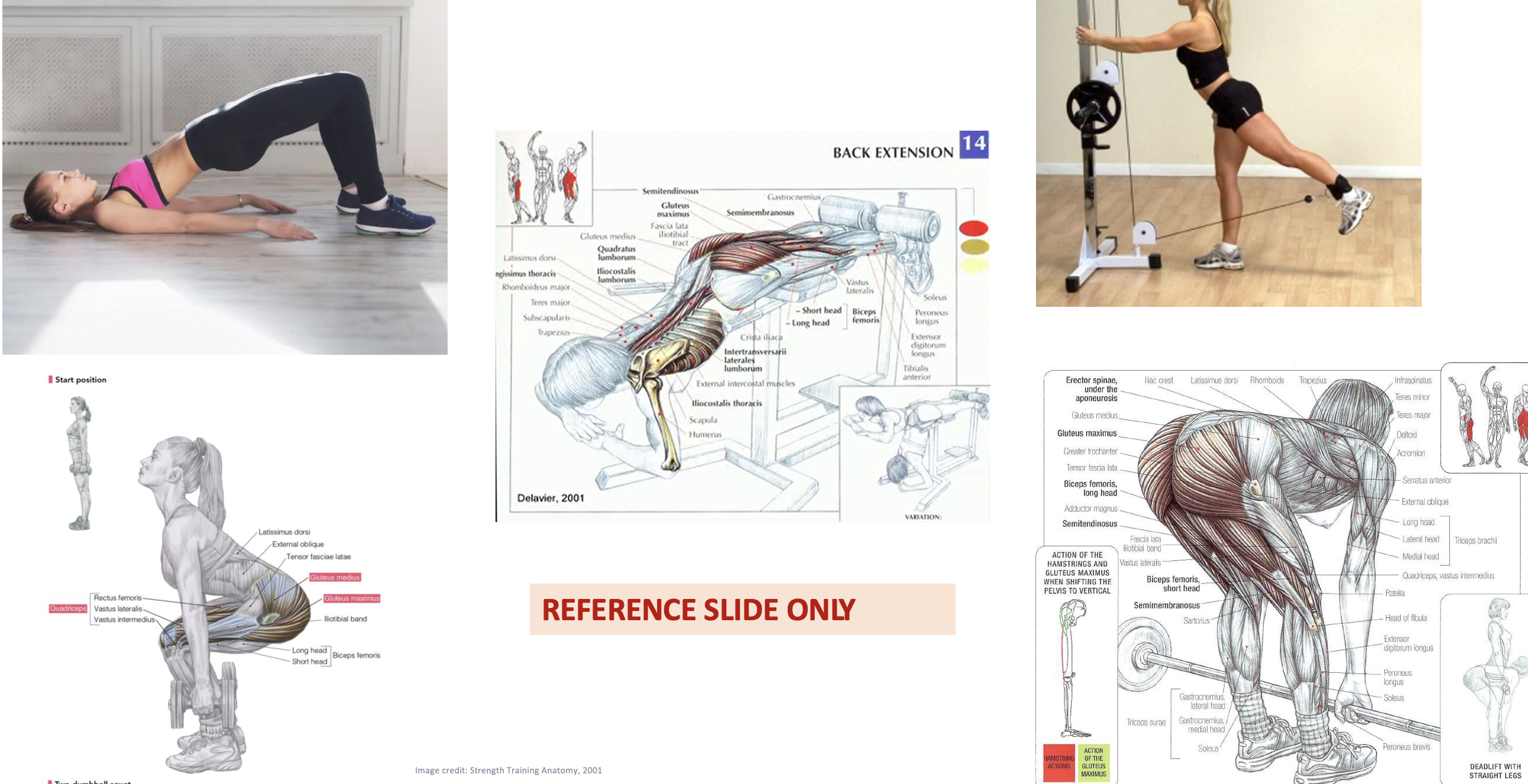
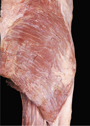
gluteus maximus
Origins:
Ilium, post glut line, sacrum & coccyx
Insertions:
ITB & gluteal tuberosity of femur
Actions:
Hip extension
Hip lateral/external rotation
Gluteus Medius
Origins:
Ilium (external surface)
Insertions:
Greater trochanter (lateral surface)
Actions:
Hip abduction
Hip medial/internal rotation
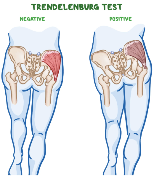
weakness of these muscles causes drooping of the pelvis to the contralateral side while walking → trendelenburg gait
Gluteus Minimus
Origins:
ilium (external surface)
Insertions:
Greater trochanter
Actions:
Hip abduction
Hip medial/internal rotation
Hip stabilisation
weakness of these muscles causes drooping of the pelvis to the contralateral side while walking → trendelenburg gait

hip extension
action of the gluteus maximus
increasing the angle between your pelvis and thigh
squatting
climbing stairs
walking, running, jumping
gluteus maximus
gluteus medius
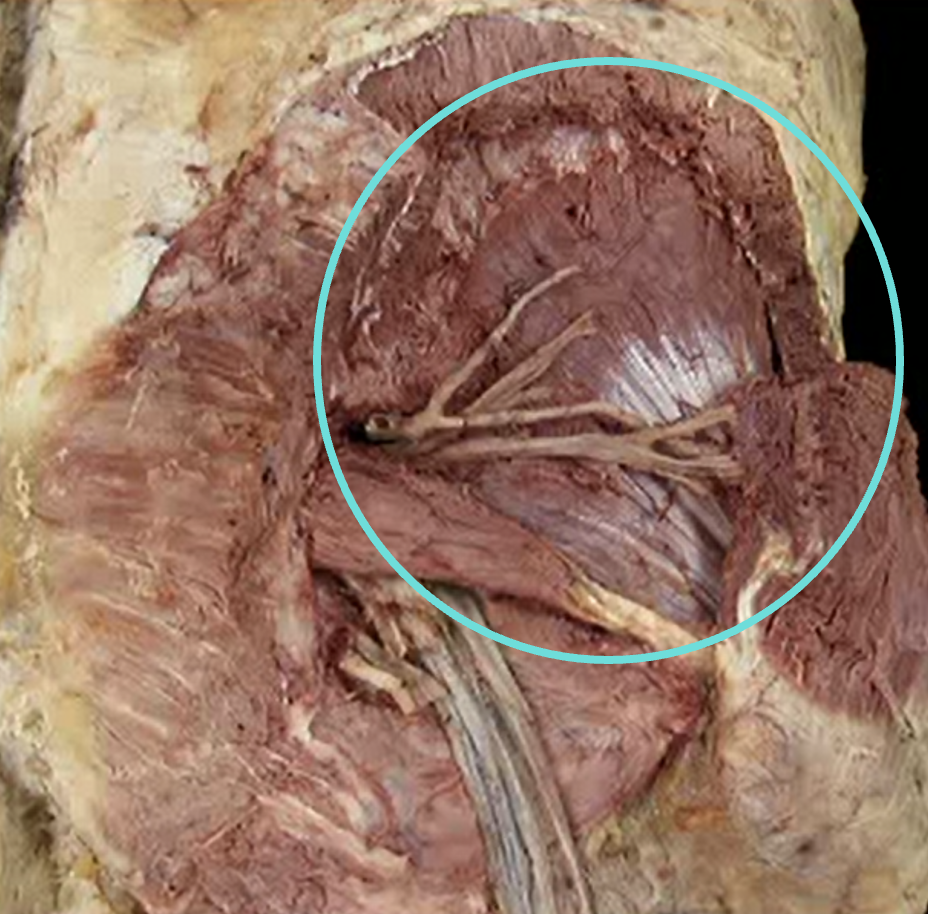
gluteus minimus (characteristic white, shiny bit)
trendelenburg test
clinical exam used to assess the strength of the hip abductor muscles, particularly the gluteus medius and minimus
patient stands on one leg
A negative (normal) test: The pelvis remains level or slightly elevates on the side of the raised leg
A positive Trendelenburg sign: The pelvis drops on the side of the lifted leg, indicating weakness of the hip abductors (gluteus medius and minimus) on the weight-bearing leg
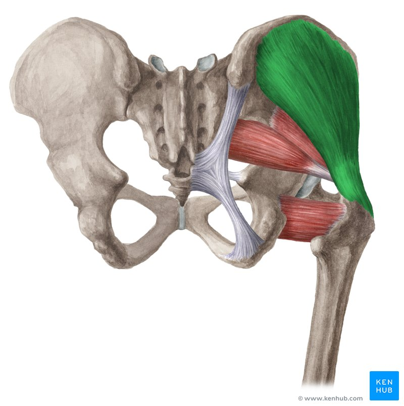
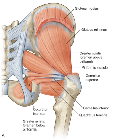
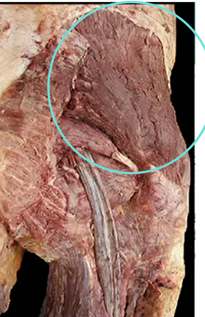
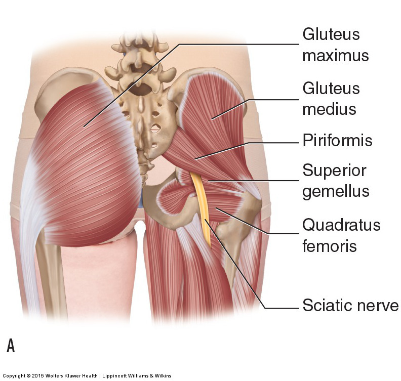
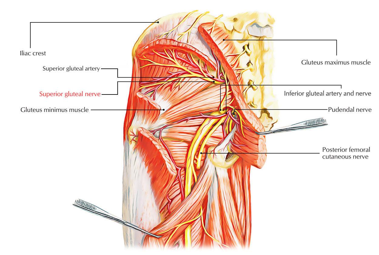
middle and superficial muscles of the hip posterior compartment
gluteal muscles:
gluteus maximus (extensor)
gluteus medius (abductor)
gluteus minimus (abductor)
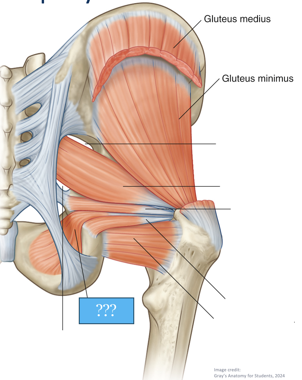
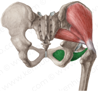
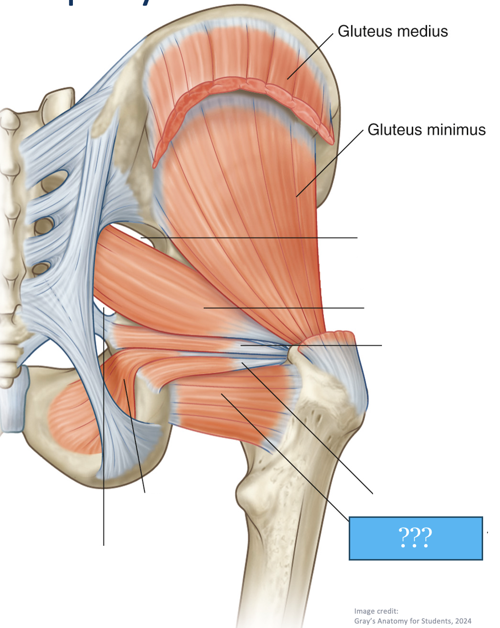
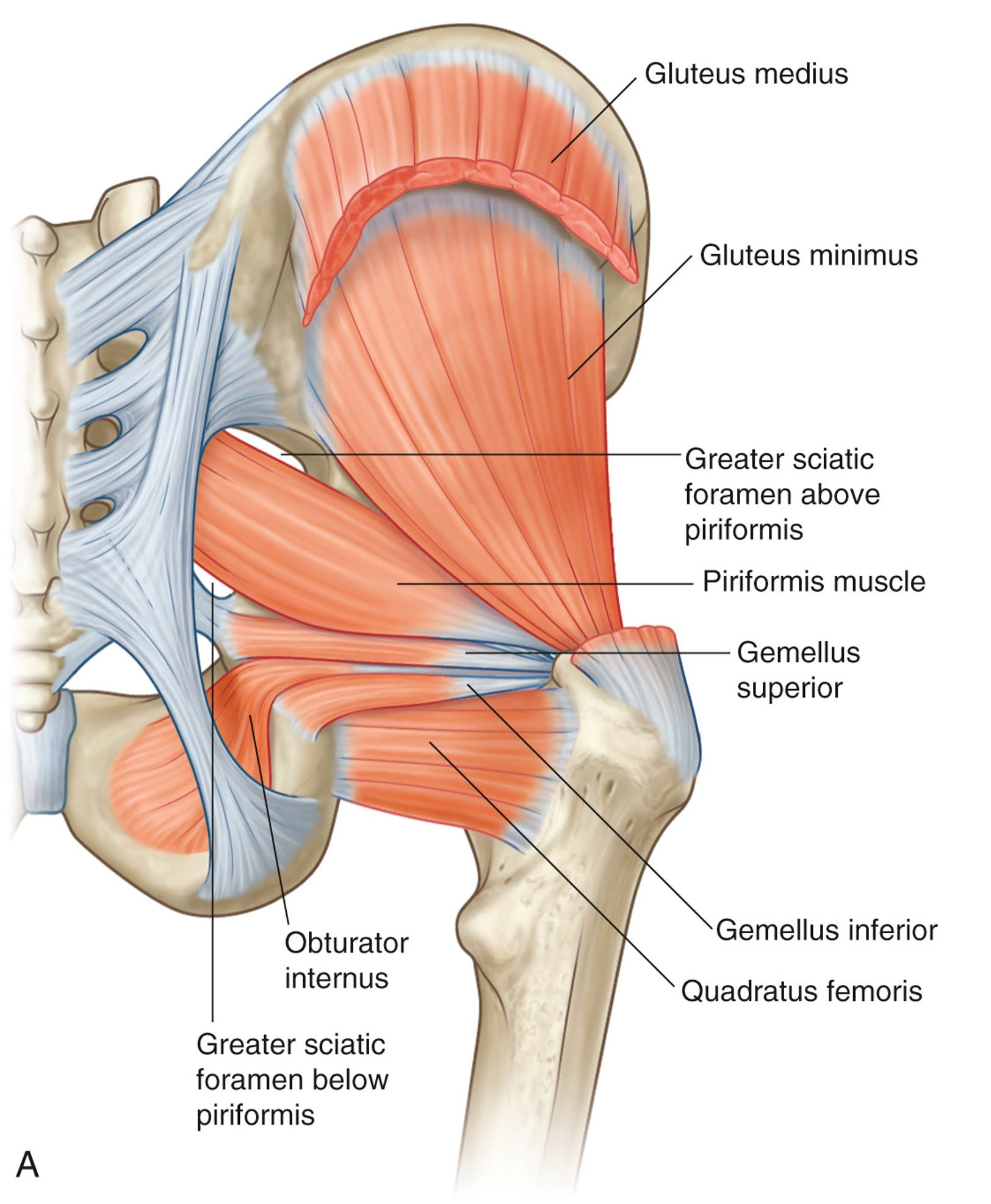
deep muscles of the hip posterior compartment
All lateral/external rotators
all originate from the sacrum or pelvic bone
all attach at greater trochanter
P GO GO Q (order)
Piriformis
Gemellus (“twin) superior
Obturator internus
Gemellus (“twin”) inferior
Obturator externus
Quadratus femoris
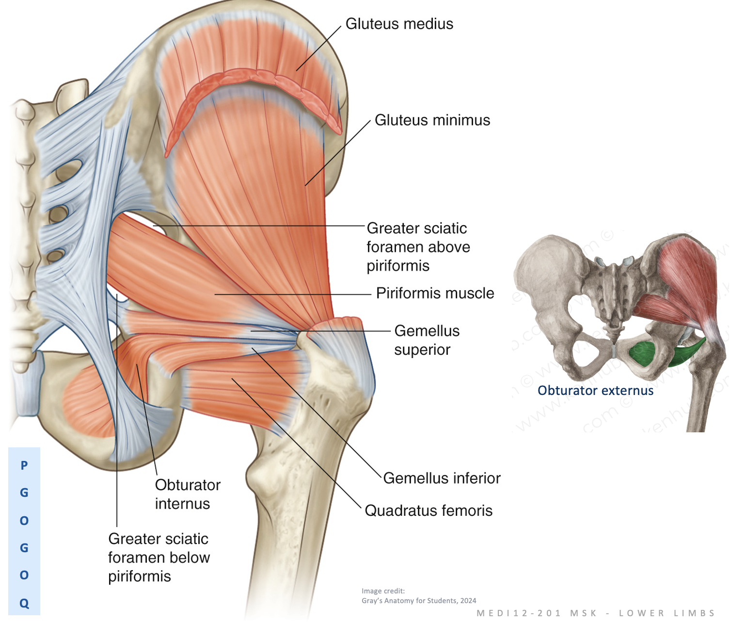
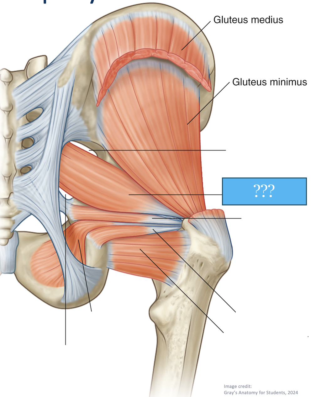
piriformis
deep muscle of the hip posterior compartment
lateral/external rotator
Origin:
sacrum
Attachment:
greater trochanter


Gemellus (“twin”) superior
deep muscle of the hip posterior compartment
lateral/external rotator
Origin:
ischium
Attachment:
greater trochanter

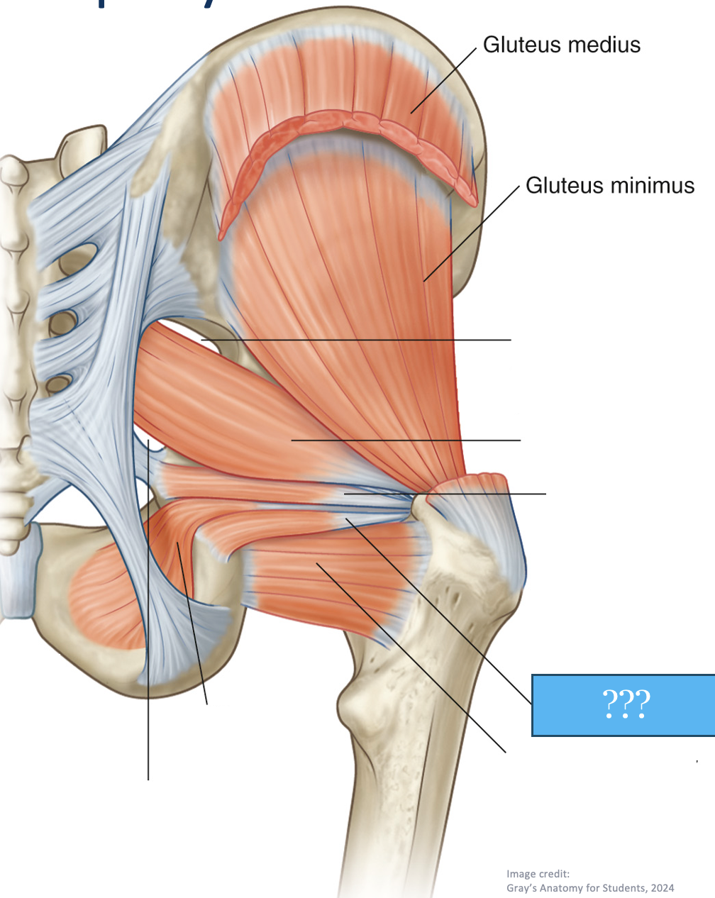
Gemellus (“twin”) inferior
deep muscle of the hip posterior compartment
lateral/external hip rotator
Origin:
ischium
Attachment:
greater trochanter

Obturator internus
deep muscle of the hip posterior compartment
lateral/external rotator
Origin:
inner obturator membrane
Attachment:
greater trochanter
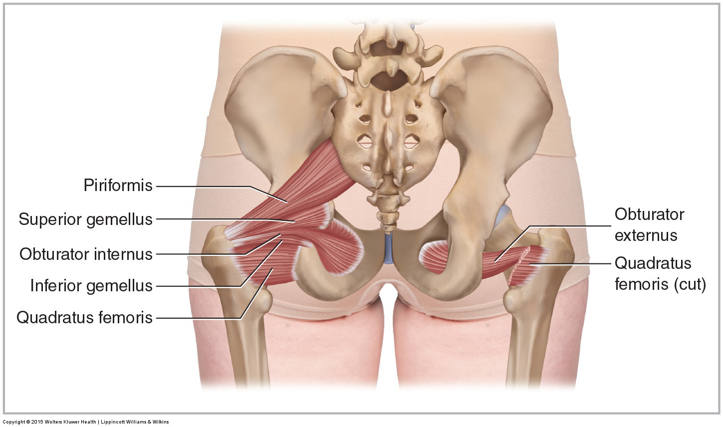
Obturator externus
deep muscle of the hip posterior compartment
lateral/external rotator
Origin:
outer obturator membrane
Attachment:
greater trochanter

Quadratus femoris
deep muscle of the hip posterior compartment
lateral/external rotator
Origin:
ischium
Attachment:
greater trochanter (ish)
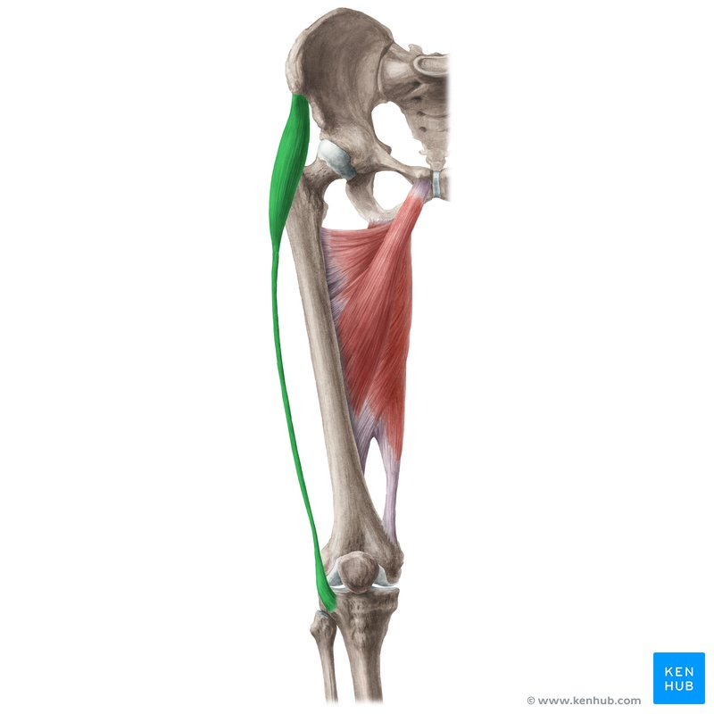
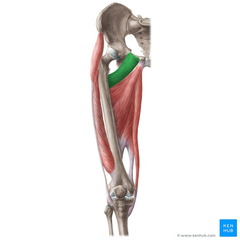
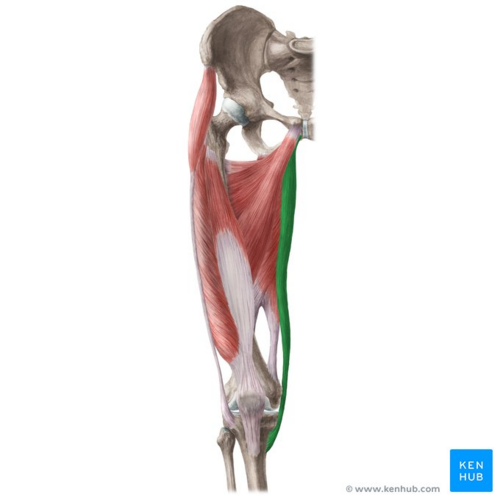
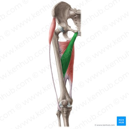
Tensor Fasciae Latae (TFL)
Actions:
Most important: synergist: acts as a dynamic ligament
contributes to knee extension
resists varus
Origin:
ASIS
Attachment:
iliotibial tract → lateral epicondyle
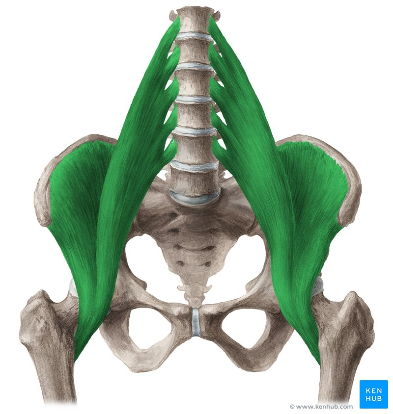
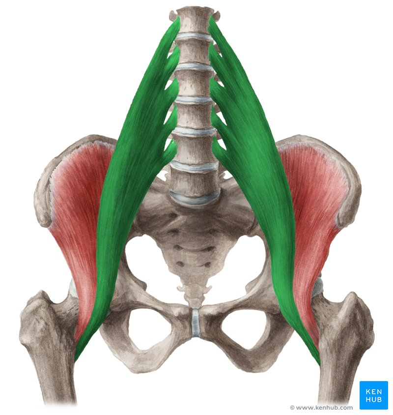
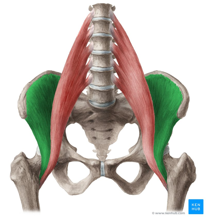
Iliopsoas
prime mover for hip flexion
combination of two muscles - therefore a biceps
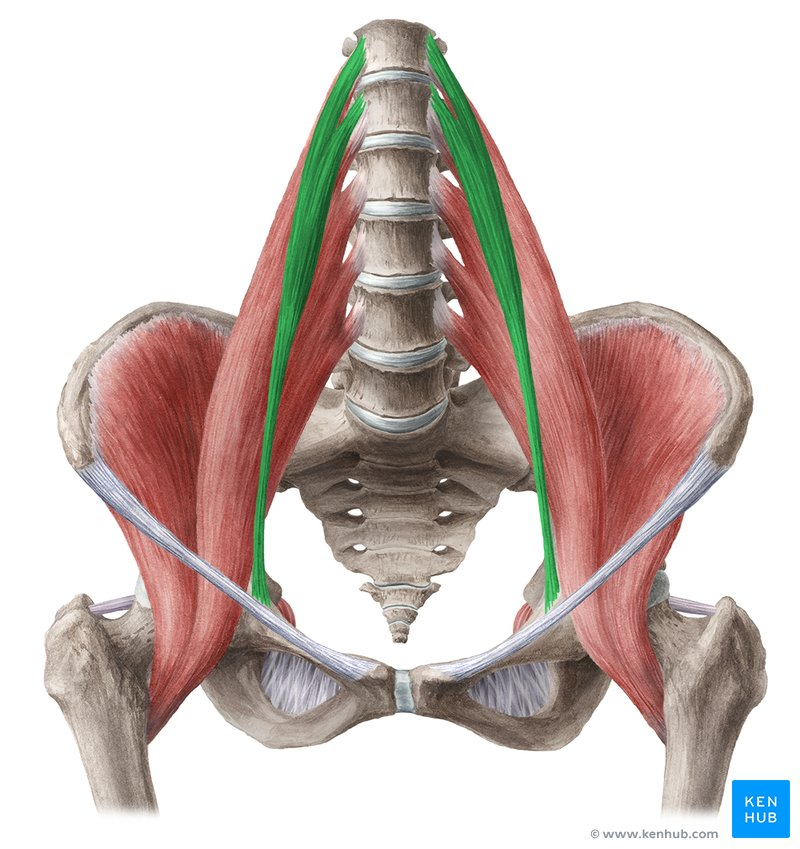
Psoas Major
Iliacus
inserts at lesser trochanter

Psoas major
Actions:
Hip flexion
Lumbar (spine) flexion
Origin:
T12-L5 vertebrae
Insertions:
Lesser trochanter
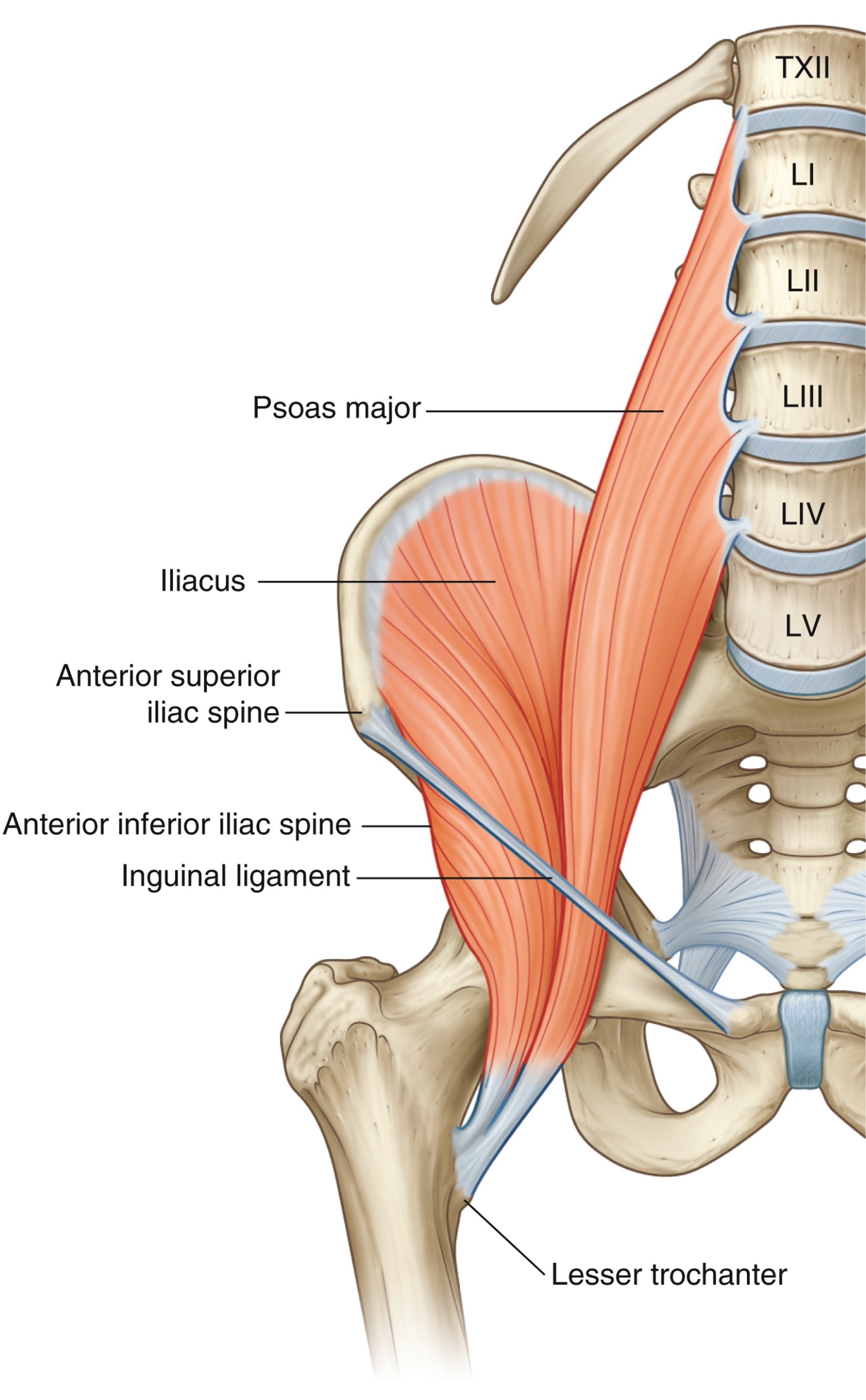
Iliacus
Action:
Hip flexion
Origin:
iliac fossa + AIIS
Insertion:
lesser trochanter

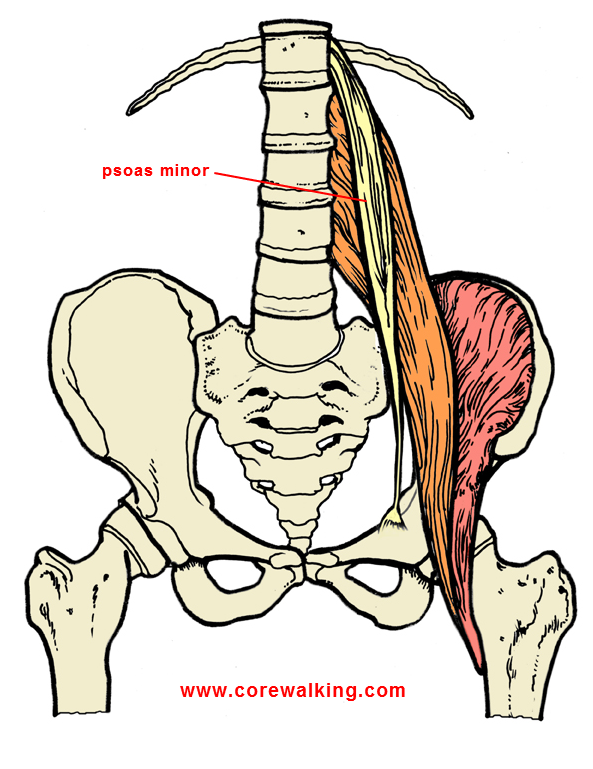
Psoas Minor
Actions:
Synergist: assists in hip & spine flexions (unnecessary muscle, half of people don’t have it)
Origin:
T12-L1 vertebrae
Insertion:
pectineal line (pubic bone/ramus)
anterior thigh compartment
hip flexors/knee extensors
nerve supply: femoral nerve
Muscles:
Quadriceps Femoris
Vastus Medialis
Vastus Intermedius
Vastus Lateralis
Rectus Femoris
Articularis Genu
Sartorius
Pectineus
femoral anterior (knee extensors), medial obturator (adductors), posterior sciatic (knee flexors)
think: females have vaginas, pecs and sass
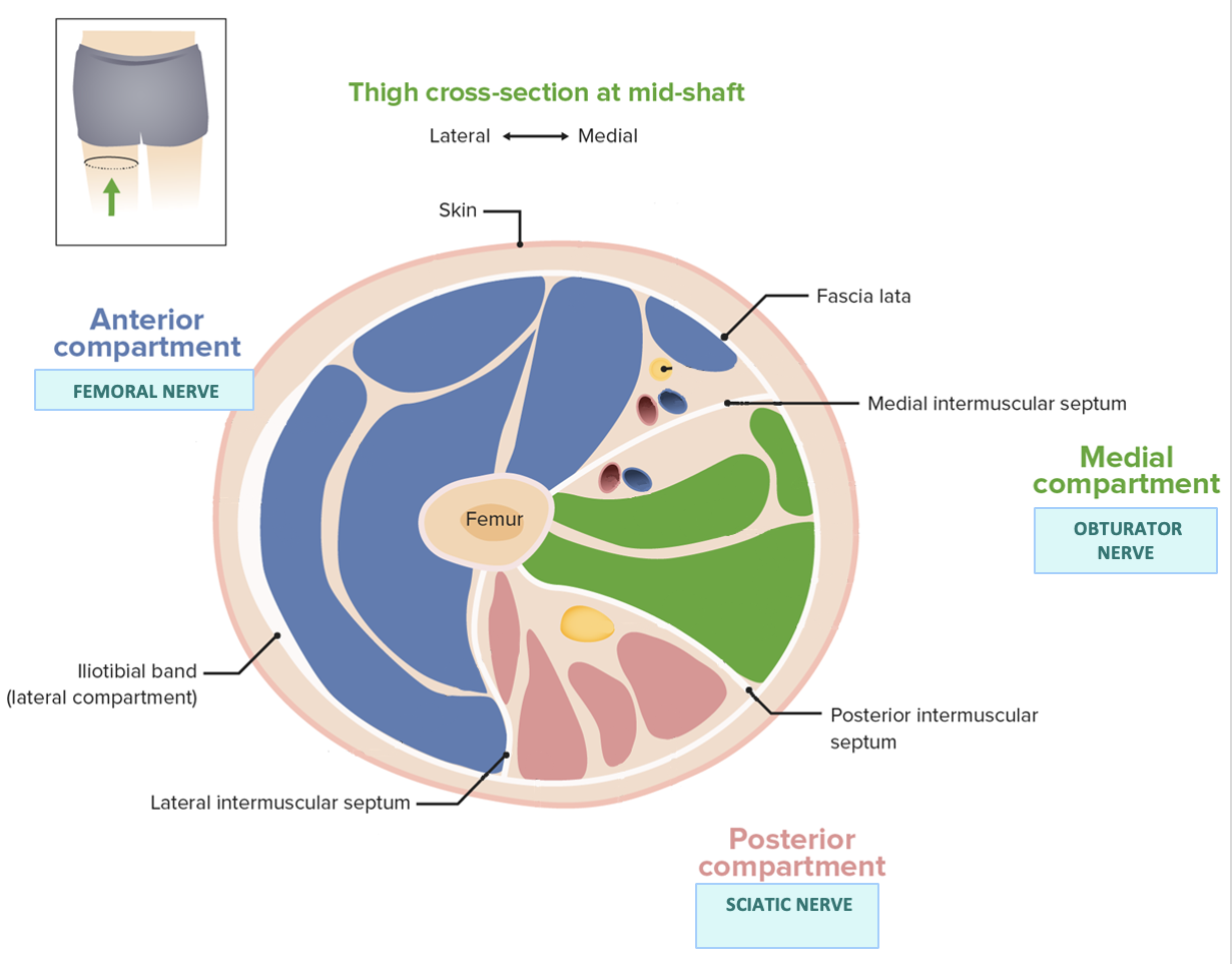
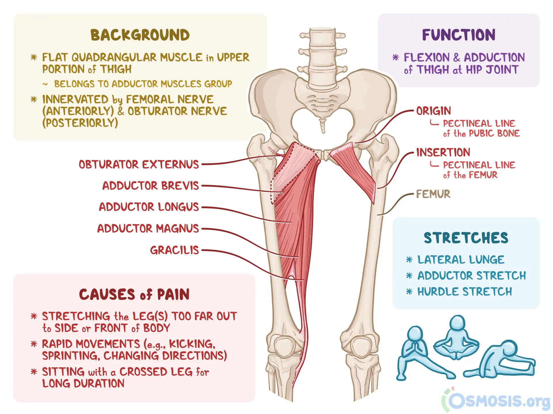
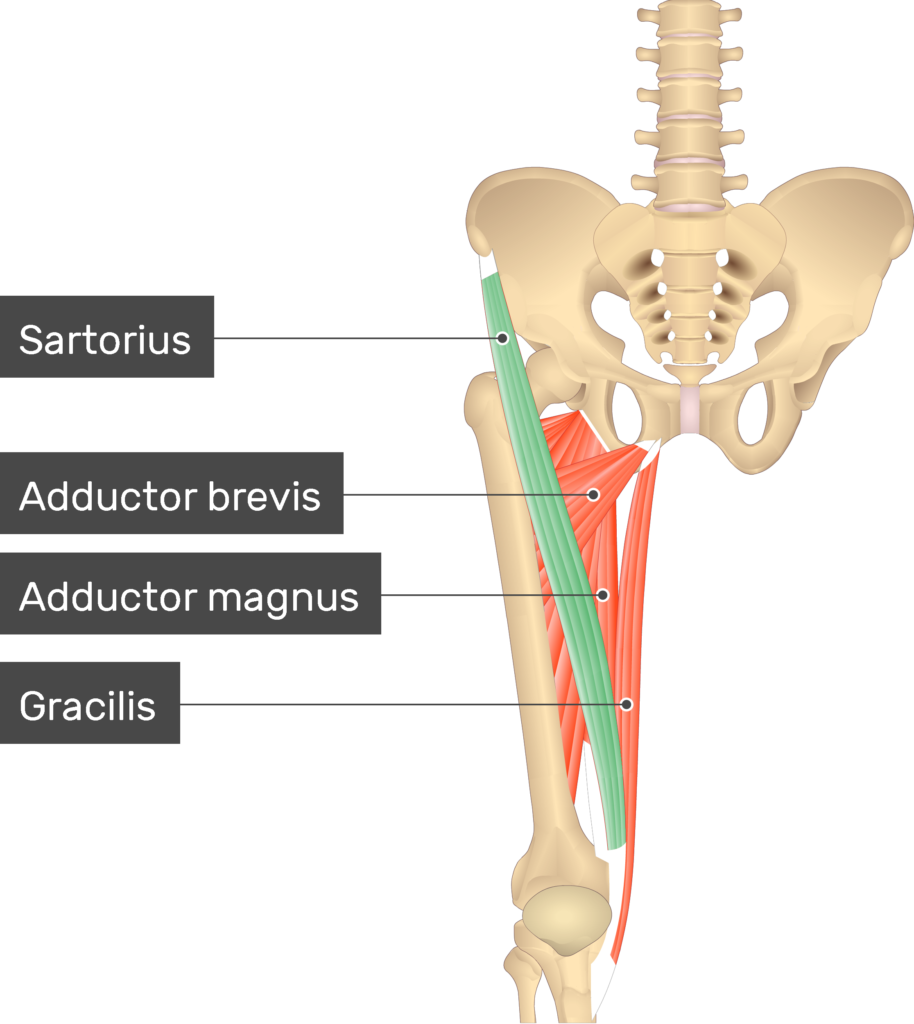
medial thigh compartment
hip adductors
nerve supply: obturator nerve
Muscles:
Adductor Longus
Adductor Brevis
Adductor Magnus
Gracilis
anterior femoral (knee flexors), medial obturator (adductors), posterior sciatic (knee extensors)
think: adductors are obtrusive and ungrateful, refuse to lift (help)

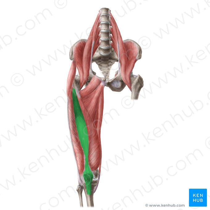
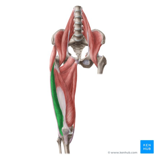
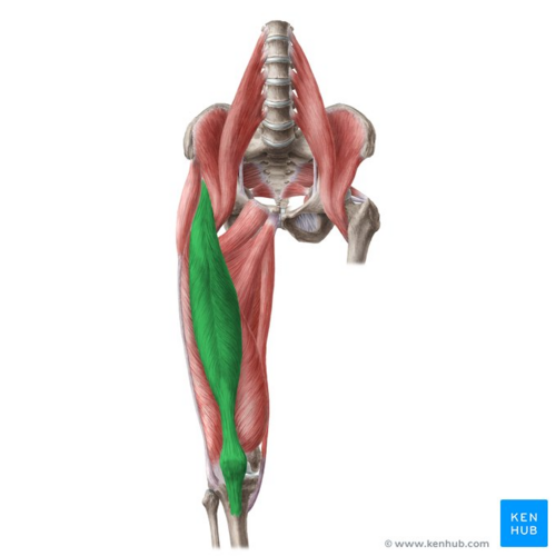
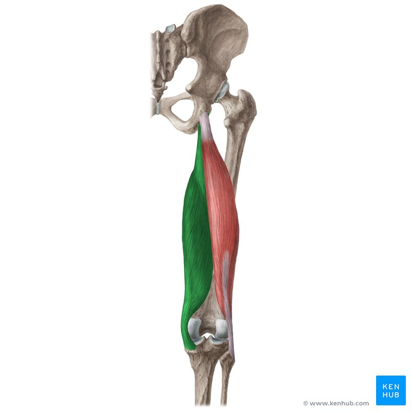
posterior thigh compartment
hamstrings
hip extensors/knee flexors
nerve supply: sciatic nerve
Muscles:
Biceps Femoris
Semitendinosus
Semimembranosus
think: femoral anterior (adductors), medial obturator (knee flexors), posterior sciatic (knee extensors)
ss = sciatic

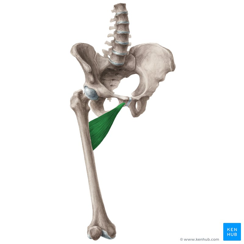
pectineus
Origins:
pectineal line (pubis bone/superior ramus)
Insertions:
Femur - pectineal line
Actions:
Hip flexion
Hip adduction
Sartorius
Origins:
ASIS
Insertions:
tibia (medial surface)
Action:
Knee flexion
Hip flexion (weak)
Hip abduction (weak)
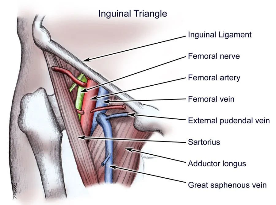
femoral triangle
triangle bordered by:
sartorius
adductor longus
inguinal ligament
contains important vessels:
femoral nerve
femoral artery
femoral vein
clinically relevant
quadriceps femoris muscles
All knee extensors
All originate from femur except Rectus Femoris
All attach to quadriceps femoris tendon → tibial tuberosity
Vastus Medialis
Vastus Intermedius
Vastus Lateralis
Rectus Femoris
also a hip flexor
origin: ilium
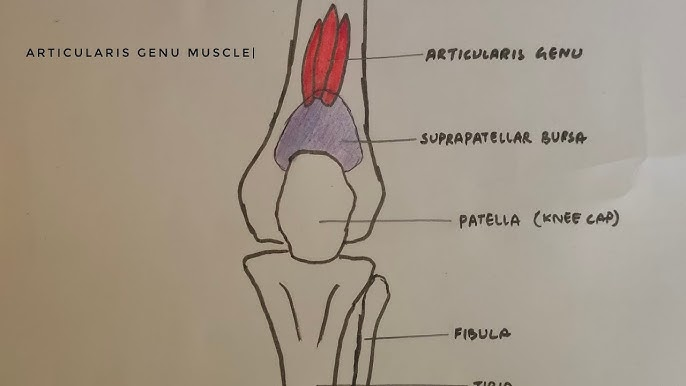
Articularis Genu (“fifth head”)
also attaches to patella

vastus medialis
Origins:
Femur
Insertions:
Quadriceps femoris tendon → tibial tuberosity
Action:
Knee extension

Vastus intermedius
Origins:
Femur
Insertions:
Quadriceps femoris tendon → tibial tuberosity
Action:
Knee extension

Vastus Lateralis
Origins:
Femur
Insertions:
Quadriceps femoris tendon → tibial tuberosity
Action:
Knee extension
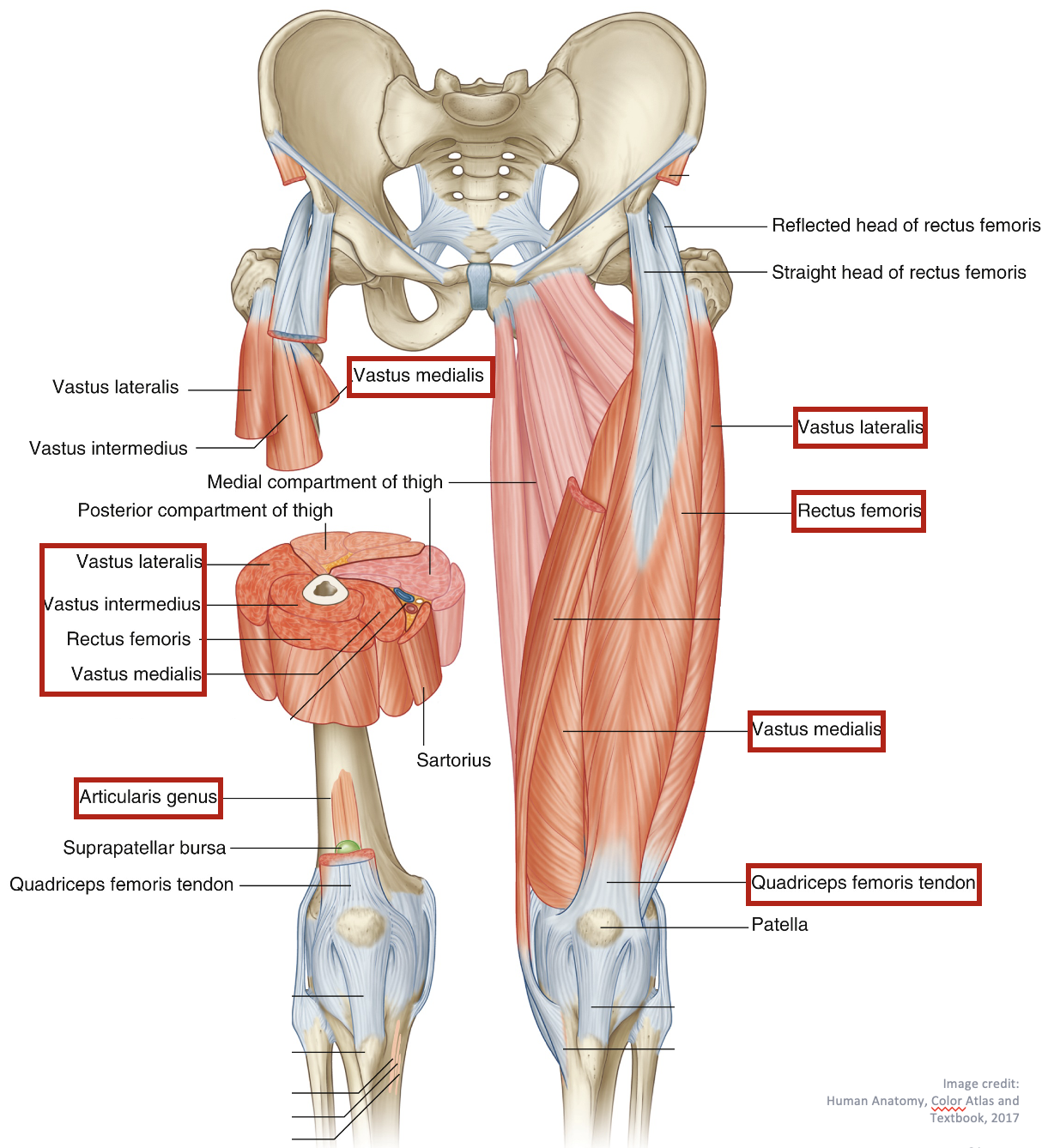
Rectus femoris
Origins:
Anterior Inferior Iliac Spine (AIIS)
Insertions:
Quadriceps femoris tendon → tibial tuberosity
Action:
Knee extension
Hip flexion

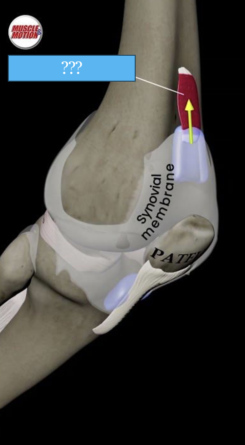
Articularis genu (“knee”)
Origins:
Femur
Insertions:
Quadriceps femoris tendon + patella
Action:
reduces friction between femur and bursa (fluid-filled sac, cushions and reduces friction between bones and soft tissue)
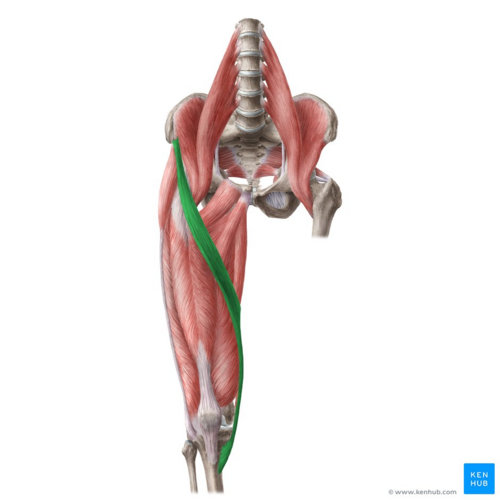
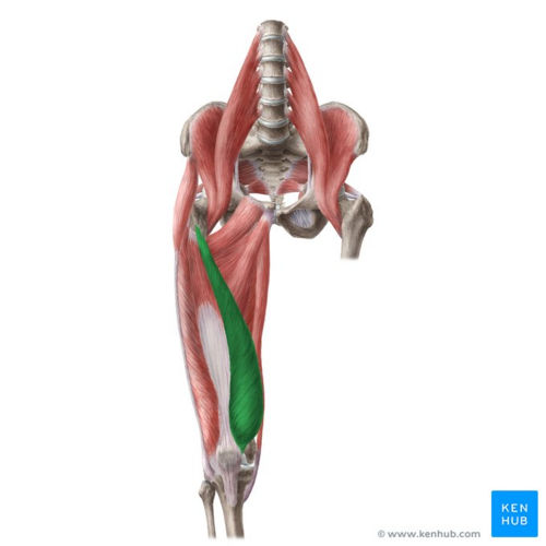
Gracilis
Origins:
Pubis
Insertions:
tibia (medial surface)
Actions (not a very strong muscle):
Hip adduction
Knee flexion (weak)

Adductor longus
Origins:
Pubis
Insertions:
Femur (linea aspera)
Actions:
Hip adduction
Hip medial rotation

Adductor Brevis
Origins:
Pubis
Insertions:
Femur (linea aspera)
Actions:
Hip adduction
Hip medial/internal rotation
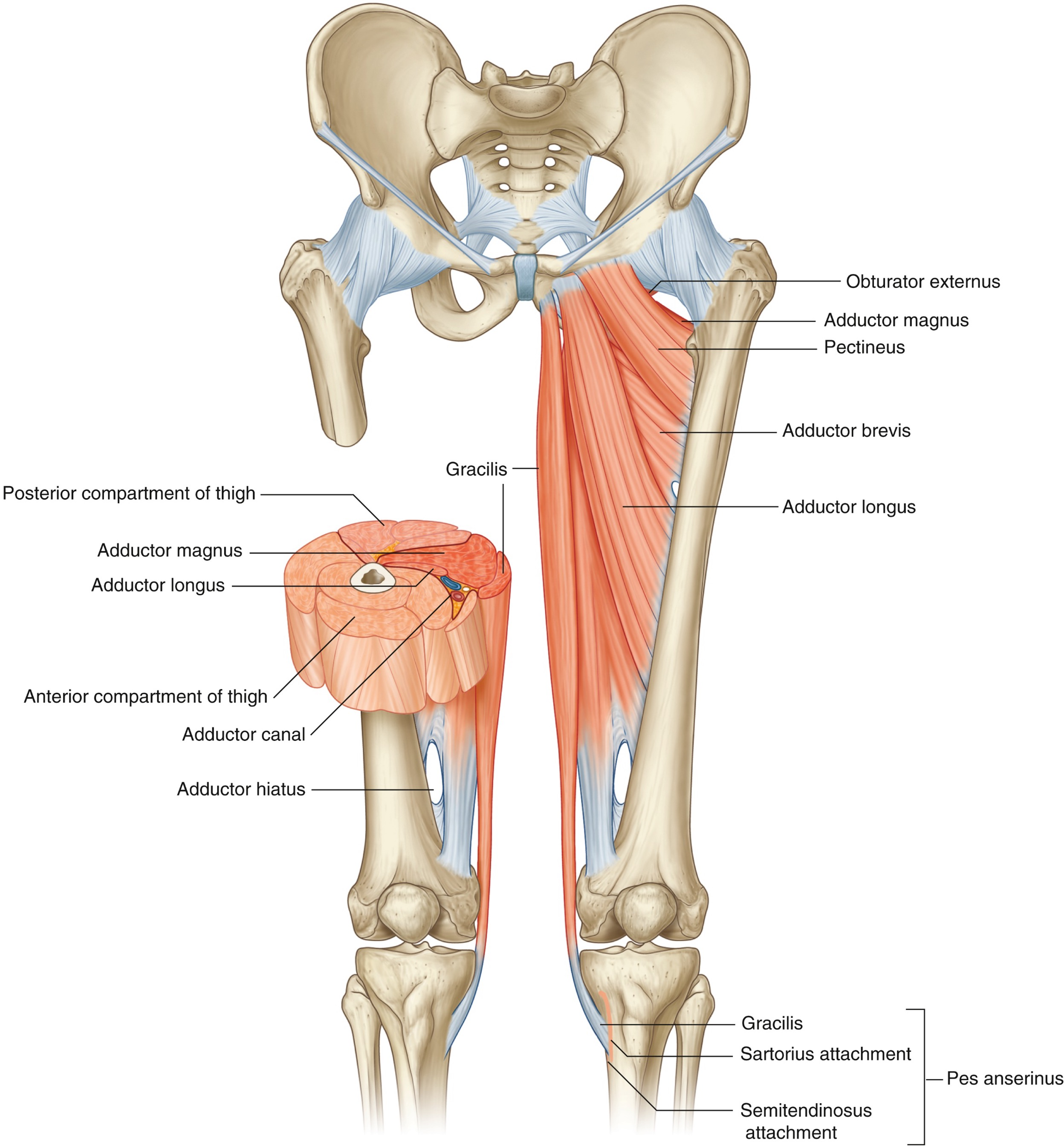
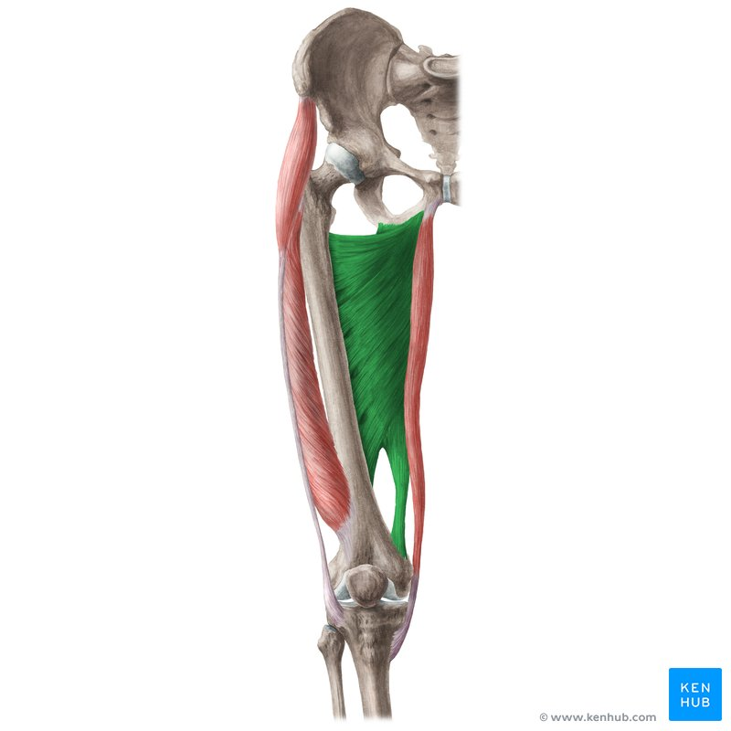
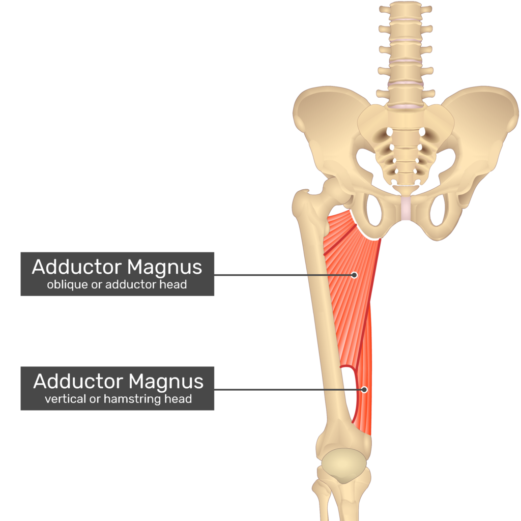
Adductor magnus
Two parts: hamstring and adductor
Forms a hiatus/canal/gap allowing femoral nerve and artery to pass through
Origins:
ischiopubic ramus
ischial tuberosity
Insertions:
Femur (linear aspera)
Femur (adductor tubercle)
Actions:
Hip adduction
Hip medial rotation
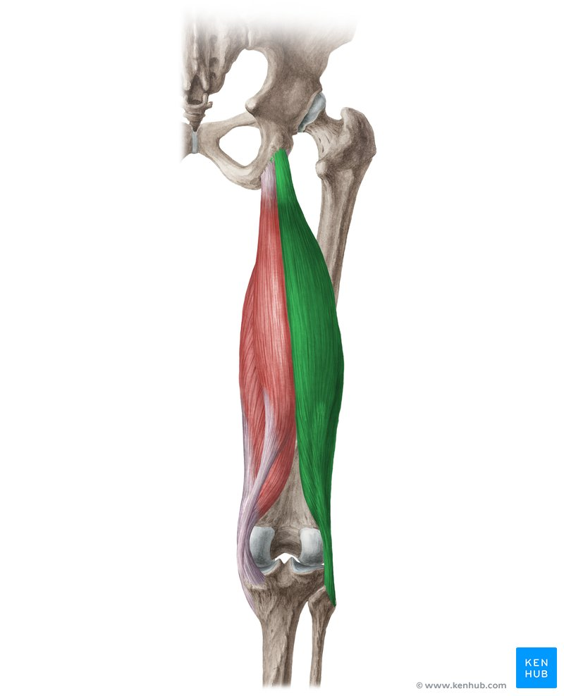
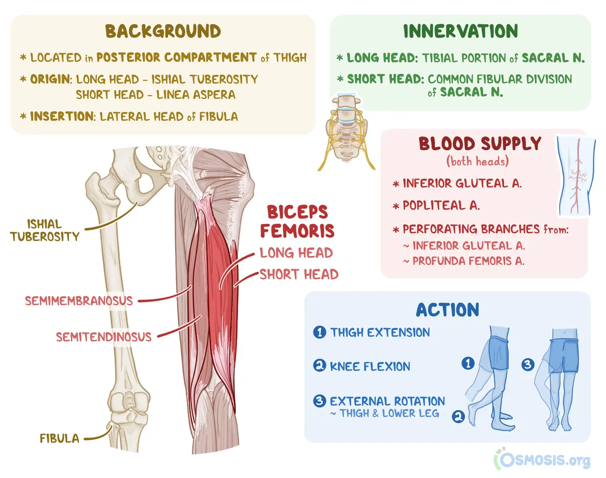
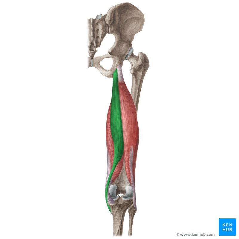
Biceps femoris
Origins:
Long head: ischial tuberosity
Short head: femur (linea aspera)
Insertions:
Head of fibula
Actions:
Hip extension
Knee flexion
Semitendinosus (thinner and more superifical)
Origins:
Ischium
Insertions:
medial tibia (surface)
Actions:
Hip extension
Knee flexion
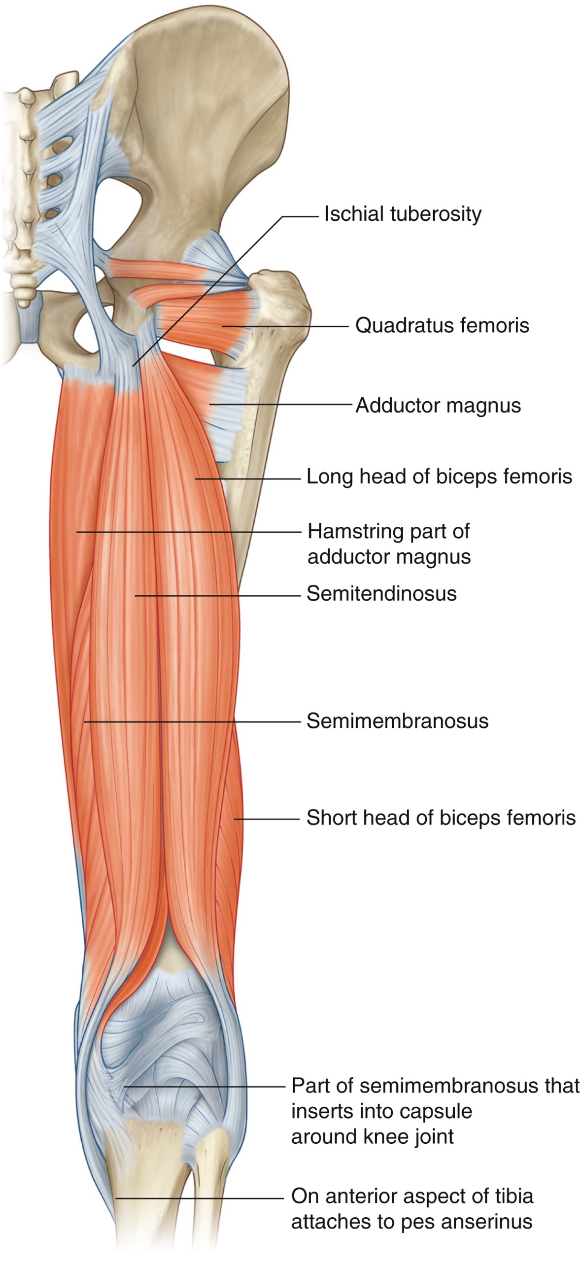
Semimembranosus (deeper and wider than semitendinosus)
Origins:
Ischium
Insertions:
medial tibia (condyle)
Actions:
Hip extension
Knee flexion

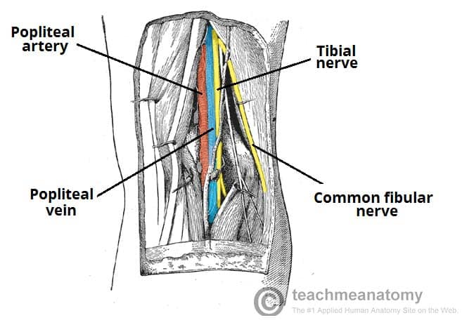
Popliteal Fossa
shallow depression in the back of the knee that contains many nerves, blood vessels, and lymph nodes
Clinical Significance:
An injury at the popliteal fossa affects the innervation of lower leg and foot.
Presentations:
Weakness in plantar flexion, inversion, and toe flexion of the foot, due to decreased innervation to the muscles in the deep compartments of the lower leg.
posterior compartment of leg: superficial group
gastrocnemius
soleus
plantaris
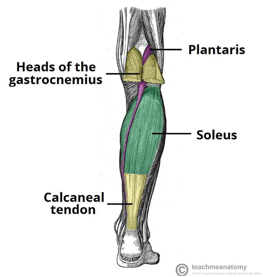
gastrocnemius
Origins:
Insertions:
Actions:
soleus
Origins:
Insertions:
Actions:
plantaris
Origins:
Insertions:
Actions:
posterior compartment of the leg: deep group
popliteus
tibialis posterior
flexor digitorum longus
flexor hallucis longus
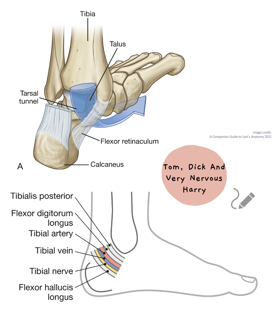
(Tiny Dick Harry, Harry is lazy and doesn’t move) (he’s posing with his leg like o->-/ bc he’s a popstar)
nerve supply: tibial nerve (bc it’s tiny and he’s not afraid to admit it)

popliteus
Origins:
femur (lateral condyle)
Insertions:
tibia (posterior surface)
Actions:
unlocks the knee by laterally rotating femur 5º on fixed tibia
Stabilises knee by resisting lat rotation of tibia on femur
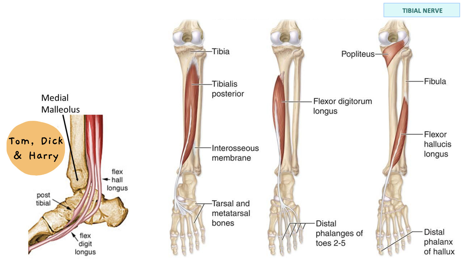
tibialis posterior
Origins:
tibia (posterior surface)
fibula (posterior surface)
Insertions:
Tuberosity of:
navicular
cuneiform
metatarsals
Actions:
ankle plantar flexion
foot inversion

flexor digitorum longus
Origins:
Insertions:
Actions:

flexor hallucis longus
Origins:
Insertions:
Actions:

anterior compartment of leg
tibialis anterior
extensor hallucis longus
extensor digitorum longus
fibularis tertius
nerve supply: deep fibular nerve
(tiny dick harry fibs bc he’s anti-small dicks)
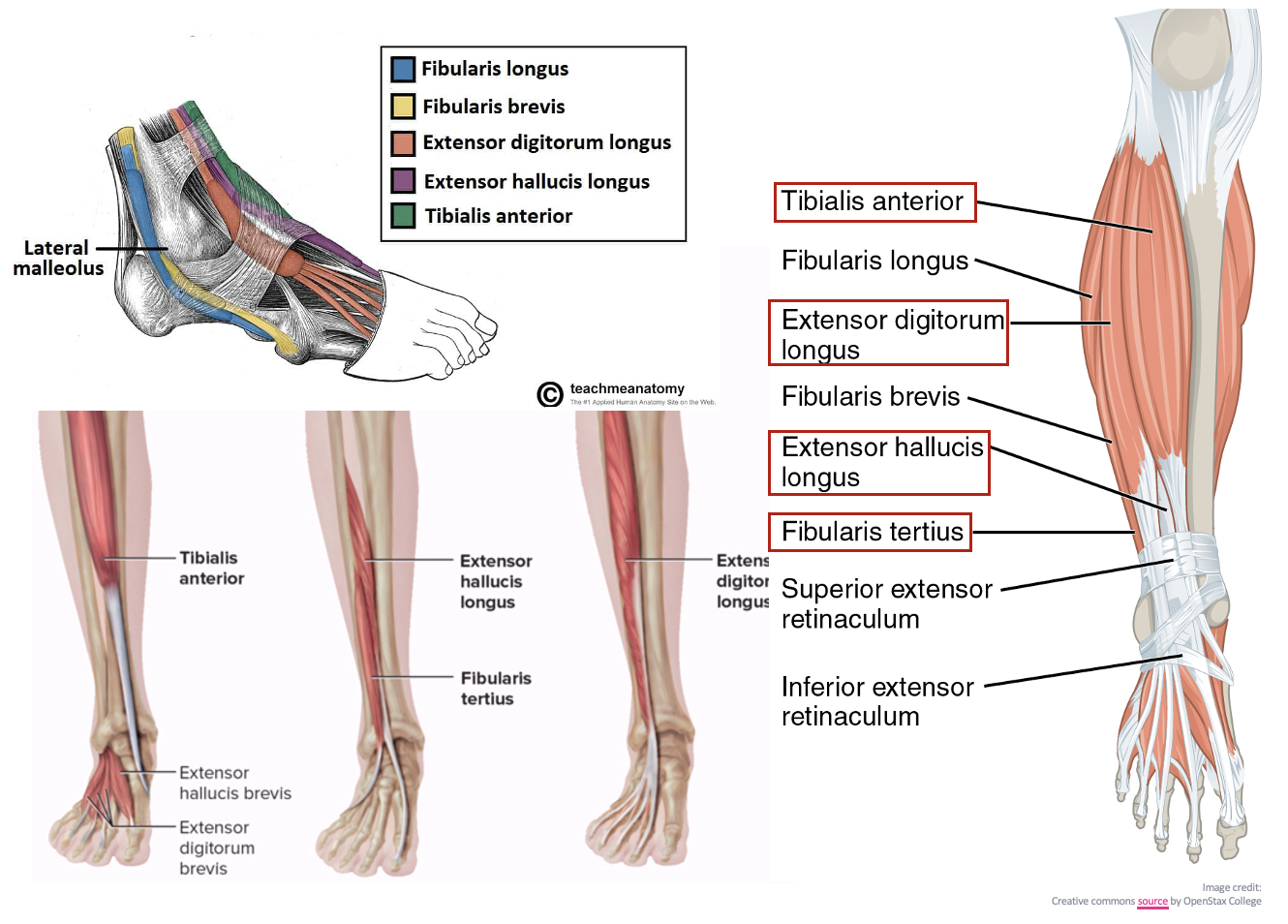
tibialis anterior
Origins:
Insertions:
Actions:

extensor hallucis longus
Origins:
Insertions:
Actions:

extensor digitorum longus
Origins:
Insertions:
Actions:

fibularis tertius
Origins:
Insertions:
Actions:

lateral compartment of leg
fibularis longus
Fibularis Brevis
nerve supply: superficial fibular nerve
actions:
foot eversion
ankle plantarflexion

fibularis longus
Origins:
Insertions:
Actions:
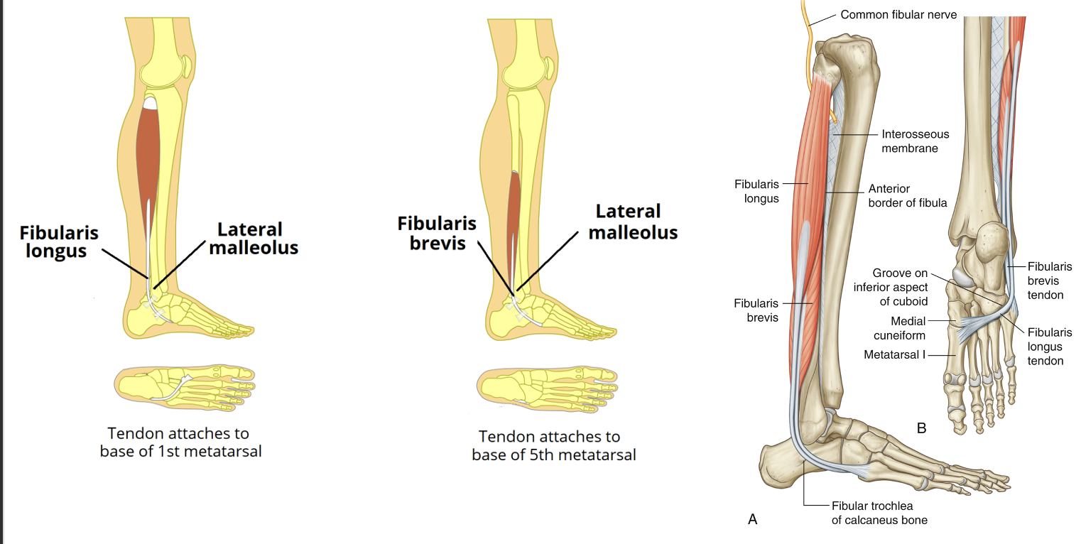
Fibularis Brevis
Origins:
Insertions:
Actions:

fibularis vs tibularis actions
fibularis = EVERT
tibialis = INVERT
tarsal tunnel
houses essential structures:
Tibialis posterior, flexor digitorum longus (FDL), and flexor hallucis longus (FHL) tendons.
Posterior tibial artery and vein.
Posterior tibial nerve
Clinical Significance
Tarsal Tunnel Syndrome: an entrapment neuropathy that is associated with the compression of the structures within the tarsal tunnel