BIO1411L Chapter 4: The Tissue Level of Organization
1/34
There's no tags or description
Looks like no tags are added yet.
Name | Mastery | Learn | Test | Matching | Spaced | Call with Kai |
|---|
No analytics yet
Send a link to your students to track their progress
35 Terms
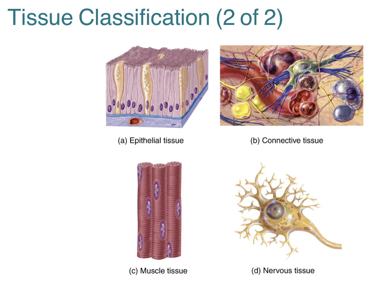
What is a tissue?
A tissue is a group of similar cells that have the same specialized function and embryonic origin.
What are the four types of tissues?
1.) Epithelial tissue: covers the body surfaces, lines cavities, and forms glands
2.) Connective tissue: binds organs together, stores energy, and participates in body defenses
3.) Muscle tissue: produces the physical force for body movements
4.) Nervous tissue: detects and responds to change in internal and external body conditions
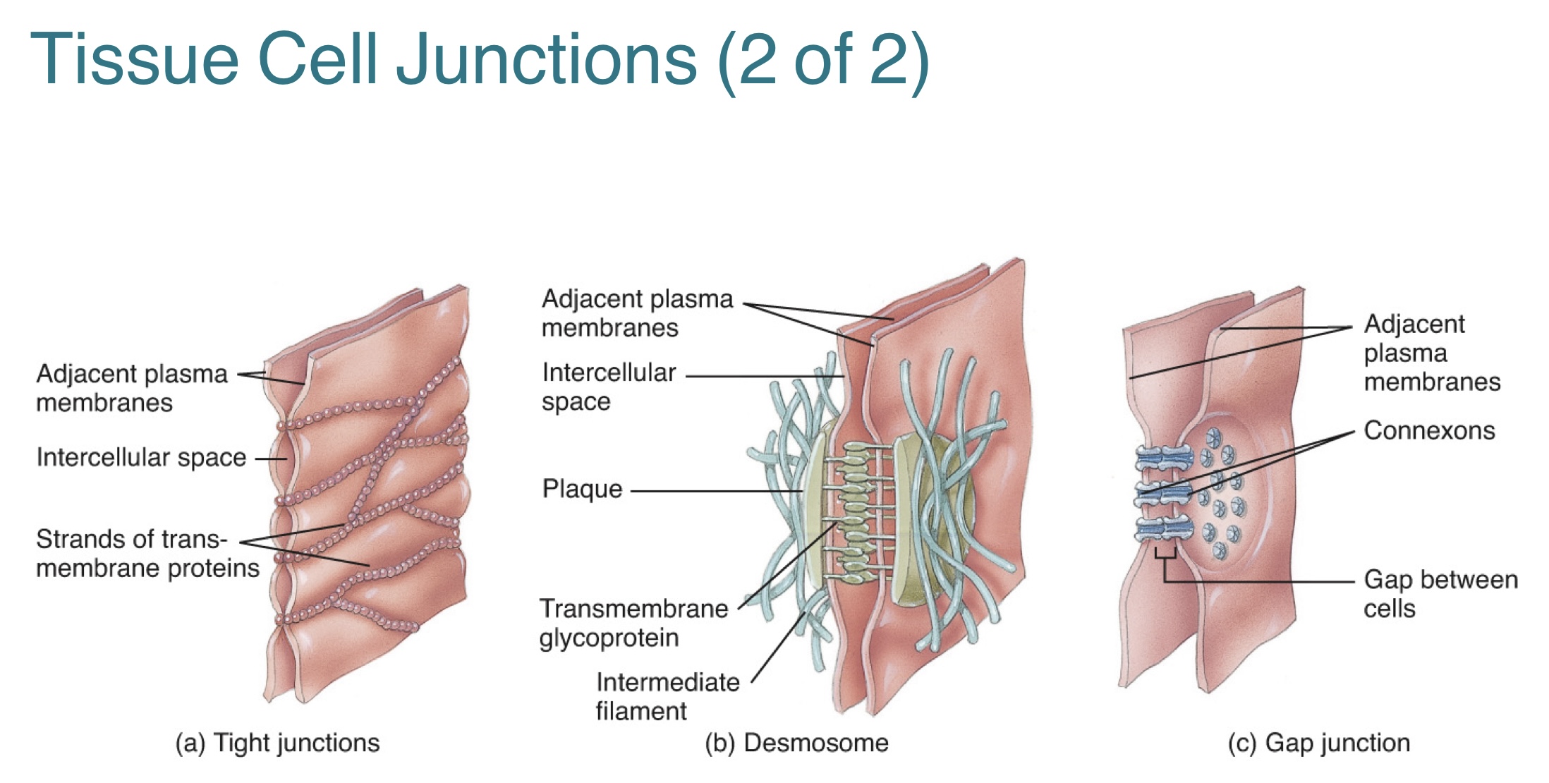
What are tissue cell junctions? What are the three types?
Tissue cell junctions are points of contact between adjacent plasma membranes of cells within a tissue?
[CHATGPT]
-Their function is to help cells stick together, which is vital for tissue stability. They ensure that cells within a tissue form a cohesive unit.
The three types of tissue cell junctions include:
1.) Tight junctions
2.) Desmosomes
3.) Gap junctions
![<p><strong>Tissue cell junctions</strong> are points of contact between adjacent plasma membranes of cells within a tissue?</p><p>[CHATGPT]<br>-Their function is to help cells stick together, which is vital for tissue stability. They ensure that cells within a tissue form a cohesive unit. <br><br>The three types of tissue cell junctions include:<br>1.) Tight junctions<br>2.) Desmosomes <br>3.) Gap junctions </p>](https://knowt-user-attachments.s3.amazonaws.com/79816c1d-a4e3-4c02-b3a6-635a9a2782da.jpg)
Provide a summary of tight junctions, desmosomes, and gap junctions. (function and where they can be found)
Tight junctions are junctions composed of proteins like occludins and claudins, forming a tight seal between adjacent cells.
-prevent substances from leaking between the cells
-can be found in the stomach, urinary bladder, intestines
Desmosomes are junctions made of cadherin proteins, linking the cytoskeletons of adjacent cells via intermediate filaments.
-anchor cells together (so that they cannot be pulled apart)
-important in tissues that undergo significant stretching
-can be found in the epidermis (eg. elbow) and cardiac muscle
Gap junctions are junctions formed by connexin proteins, that create channels between adjacent cells.
-allow the direct exchange of small molecules and electrical signals between cells
-important for facilitating coordinated activities
-can be found in cardiac muscle (to synchronize heartbeats) and in neurons (for electrical signaling)
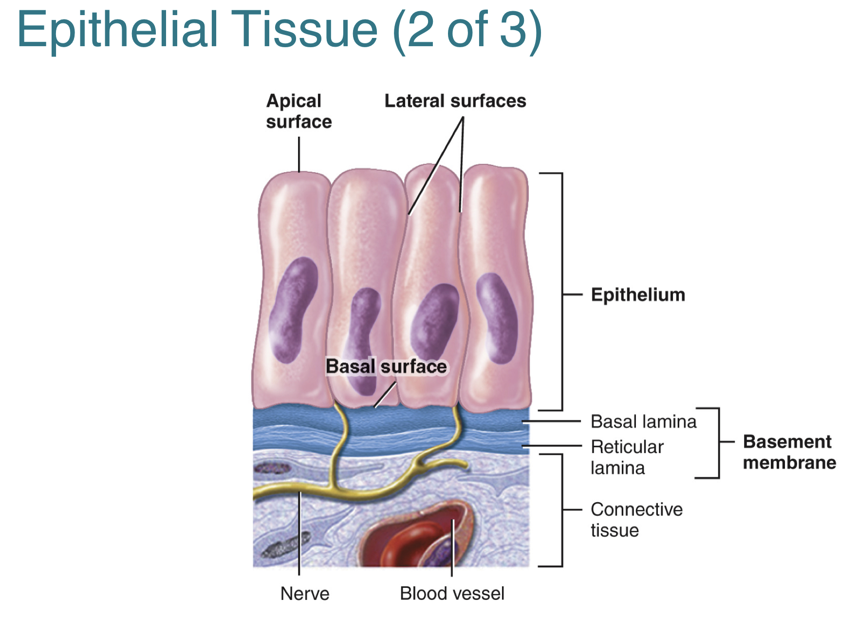
What is the epithelial tissue? What are its three structural components?
The epithelial tissue is a continuous sheet of cells that form protective barrier on body surfaces and passageways.
The three structural components of the epithelial tissue include:
1.) Apical surface: free surface facing outwards towards the body cavity, lumen, or surface
2,) Basal surface: Opposite side of the apical surface, that is deep into the cell and is connected to the basement membrane
3.) Basement membrane: extracellular material that attaches the epithelial tissue to underlying connective tissue
List three characteristics of the epithelial tissue.
1. The epithelial tissue is the only avascular body tissue.
--this means that they rely on diffusion from underlying connective tissue blood vessels for obtaining nutrients and removing wastes.
2. The functions of the epithelial tissue include protection, filtration, secretion, absorption, and excretion.
3. The epithelial tissue is classified under two types, covering and lining & glandular.
-covering and lining: forms outer part of skin and some organs
-glandular: forms secreting portion of glands
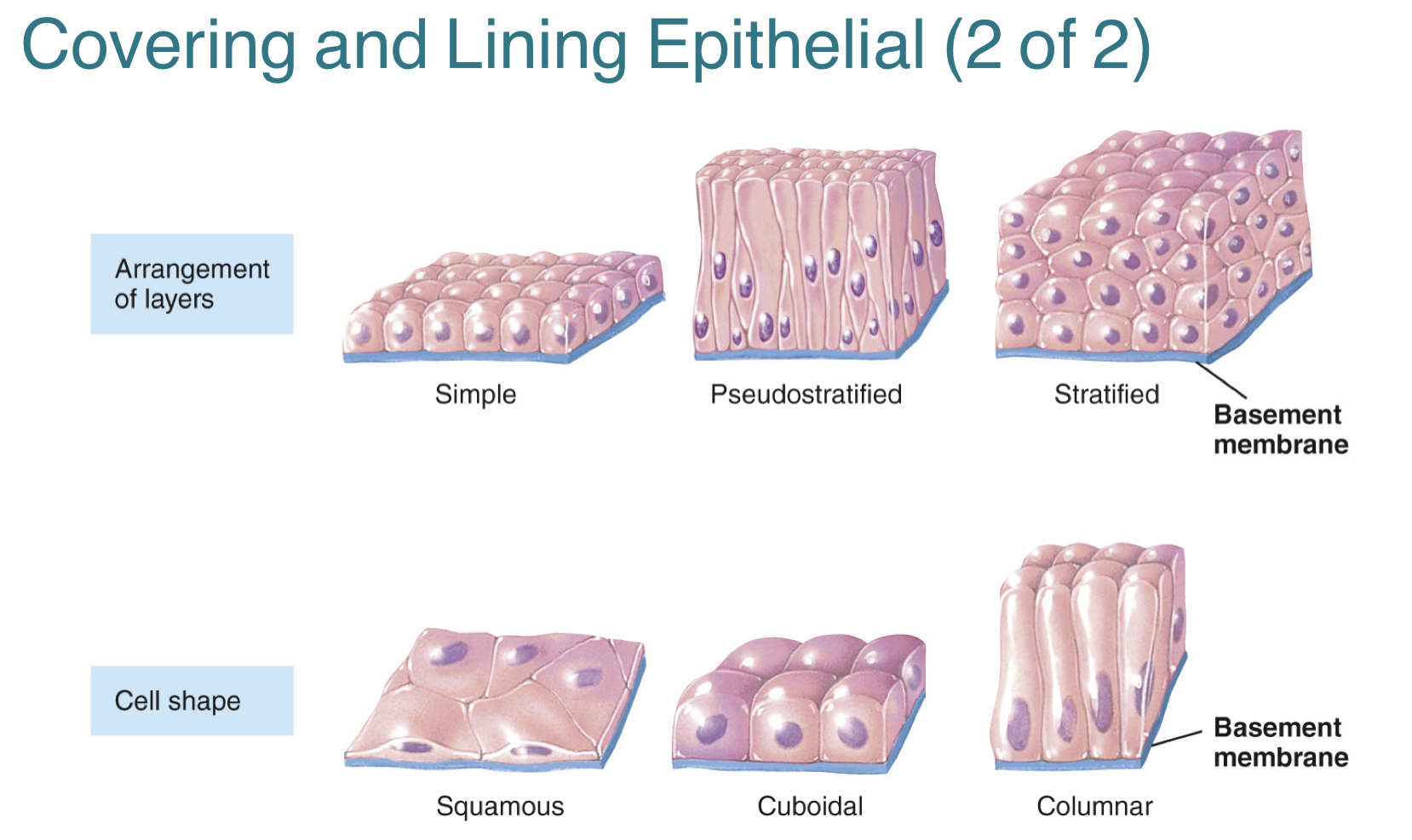
What are the two structural characteristics of the covering and lining epithelial tissue?
1.) Specific arrangement of cells in layers
-includes simple epithelium, pseudostratified epithelium, stratified epithelium (has layers)
2.) Specific cell shapes
-includes squamous cells, cuboidal cells, columnar cells, and transitional cells
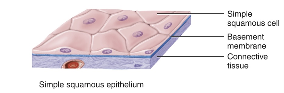
Provide a summary of the simple squamous epithelium. (its layers, functions, and where they can be found)
The simple squamous epithelium is a single layer of broad, flat cells.
-involved in filtration and diffusion
-can be found in the peritoneum of the small intestine
The two layers of the simple squamous epithelium include:
1.) endothelium: the lining for blood/lymph vessels and heart
2.) mesothelium: the serous membrane
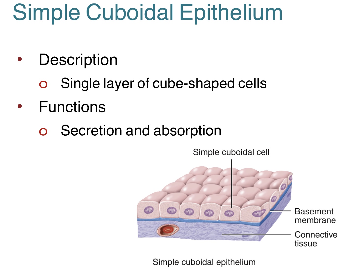
Provide a summary of the simple cuboidal epithelium. (its function and where they can be found)
The simple cuboidal epithelium is a single layer of cube-shaped cells.
-involved in secretion and absorption
-can be found along urinary tubules
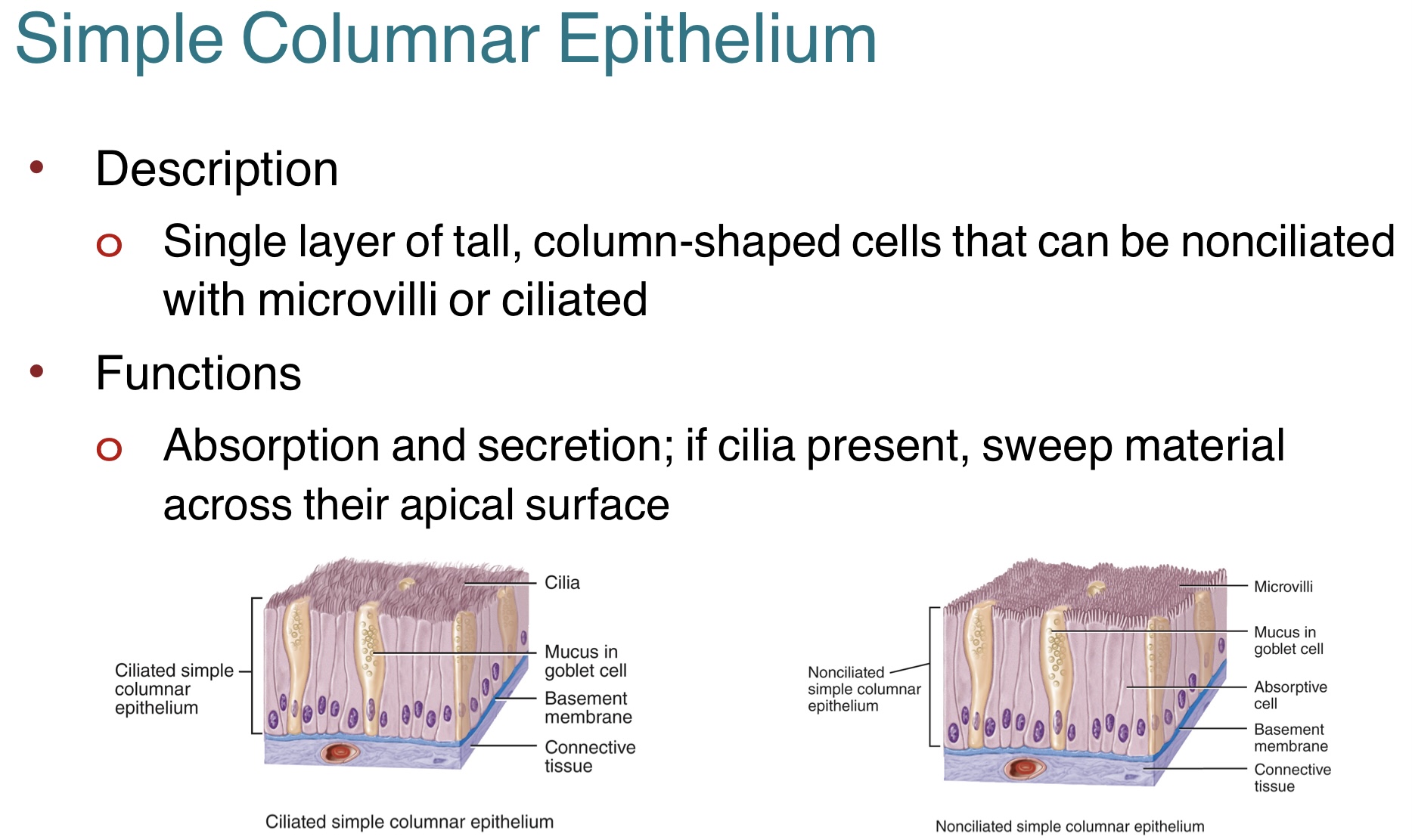
Provide a summary of the simple columnar epithelium. (its functions and where they can be found)
The simple columnar epithelium is a single layer of tall, column-shaped cells that can either be lined with cilia or microvilli
-involved in absorption and secretion
-if cilia is present, sweep material across their apical surface
-can be found in the jejunum lining of the small intestine or uterine tube
Provide a summary of the pseudostratified epithelium. (its functions and where they can be found)
The pseudostratified epithelium a single layer of nonciliated or ciliated cells attached to basement membrane.
-appears falsely stratified b/c nuclei are at different depths and not all have apical surface
-can be found in the trachea
The difference in functions between ciliated and nonciliated epithelium include:
-ciliated: sweeps away mucus w/ trapped foreign particles
-nonciliated: involved in absorption and protein
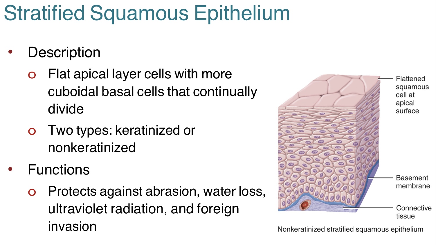
Provide a summary of the stratified squamous epithelium. (its functions, types, and where they can be found)
The stratified squamous epithelium is flat with flattened cells in the apical layer and cuboidal cells in the basal cells.
-these cells continually divide
-protect against abrasion, water loss, ultraviolet radiation, and foreign invasion
-can be found in the vagina lining (nonkeratinized) and epidermis (keratinized)
The two types of stratified squamous epithelium includes:
1.) keratinized
2.) nonkeratinized
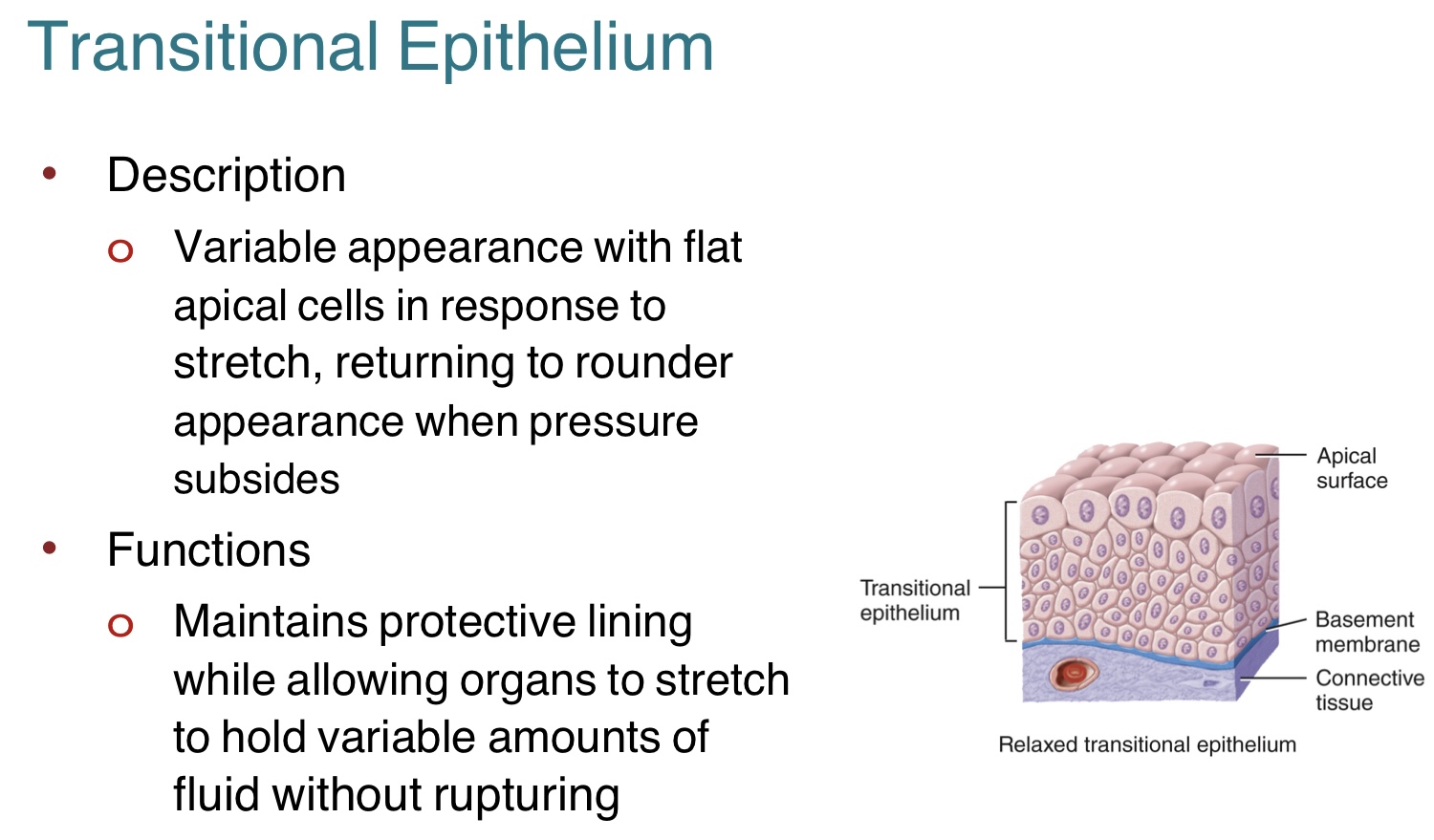
Provide a summary of the transitional epithelium. (its function and where they can be found)
The transitional epithelium has a variable appearance w/ flat apical cells in response to stretch, and returns to rounder appearance when pressure subsides.
-maintains protective lining while allowing organs to stretch and hold variable amounts of fluid w/o rupturing
-can be found in the urinary bladder
What is a glandular tissue? Provide a summary of the two types of glandular tissues, endocrine and exocrine. (its function and examples)
Glandular tissue is a type of epithelial tissue that specializes in producing and secreting substances
(like hormones, enzymes, and milk).
The two types of glandular tissue include:
1.) Endocrine: secrete hormones to interstitial fluid then to blood
-secreted hormones regulate metabolism and maintain homeostasis
-eg. thyroid gland
2.) Exocrine: secrete substances, through ducts, that empty onto skin surfaces or into lumen of an organ
-produces substances such as sweat, oil, earwax, saliva, and digestive enzymes
-eg. sweat gland
List three characteristics of the connective tissue.
1. The connective tissue consists of two basic elements, which are the extracellular matrix and its cells.
-extracellular matrix: material between cells
2. Most of the connective tissue types are vascular, with good blood supply.
-the exceptions include cartilage and tendons
3. The connective tissue has two main functions.
-function 1: binding, supporting, strengthening other body tissues
-function 2: protecting, insulating, and compartmentalizing structures
What is the extracellular matrix of the connective tissue made of?
The connective tissue extracellular matrix is made up of ground substance and the protein fibers embedded in the ground substance.
Ground substance:
-is composed of water, proteins, and gycosaminoglycans (polysaccharides)
-supports embedded cells while allowing for material exchange
Protein fibers:
1.) collagen: strong, flexible, often packed in parallel bundles
2.) elastic: elastin protein that can stretch and recoil back into shape
3.) reticular: fine, branching collagen network that forms supporting framework for soft organs
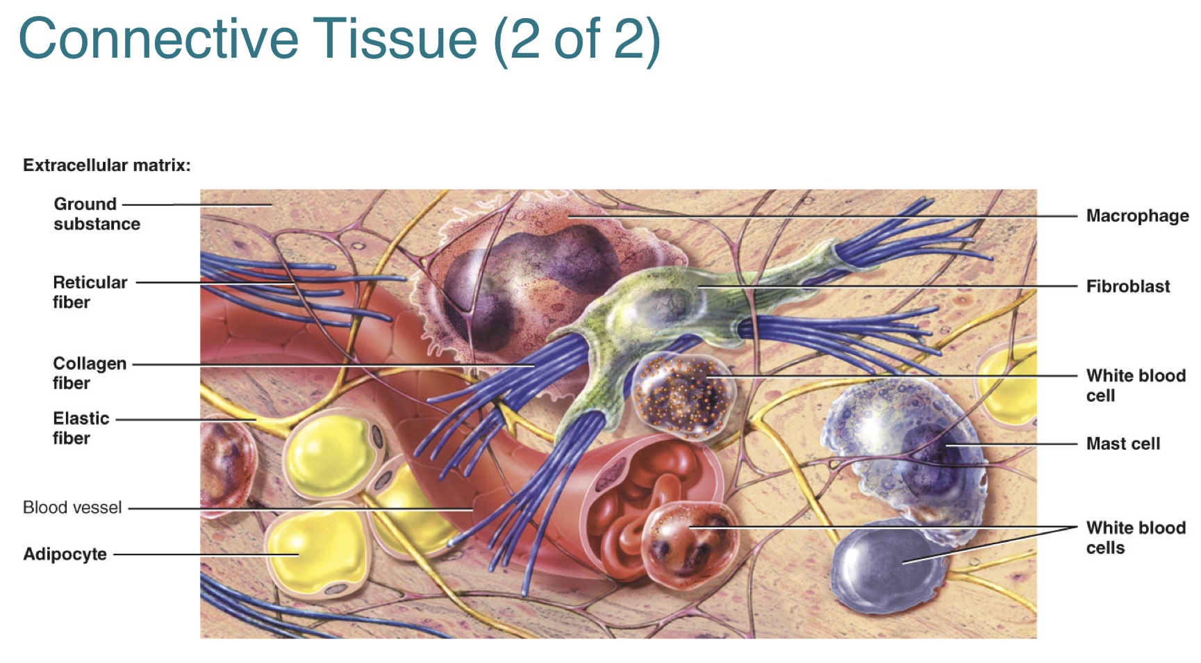
What are the different types of connective tissue cells and their functions?
1.) Fibroblasts: most common type of cell found, large flat cells with branching processes
-immature (“-blast”) cells produce ground substance and fibers, and undergo cell division
-some mature (“cyte”) cells differentiate and maintain matrix
2.) Macrophage: phagocytic cells
3.) Mast cell: inflammatory cells
4.) Adipocytes: fat-storing cells
5.) White blood cells: immune defense cells
What are the five types of mature connective tissue?
1.) Loose connective tissue includes:
-areolar, adipose, reticular
2.) Dense connective tissue includes:
-dense regular, dense irregular, elastic connective
3.) Cartilage connective tissue includes:
-hyaline, fibrocartilage, elastic cartilage
4.) Osseous connective tissue includes:
-bones
5.) Liquid connective tissue includes:
-blood, lymph
Provide a brief summary of the three types of loose connective tissue- areolar, adipose, reticular. (its function and where they can be found)
1.) Areolar connective tissue: fibroblasts + other types of cells w/ loosely intertwined collagen, elastic, and reticular fibers in semi-fluid ground substance
-provides strength, elasticity, support
-can be found in the hypodermis
2.) Adipose connective tissue: large triglyceride (fat) droplets w. cytoplasm and nucleus pushed to periphery of cell
-provides insulation, organ support and protection, storage for energy
-can be found in the fat tissue around the heart
3.) Reticular connective tissue: cells spaced between fine, interlacing reticular fibers
-forms supporting framework of organs AND a net to capture microbes/worn-out blood cells
-can be found in a lymph node
Provide a brief summary of the three types of dense connective tissue- dense regular, dense irregular, elastic connective. (its function and where they can be found)
1.) Dense regular connective tissue: regularly arranged parallel bundles of collagen fiber w/ few fibroblasts
-provides strong attachment between structures
-can be found in the tendon
2.) Dense irregular connective tissue: irregularly arranged bundles of collagen fiber w/ few fibroblasts
-provides tensile (pulling) strength in many directions
-can be found in the reticular region of the dermis
3.) Elastic connective tissue: predominantly elastic fibers w/ fibroblasts between them
-allows stretching and recoil to original shape, after being stretched
- can be found in the aorta of the heart and elbow
Provide a brief summary on the three types of cartilage connective tissue- hyaline , fibrocartiilage, elastic cartilage. (its functions and where they can be found)
1.) Hyaline cartilage connective tissue: chondrocytes found in lacunae (spaces) within resilient gel ground substance; fine collagen fibers not visible
-reduces friction, absorbs shock at joints
-provides flexibility and support
-can be found in the nose or a developing fetal bone
2.) Fibrocartilage connective tissue: chondrocytes found in lacunae (spaces) within resilient gel ground substance and thick bundles of collagen fibers
-provides strong and resilient support
-joins structures together
-can be found in the vertebrae, knee, hip
3,) Elastic cartilage connective tissue: chondrocytes found in lacunae (space) within resilient gel ground substance w/ network of elastic fibers
-provides strength and elasticity
-helps maintain shapes of structures
-can be found in the auricle of ear and epiglottis
Provide a biref summary of the osseous tissue. (its function and where they can be found)
Osseous connective tissue: osteocytes found in lacunae (spaces) within solid ground substance w/ mineral salts and collagen fibers
-provides support, protection, storage
-provides leverage for movement of body by muscles
-can be found in the bones (including thigh bones)
Provide a brief summary of the blood connective tissue. (its function and where they can be found)
Blood connective tissue: various types of cells suspended in liquid extracellular matrix (plasma)
-transport and protection, by phagocytosis and immunity
-can be found in blood vessels
List three differences between the epithelial tissue and connective tissue.
Epithelial tissues:
-cells are tightly packed with intracellular junctions
-avascular
-typically form surface layers toward inside or outside space
Connective tissues:
-relatively few cells embedded in extracellular material
-vascular (except cartilage and tendon)
-typically fill spaces, support, and connect structures
What are the four different types of epithelial membranes?
Epithelial membranes are membranes that are composed of an epithelial layer and underlying connective tissue layer.
-flat sheets of flexible tissue that cover or line a body structure
The four types of epithelial membranes include:
1.) Mucous membrane: consists of mucosa and lamina propria
-lines body cavities that open to the outside
-produces mucous to protect surfaces
2.) Serous membrane: consists of mesothelium and areolar
-forms parietal layer and visceral layer
-lines body cavities that do not open directly to the outside
-produces serous fluid to reduce friction between organs
3,) Cutaneous membrane: consists of the epidermis and dermis
-the skin that covers the surfaces of the body
-protective barrier against external threats
4,) Synovial membrane: lacks epithelium
-lines joint cavities
-produces synovial fluid to lubricate joints and reduce friction during movement

What is the muscle tissue?
The muscle tissue is composed of elongated cells called muscle fibers.
-function: generate force to produce body movements, maintain posture, and generate heat
The three types of muscle tissue includes:
1.) Skeletal muscle tissue: has striations, contains many nuclei, produces voluntary movements, control motion/posture/heat production/and provide protection (can be found in the bone)
2.) Cardiac muscle tissue: has striations, (heart) produces involuntary movement,
3.) Smooth muscle tissue: (arteries) produces involuntary movements, controls motion of substances in hollow organs, and contraction of iris of eye
What is the nervous
Nervous tissue is composed of neurons.
-neurons: cells that convert stimuli to electrical signals
Three part of a neuron:
1.) Cell body
2.) Dendrite: extensions
3.) Axon: single, elongated extension
What is the tissue repair process?
-Epithelial cells can be constantly replaced
-Connective tissue and muscle tissue have difficulties with being replaced.
-Nervous tissue is almost impossible to be replaced.
What is the physiology behind muscle relaxation?
-When motor neuron impulses stop, there is no more release of acetylcholine molecules
-Ca2+ active transport pump uses ATP to move Ca2+ ions back into SR cisterns
-Troponin-tropomyosin complex slides back to cover myosin-binding sites
-Actin filaments slide, returning back to relaxed position
What is a motor unit
A motor unit consists of one motor neuron and all the skeletal muscle fibers that the motor neuron stimulates.
-a single nerve impulse elicits a single muscle twitch contraction, in all muscle fibers it innervates
Frequency of stimulations
-is the number of impulses per second
-governs total tension produced by a single muscle fiber when stimulated
Contraction control
1.) many small motor units control small precise movements (10-20 muscle fibers per motor unit)
2.) fewer, larger motor units control large, powerful movements (up to 3000 muscle fibers per motor unit)
Contraction strength
-size of a skeletal muscle’s motor unit
-number of motor units activated
What is a twitch contraction?
A twitch contraction is the response of a motor unit to a single nerve impulse in its motor neuron
-contractions can be recorded through a myogram, which shows the duration of the twitch
-duration varies with type of muscle fiber (10 to 100 msec)
1.) Latent period: cell events leading up to contraction, brief delay between stimulus and contraction
2.) Contraction period: power strokes generate tension
3.) Relaxation period: cell events allowing muscle to resume original length
Wave summation: second stimulus arrives before muscle fiber has fully relaxed, causing second, stronger contraction
Unfused tetanus: frequency of multiple stimuli allows only partial muscle relaxation between each stimulus
-occurs when muscle fibers are stimulated at a moderate frequency, allowing for brief period of relaxations between contractions
Fused tetanus: sustained contraction caused by rapid frequency of stimulation
-occurs when muscle fibers are stimulated at a very high frequency, eliminating the relaxation phase between contractions
What happens during motor unit recruitment?
(VOLUNTARY) Motor unit recruitment is the process of increasing the number of contracting motor units, so that the force of muscle contraction becomes greater.
-smooth, voluntary movement from asynchronously stimulated motor units of a skeletal muscle
(INVOLUNTARY) Muscle tone is the small amount of tension due to involuntary, alternating contractions of a small number of motor units of a skeletal muscle, even at rest
-does not produce movement
The two types of contractions include:
1.) Isotonic contraction, which involves a change in muscle length without a change in its tension
-concentric: when muscle shortens
-eccentric: when muscle lengthens
2.) Isometric contraction: muscle does not change length
-the load equals or exceeds the muscle tension created
What is the energy for muscle contraction?
ATP is the only direct source of energy for muscle fiber contraction
There are three ways for muscles to produce ATP:
1.) Creatine phosphate
2.) Anaerobic cellular respiration
3.) Aerobic cellular respiration
(1) Creatine phosphate: an energy-rich molecule found only in muscle fibers
-synthesized in liver, kidneys, and pancreas; THEN transported to muscle fibers
-creatine kinase transfers high-energy phosphate group from excess ATP to creatine when muscle is relaxed
-first source of energy when contraction begins; sufficient only for short bursts of activity (about 15 seconds)
-during contraction, transfer of phosphate group to ADP, to quickly form new ATP
(2) Anaerobic cellular respiration: series of ATP-producing reactions in sarcoplasm that do not require oxygen
-glycolysis: breakdown of glucose from blood stream or from breakdown of glycogen stores in sarcolemma; yields two ATP molecules and two pyruvic acid molecules
-pyruvic acid: converted to lactic acid in absence of oxygen
-lactic acid moved into blood and carried to liver for reconversion to glucose
**sufficient energy for 30-40 seconds of activity
(3) Aerobic cellular respiration: series of oxygen-requiring reactions in mitochondria
-oxygen sources: diffuse into muscle fibers
-duration of energy provided: several minutes to hours
After prolonged exercise…
Muscle fatigue: inability to muscle to contract forcefully after prolonged activity
Recovery oxygen uptake (oxygen debt)
-breating rate and oxygen consumption remains above resting level during recovery period
-helps restore metabolic conditions
Muscle fibers vary in amount of red myoglobin
-white muscle fibers are pale w/ low myoglobin
-red muscle fibers are dark red w/ high myoglobin
Most skeletal muscles are mixture of three structural and functional classes of muscle fibers.
1.) Slow-oxidative (SO): slowest contraction, fatigue resistant, aerobic respiration
-used to maintain posture and endurance
-mostly red muscle fiber
2.) Fast oxidative-glycolytic (FOG): intermediate contraction, moderate fatigue resistance, aerobic respiration and glycolysis
-used for walking and sprinting
-red pink muscle fiber
3.) Fast glycolytic (FG): fastest strongest contraction, use glycolysis, fatigue rapidly
-used for rapid, intense movements of short duration
-mostly white muscle fibers
(1) Cardiac muscle fibers: muscles found in the wall of the heart
-one nucleus, branched, striated, and involuntary
-interconnected by intercalated discs (desmosomes and gap functions), so action potential spreads to cell to cell
Similarities between cardiac muscle fibers and skeletal muscle fiber:
1.) Same sarcomere arrangement of actin and myosin
2.) Long, sustained contractions supported by Ca2+ ion influx from both SR and interstitial fluid
3.) Atuor
-autorhythmicity refers to the cardiac muscle’s ability to initiate their own electrical impulses w/o external stimulus; involuntary movements)
4.) Can use lactic acid from skeletal muscle anaerobic respiration, to make ATP during exercise
(2) Smooth muscle fibers:
There are two types of smooth muscle tissue:
1.) Visceral (single-unit): most common type; found in skin, walls of blood vessels, and hollow organs, autorhythmic fibers connected by gap junctions, action potential spreads so all cells contract as single unit
2.) Multiunit: found in walls of large arteries, lung airways, arrector pili of hair follicles, and internal eye muscles, individual fibers has their own motor neuron, few gap junctions and lack autorhythmicity
Differences between cardiac muscle fibers and smooth muscle fibers:
-Cardiac muscle is a single-unit muscle, as the muscle fibers are interconnected by gap junctions, allowing the heart to contract as a coordinated unit
-Smooth muscle can be either single-unit (like in the walls of organs, where muscle fibers contract as a unit) or multi-unit (like in the iris of the eye)
Differences between smooth muscle fibers and skeletal muscle fiber:
1.) Lack sarcomere; actin and myosin slide within bundles formed by intermediate fibers, between dense bodies instead of Z discs
2.) Contraction starts slowly and lasts an extended time, twisting in a helix to length