Physiological Psych Part 1
 Call Kai
 Learn
Learn Practice Test
 Spaced Repetition
 Match
Match Flashcards
 Knowt Play
1/86

There's no tags or description
Looks like no tags are added yet.
Name | Mastery | Learn | Test | Matching | Spaced | Call with Kai | Chat |
|---|
No analytics yet
Send a link to your students to track their progress
87 Terms
Gall
Theory of organology- proposed brain was made up of different organs that drives particular traits, led to phrenology and localization of function that’s not true
Phineas Gage
Major changes to personality and behavior after damage to frontal lobe, first documented case relating brain with personality leading to localization of function, example of learning from loss
Moniz
Developed way to visualize artery structure in brain, earliest example of brain imagery, believed mental illness was due to malfunctioning synapses in frontal cortex which led to severing connections to frontal lobe which is leucotomy and then lobotomy which resulted in issues in patients but was still widely used
Hebb
Wrote about neural basis of learning which is the internal part of the brain, this year emphasized Skinner’s findings
Golgi
Led to advancements in knowledge about brain tissue, promoted concept of syncytium (fused axons) and reticular theory
Reticular theory
Neurons share direct connection, continuous network with no gaps/synapse
Cajal
Created detailed drawings and found that neurons were individual entities with signal transmission, neuron doctrine (nervous system made up of individual cells) which goes against syncytium, Cajal was right
Neuron doctrine
Neurons are individual cells with small gaps between (synapse), connections not random but specific pathways, dynamic polarization (parts of neurons take in information and some parts send information)
Henry Head
Charted sensory progress during regeneration of severed sensory nerves, introduced concept of different types of sensory fibers regenerating at different speeds
Loewi’s experiment
Tested if direct connection is needed for neurons, stimulated heart in container that shared fluid with another heart with no direct connection
Neurons
Communicate by electrochemical signals (neurotransmitters)
Around 100 billion neurons in the brain with each neuron making around 1000 connections
Every connection collects input, process/decide, and produce output
Can transmit information over distance
Critical parts for communication are dendrites and axons
Glia cells
Critical non-neural cells playing supportive function
Supports neurons in structure, waste removal, creation of myelin sheath, promote neuron growth and guidance
Dendrite
Receive information
Axon
Send information
Dendritic Spines
Points of contact with other neurons, spines change as they grow and retract
Plasma membrane
Lipid bilayer (2 layers of fats)
Semi-permeable (only some compounds get through, so electric charges can’t get through)
Limit of what can pass through is critical for action potential
Motor neurons
Connects to muscle or organ
System output component
Many dendrites, only one axon, axon hillock, myelin sheath, terminal bouton
Efferent
Sensory neurons
Brings sensory information to brain
System input component
No true axons or dendrites
Each end combination of myelinated and fast conduction with cell body located off to side
Afferent
Interneuron
Communicates within nervous system, occurs in between input and output
Multipolar
Common, more complex processing, one axon and many dendrites, spinal motor neurons
Afferent neuron
Approach and coming toward
Sensory neurons bring sensory information to brain
Efferent neuron
Exit and going away from
Motor neurons connect to muscle or organ
Astrocytes
Type of glia
Surround neuron
Removes waste
Store and release glucose and potassium regulating ion concentrations
Contact with vasculature at end feet which helps form blood brain barrier

Microglia
Type of glia
Assist in repair and proliferate (multiply) in damaged region
Phagocytic function which removes waste and releases nitric oxide to kill damaged neurons
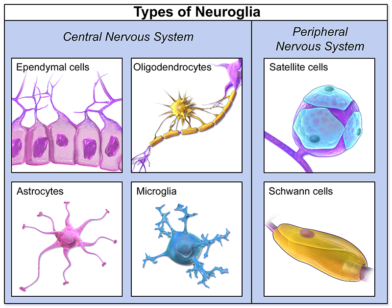
Oliogodendrocytes
Type of glia
Produce myelin and wrap sheath concentrically
In CNS, oliogodendrocytes myelinate up to 50 axons at once, inhibit regrowth, and axons don’t regenerate
In PNS, Schwann cell wrapping and myelinating only single axon, promote regrowth, and axons regenerate
Interruption at nodes of Ranvier needed for saltatory conduction

Gliosis
Glia fill space left by dead neurons, seen after injury, may be combination of astrocytes and microglia, white here is glial cells not white matter
Dorsal (superior)
Top part of brain

Ventral (inferior)
Bottom part of brain
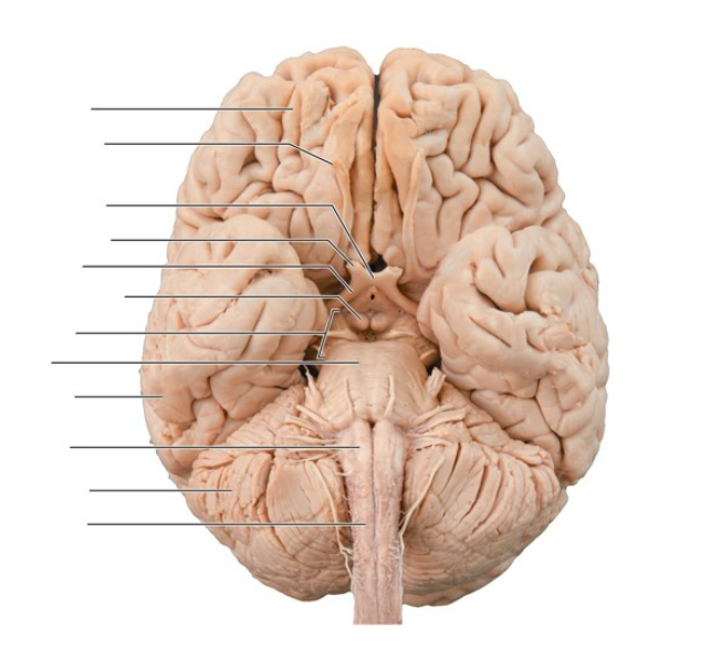
Lateral
Outer side of brain
Medial
Middle part of brain
Rostral (anterior)
Front part of brain

Caudal (posterior)
Back part of brain

Coronal
Slices front to back

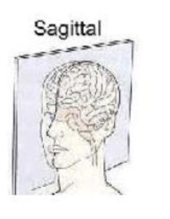
Sagittal
Slides from side to other side
Transverse
Slices from top to bottom

Nuclei
Neuron cell bodies located in central nervous system
Make up gray matter
Process and initiate signals and serves as origin point for cranial nerves
Nerve
Bundles of axons located in peripheral nervous system
Composed of bundles of axons which make up white matter
Transmits electric signals rapidly over long distances to and from target
Nervous system
Composed of CNS and PNS that can be divided into sections
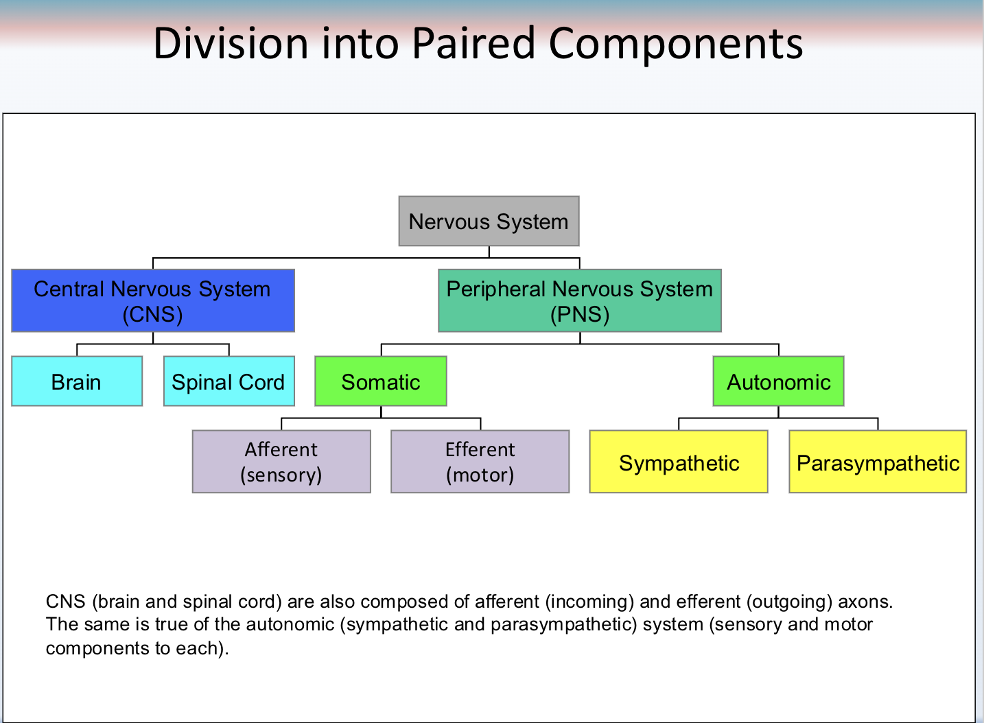
Central nervous system
Splits into brain and spinal cord
Drives behavior

Peripheral nervous system
Splits into somatic and autonomic
Series of nerves that connect CNS to rest of body
31 pairs of spinal nerves with each nerve, which are axons of many neurons grouped together, each containing sensory and motor axons
12 pairs of cranial nerves with some sensory and some motor with some that are both

Autonomic
Automatic
Communicates internal body state information to and from CNS
Splits into sympathetic and parasympathetic
Innervates internal organs, most organs dually innervated where sympathetic and parasympathetic are both present
Sympathetic
Stimulates fight or flight, kicks in when there’s immediate physical threat for most animals
Parapsympathetic
Calms down from sympathetic activity and stress, conservation of energy and maintains homeostasis
Somatic
Communicates external stimuli information to and from the central nervous system
Splits into afferent (sensory information arriving in CNS) and efferent (motor information exiting CNS)
Comprised of nerves (many neurons and axons)
Gray matter
Grey matter in spinal cord surrounded by white matter while opposite for brain, lots of cell bodies and dendrites processing information

White matter
Tissue that appears white because of bundle of myelinated fatty axons, acts as communication network as information flows from gray matter to white matter

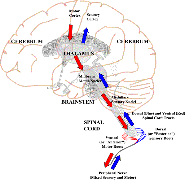
Bell Magendie Law
Motor nerves faces and attaches to ventral of spinal cord while sensory nerve faces and attaches to dorsal for everyone
Cranial nerves
Not all combination of both motor and sensory nerves, 12 cranial nerves
If there’s damage to olfactory (smell) nerve it’ll produce inability to smell
Bell’s palsy is damage to facial nerve which creates paralysis of one half of face
Novocain given by dentist directed at trigeminal nerve which has both sensory and motor components to stop action potential of neurons so there’s loss of sensation and motor control
Vagus nerve is the wandering nerve going throughout body and major part of the parasympathetic nervous system
Norepinephrine
Neurotransmitter for sympathetic system, brain messenger for attention, stress hormone
Acetylcholine
Neurotransmitter for parasympathetic, comes out of cranial nerves and sacral spinal cord
Protection of the CNS
Blood brain barrier, bone, membranes, cerebrospinal fluid
Blood brain barrier
Prevents harmful substances from getting to neurons in brain and spinal cord
Located where blood vessels contact brain tissues
Endothelial cells (flat) tightly packed with no gaps
Small uncharged molecules and fat soluble molecules including many drugs can go through
Active transport required for glucose and amino acids (some hormones and vitamins)
Bones
Skull and vertebrae
Meninges
3 membranes surround brain and spinal cord, provide cushioning
Pia mater: innermost
Arachnoid mater: web like and spongy
Dura mater: outermost tough and thick
Cerebrospinal fluid
4 Ventricles: fluid filled cavities in brain
Continuously produced and absorbed by choroid plexus
Allows brain to float reducing effective weight
Found in central canal of spinal cord

Gyri and Sulci
Bumps and ridges folded to fit a lot into small space and to be closer to axonal connections
3 subdivisions of brain
Hindbrain, midbrain, forebrain
Brainstem
Midbrain, pons, medulla, doesn’t include cerebellum

Medulla (hindbrain)
Controls life sustaining functions like circulation of blood, respiration, reflexes, maintains muscle tone
Pons (hindbrain)
Controls sleep and arousal
Locus coeruleus: area in pons that produce norepinephrine for brain, can be activated by other higher regions
Dysfunction: lack of paralysis in REM state
Cerebellum (hindbrain)
Little brain, coordination of movement and sense of balance, involved in abstract pattern learning and procedural memories
Mesencephalon (midbrain)
Tectum (roof): superior colliculus (visual reflexes) and inferior colliculus (auditory reflexes)
Tegmentum (floor): includes important neurotransmitter connections to other areas, substantia nigra (dopamine), ventral tegmental area
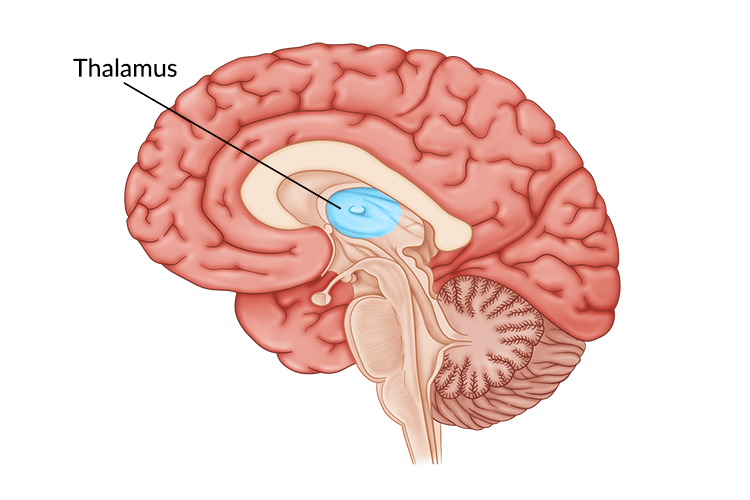
Thalamus (forebrain)
Sensory relay center, all sensory information except for smell goes to thalamus
Pathway connections very important, there’s direct connection to amygdala and to higher sensory processing centers
Hypothalamus (forebrain)
Below thalamus and important part of endocrine system
Associated with fighting, fleeing, feeding, and reproduction

Limbic system
Areas involved in learning and memory, emotions, and motivation
Doesn’t operate in isolation!
Hippocampus (limbic system)
Learning and memory including spatial memory
Amygdala (limbic system)
Emotions especially fear and anxiety
Some stimuli carry innate fear and don’t require much conditioning to learn while some stimuli must be learned to generate fear
Central region involved in innate fear while basolateral region more complex and can learn fear
Gets input from thalamus, pain from PAG in midbrain, frontal cortex connections
Has direct outward connection to motor cortex, hippocampus, and locus coerulues in pons that controls alertness
Basal ganglia
Plans movement control
Cerebral cortex
Doesn’t work in isolation
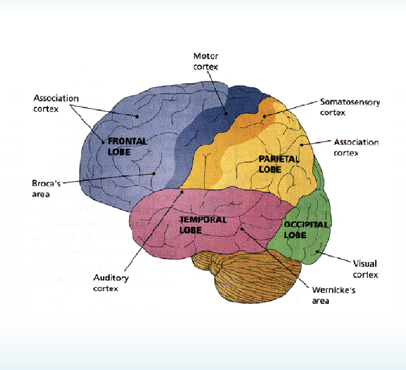
4 major lobes: frontal, parietal, temporal, occipital
Layered structure
Gyri and sulci increase surface area and reduce axonal distance with multiple layers having more dendrites than axons, more gyri and sulci allows more neurons being able to process complex information
Central sulcus
Divides frontal and parietal with motor and sensory sulci
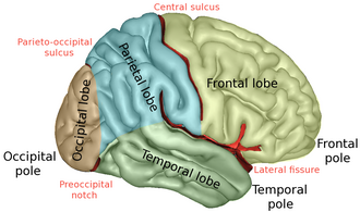
Lateral sylvian fissure
Divides temporal from frontal and parietal

Corpus callosum
Information bridge of axons between the left and right hemispheres
Occipital lobe
Very back of brain
Important for vision, visual information relayed from thalamus and sent to occipital lobe
Temporal lobe
Primary auditory cortex
There’s association areas related to memory
Visual processing with multiple regions with specificity for identifying visual stimuli, prosopagnosia result from damage to temporal lobe
Parietal lobe
Touch, pain, temperature, limb proprioception
2 main paths are pain and temperature then touch, proprioception, and movement
Primary somatoesory area
Also involved in visual perception, particularly awareness of where objects are in space, damage can lead to sensory neglect
Somatosensory cortex
Postcentral gyrus in parietal lobe
Sensory information coming from skin
Size of cortical representation not proportional to size of body part, it’s proportional to acuity of touch
Sensory homunculus
Little human representation in head
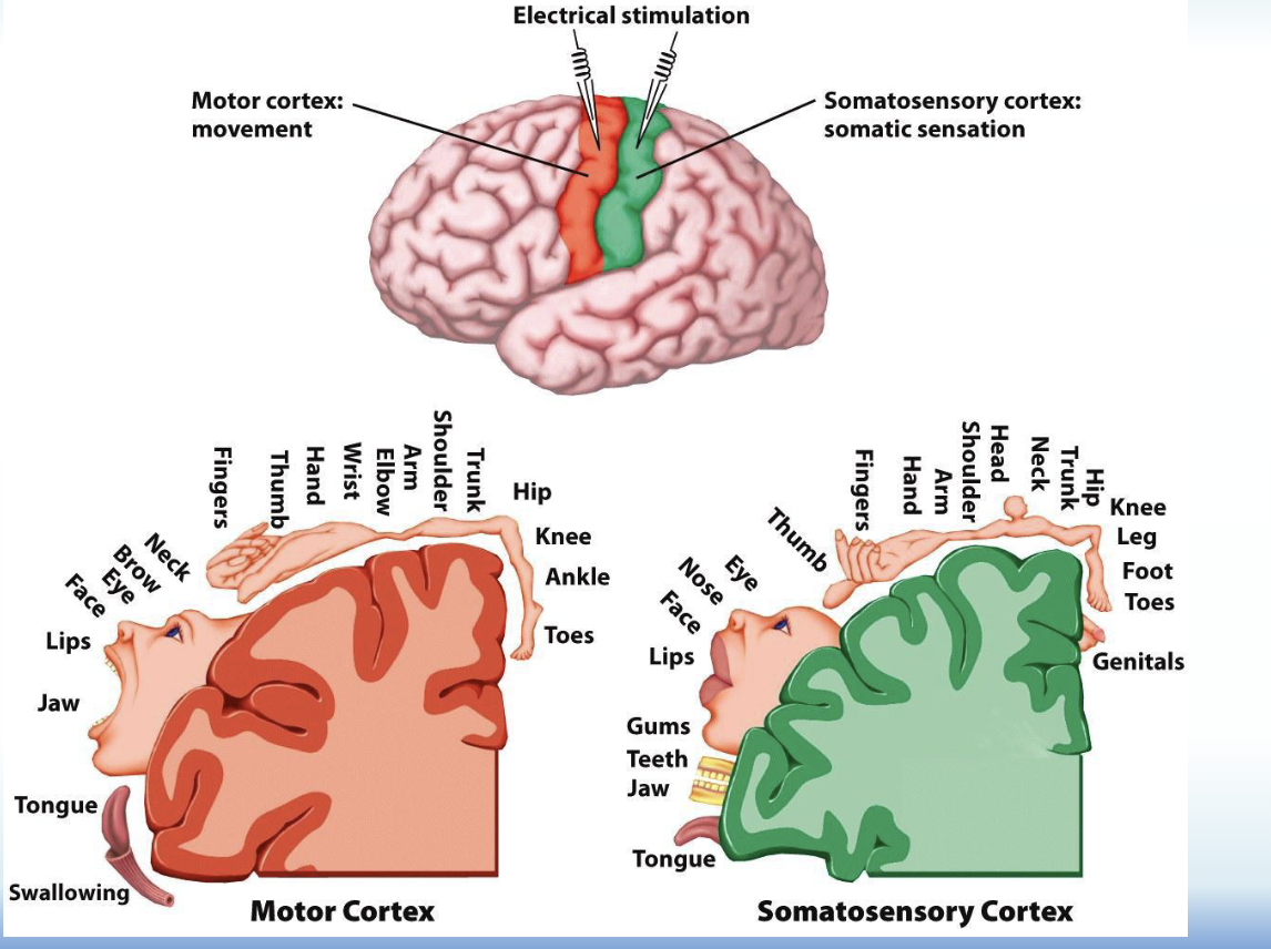
Motor cortex
Precentral gyrus in frontal lobe, movement of skeletal muscles where size of cortical area is proprotional to precision of movement
Frontal lobe
Area involved in highest and most complex cognitive functions
Planning, morality, decisions, working memory
Prefrontal cortex is most complex among the many sub regions of frontal lobe
Dorsolateral prefrontal cortex
Associated with following rules and making decisions
Decision making center
Most evolutionary recent area
Ventromedial prefrontal cortex
Has tight connections with amygdala
Emotional component of executive function
Being logical and not having emotions impact decision making
Vmpc damage individuals have poor decision making in many respects
Electrical gradients
Distribution based on electrical charge
Chemical gradients
Distribution based on molecular composition
Electrochemical gradients
Both of electrical and chemical gradients happening simultaneously
Major concepts to understand neural conduction
Resting potential, Action potential, Graded potentials
Resting potential
Inside of neuron there’s -70 millivolts compared to outside neuron
Not passive process and necessary to have an action potential since electrochemical gradient is needed to start action potential
Na+/K+ pump: necessary for resting potential, helps to restore balance of Na+ and K+ by moving 3 Na+ ions out and K+ ions in and requires ATP
Action potential
Axons send information from cell body to axon terminal
Begins at axon hillock
If add positive current to inside cell there’s depolarization (polarity moves toward zero and more positive)
If add negative current to inside cell there’s hyperpolarization (polarity moves away from resting potential and more negative)
Threshold of excitation: around 55mv when voltage gated Na+ channels open and there’s enough positive current to reach -55mv triggering action potential, when cell reaches positive the gated Na+ channels close and gated K+ channels open as inside of cell becomes more negative, lastly there’s hyperpolarization of K+ channels slow to close, all or none action potential
