Pelvic and Perineal Muscles
1/16
Earn XP
Description and Tags
i couldn't think of a witty one-liner...
Name | Mastery | Learn | Test | Matching | Spaced | Call with Kai | Chat |
|---|
No analytics yet
Send a link to your students to track their progress
17 Terms
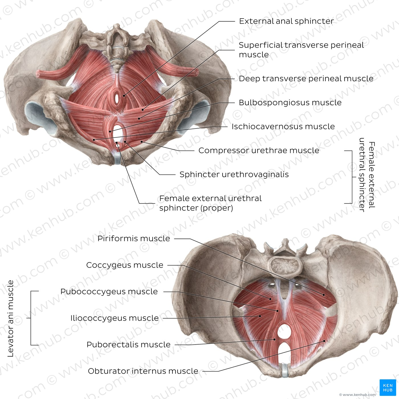
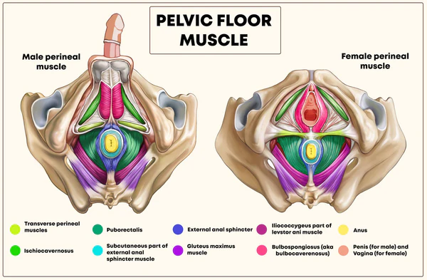
Overview of Pelvic & Perineal Muscles
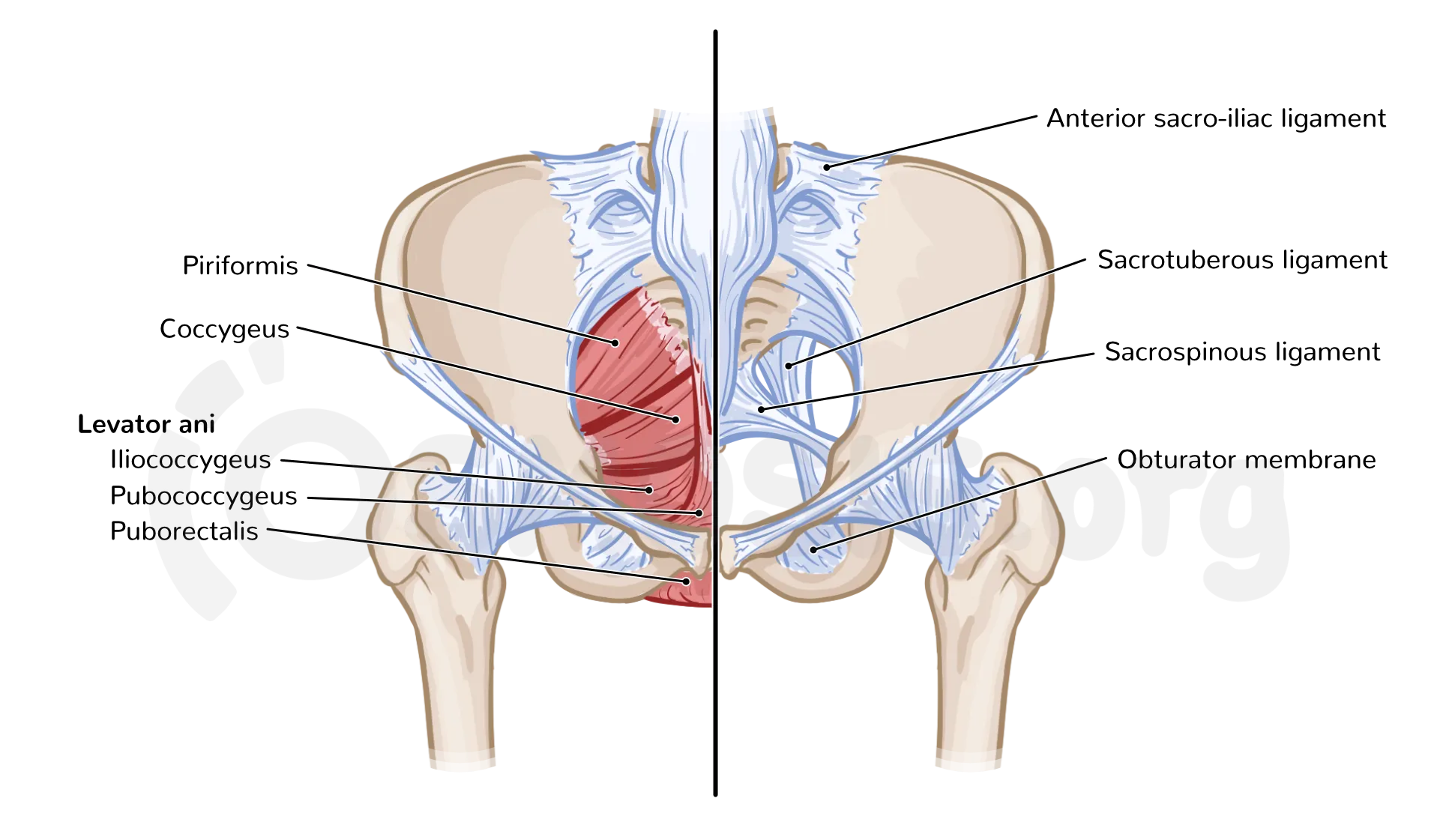
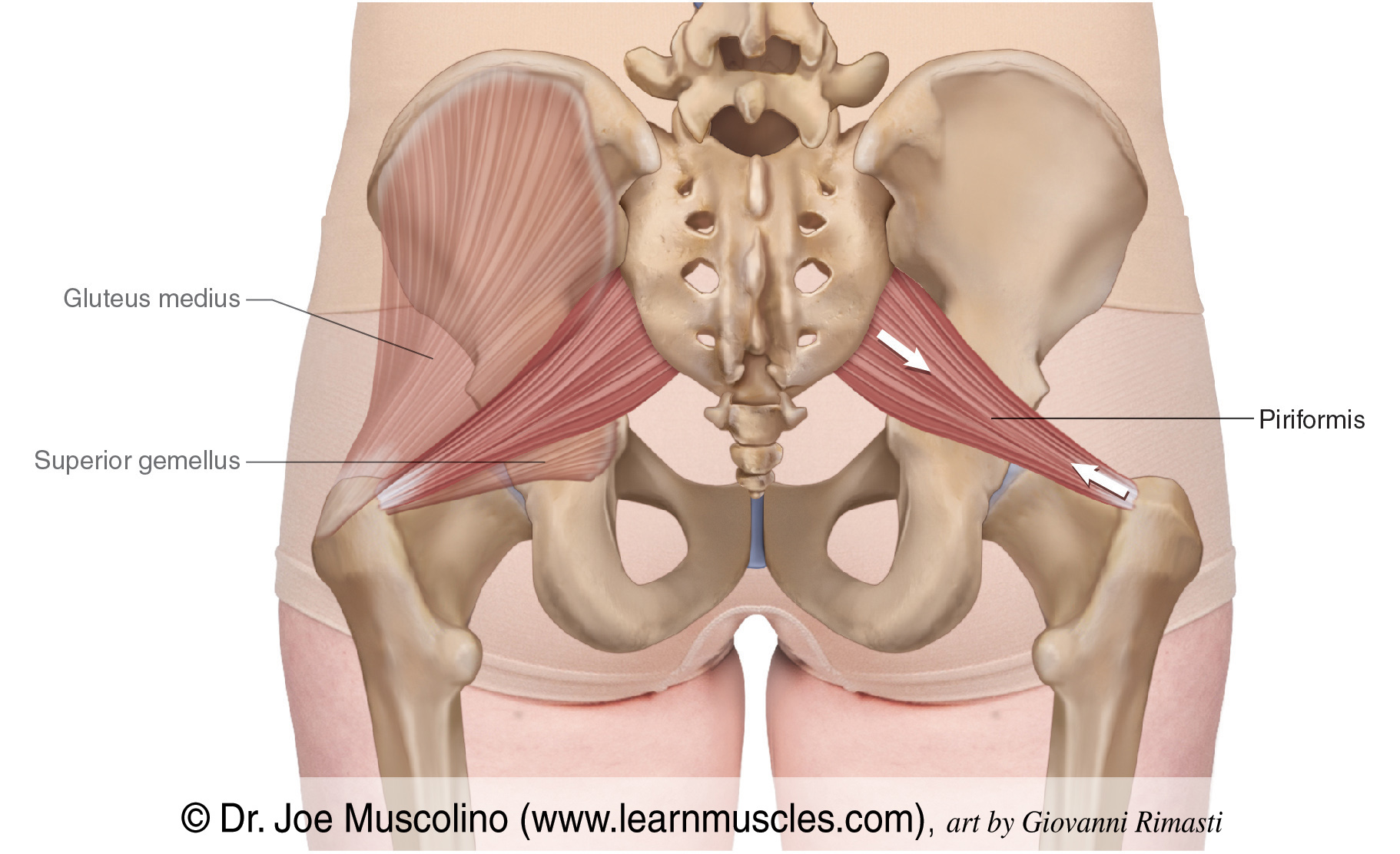
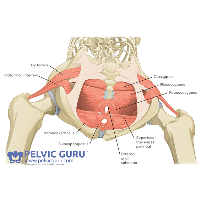
piriformis
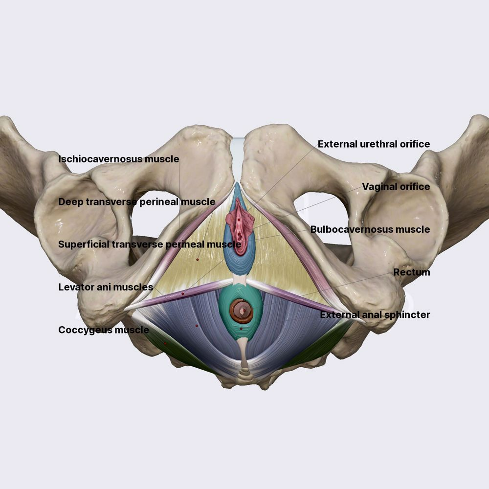
coccygeus
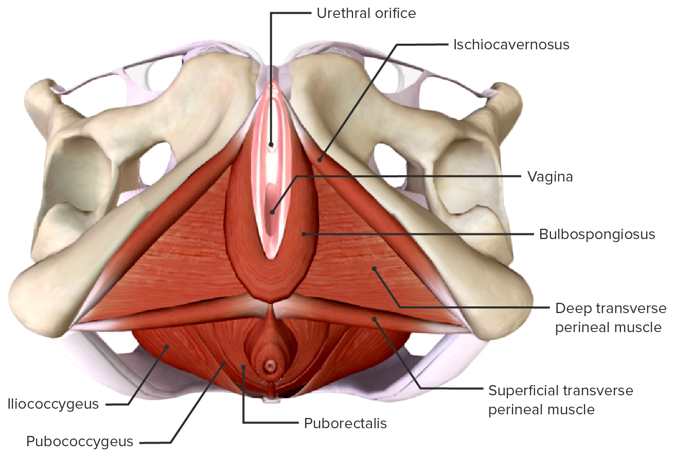
levator ani muscle
pubococcygeus
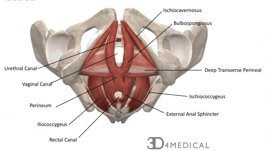
iliococcygeus
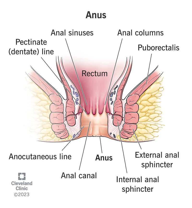
puborectalis
obturator internus
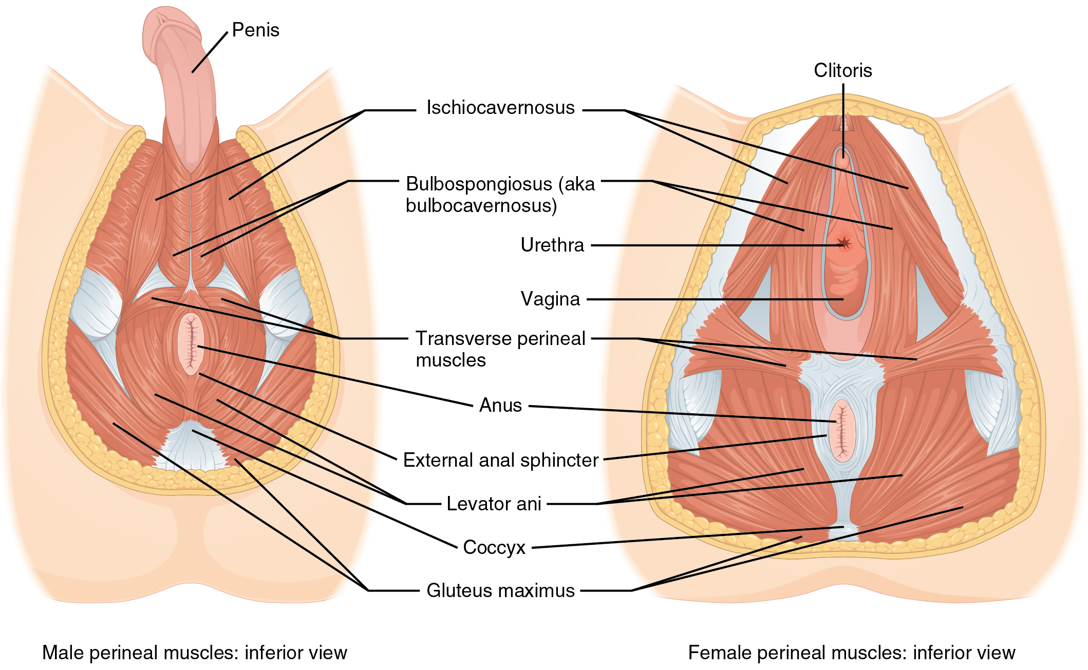
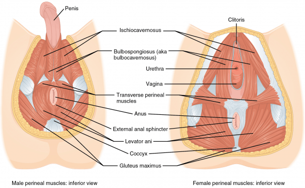
external anal sphincter
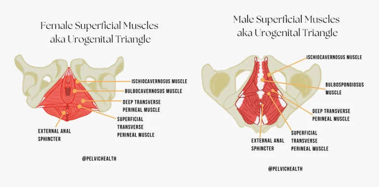
superficial transverse perineal muscle
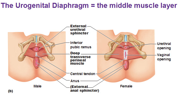
deep transverse perineal muscle
bulbospongiosus muscle
ischiocavernosus muscle
femal external urethral sphincter:
compressor urethrae
sphincter urethrovaginalis
female external urethral sphincter (proper)

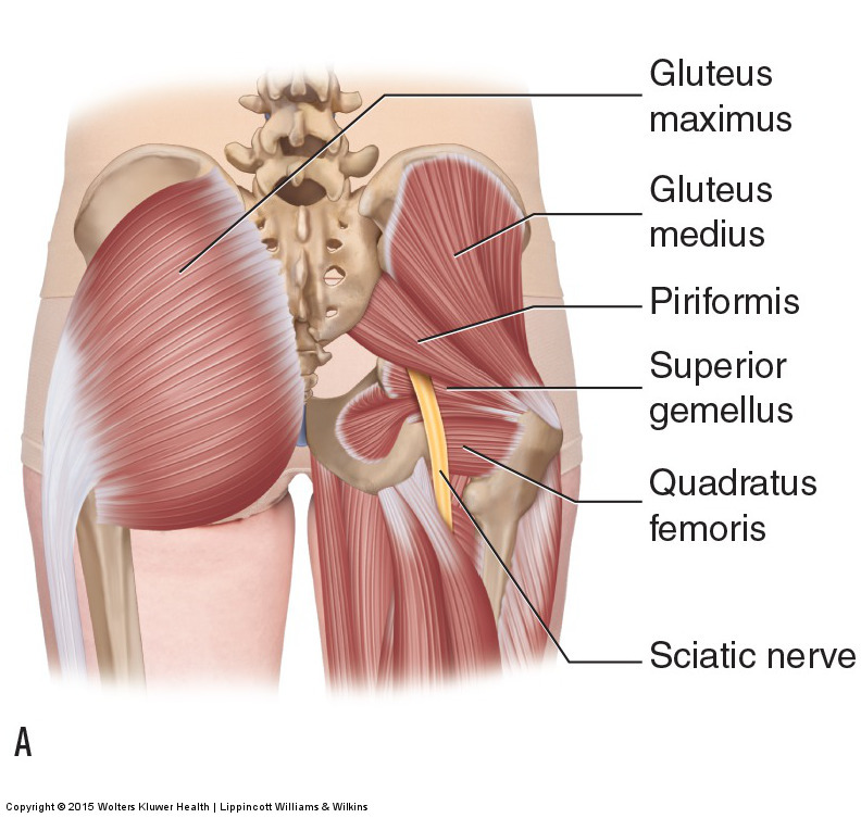
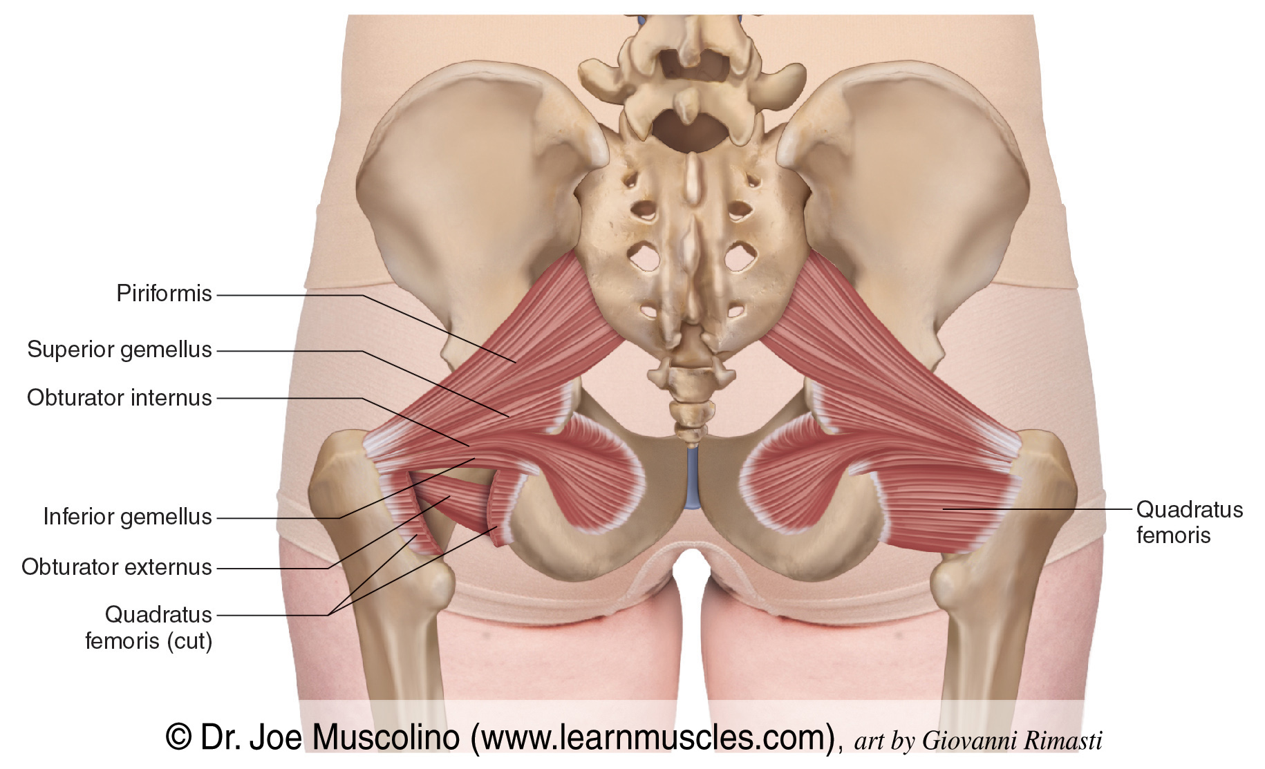
piriformis
a muscle of the gluteal region that lies deep to glute max
belongs to the short external rotators of the hip
spans between the sacrum and the greater trochanter of the femur, stabilizing the hip joint and moving the thigh
origin → anterior surface of the sacrum (between S2 & S4), gluteal surface of ilium (near posterior inferior iliac spine), sacrotuberous ligament
insertion → apex of greater trochanter of the femur
action → external rotation and abduction of the thigh at the hip joint; stabilizes head of femur in acetabulum
innervation → nerve to piriformis (S1-S2)
blood supply → superior gluteal artery, inferior gluteal artery, gemellar branches of internal pudendal artery
mnemonic:
Structures passing through the greater sciatic foramen inferior to piriformis muscle: PIN & PINS
(standing for: Posterior cutaneous nerve of thigh, Inferior gluteal vessels and nerves, Nerve to quadratus femoris, Pudendal nerve, Internal pudendal vessels, Nerve to obturator internus, Sciatic nerve)
coccygeus
a sheet of muscle and fibrous tissue that along with levator ani, comprises the pelvic diaphragm that forms the inferior wall of the true pelvis
extends between the ischium of the hip bone and the sacrum and coccyx
helps support the pelvic viscera and has a minor role in flexing the coccyx
may be described as the ischiococcygeus muscle and a part of the levator ani, but can also be referred to separately as coccygeus
origin → ischial spine
insertion → inferior end of sacrum, coccyx
action → supports pelvic viscera, flexes coccyx
innervation → anterior rami of spinal nerves S4 & S5
blood supply → inferior vesical artery, inferior gluteal artery, pudendal artery
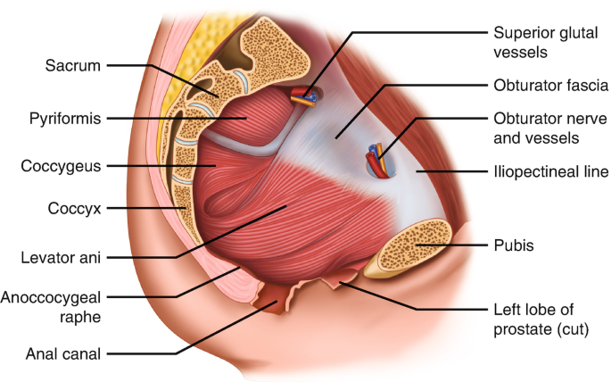
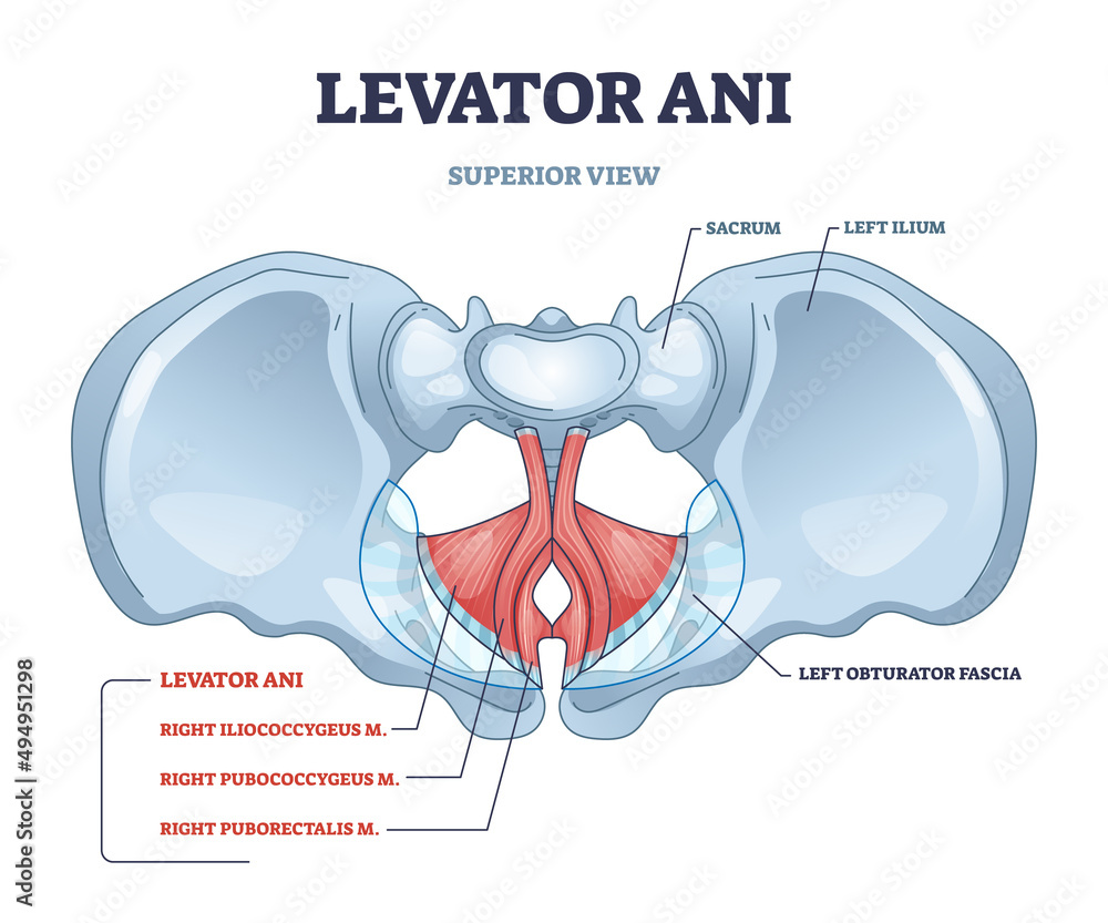
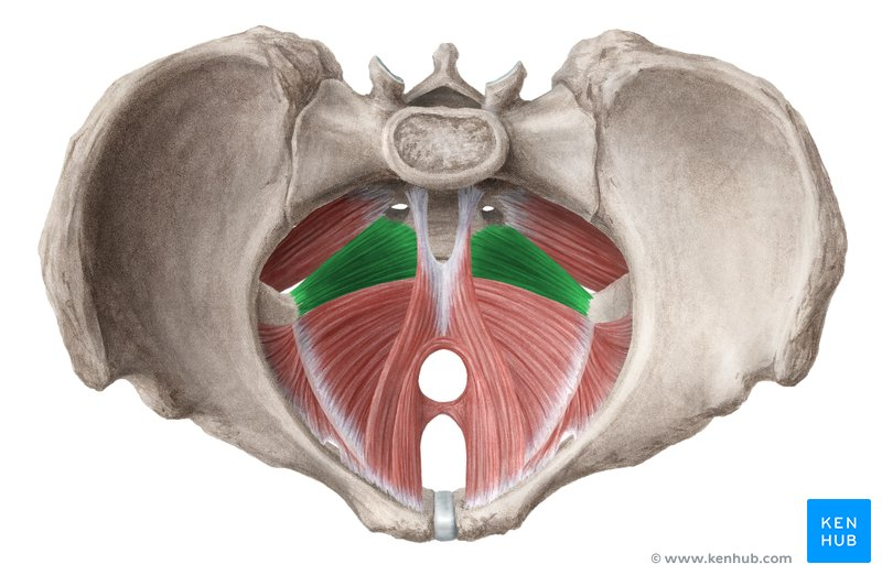
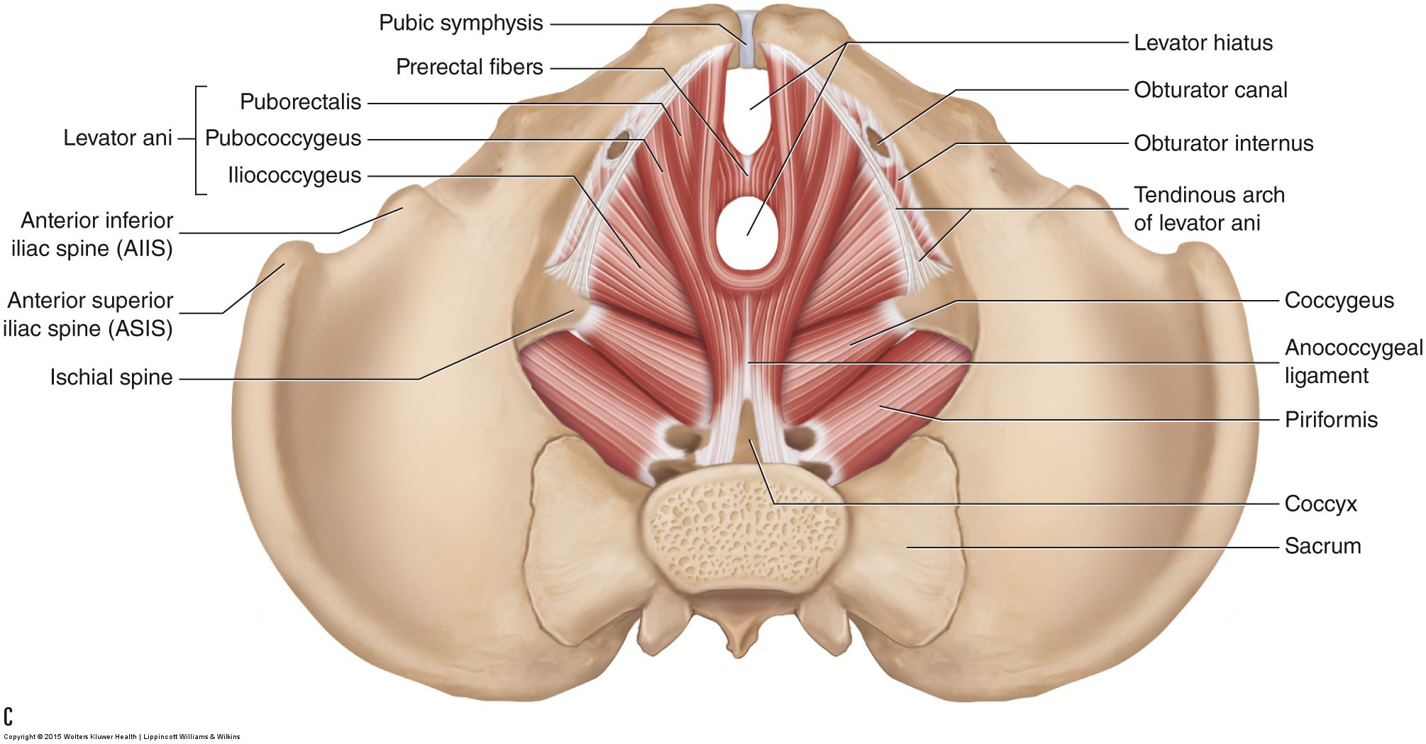
levator ani
a broad muscular sheet that forms the pelvic diaphragm along with the coccygeus
formed from 3 muscles:
puborectalis
pubococcygeus
iliococcygeus
function to stabilize the abdominal and pelvic organs and literally stop them from falling straight outta ya
origin → puborectalis: poserior surface of bodies of pubic bones
→ pubococcygeus: posterior surface of bodies of pubic bones (lateral to puborectalis)
→ ischiococcygeus: tendinous arch of internal obturator fascia, ishcial spine
insertion → puborectalis: none (forms puborectal sling posterior to rectum)
→ pubococcygeus: anococcygeal ligament, coccyx, perineal body, musculature of prostate/vagina
→ iliococcygeus: anococcygeal ligament, coccyx
action → stability and support of abdominal and pelvic organs, resistance against increased intra abdominal pressure, opening and closing of the levator hiatus
innervation → nerve to levator ani (S4); pubococcygeus also receives branches via inferior rectal/perineal branches of pudendal nerve (S2-S4)
blood supply → inferior gluteal, inferior vesical, pudendal arteries
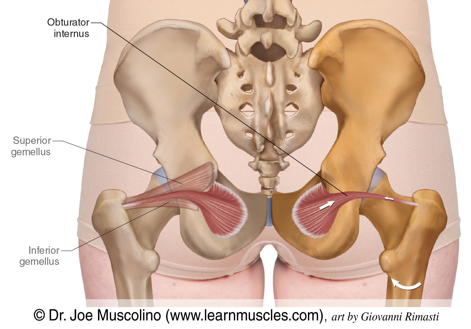
obturator internus
a bilateral, triangular shaped muscle situated deep within both the pelvic and gluteal regions
primarily considered a muscle of the lower limb (gluteal region)
referred to as the triceps coxae muscles along with the superior and inferior gemelli, which share a common tendon and insert at the greater trochanter of the femur
origin → posterior surface of obturator membrane; bony boundaries of obturator foramen
insertion → medial surface of the greater trochanter of the femur
action → external rotation of extended thigh; abduction of flexed thigh; stabilization of hip joint
innervation → nerve to obturator internus (L5 & S1)
blood supply → obturator artery, internal pudendal artery
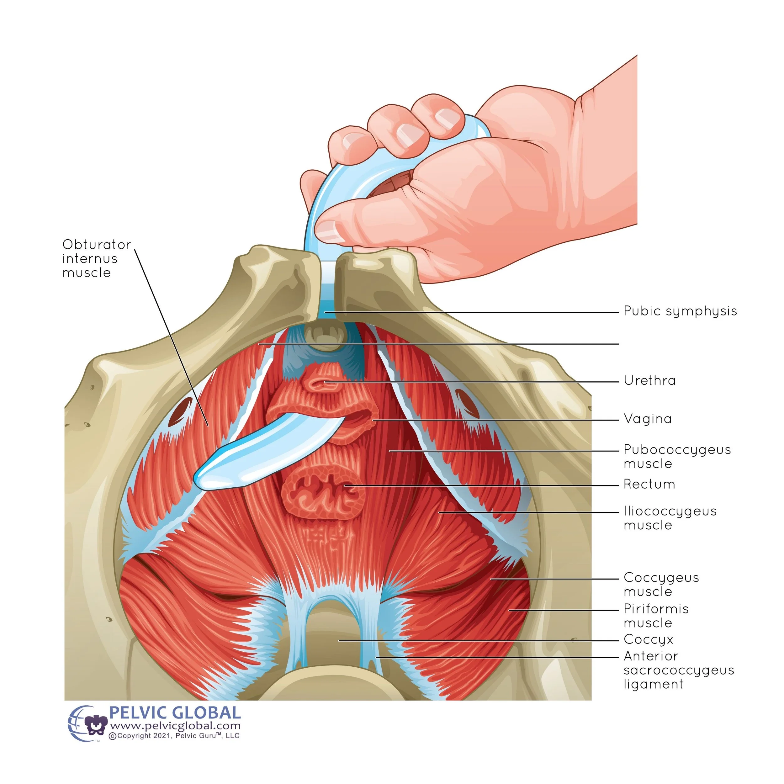
internal obturator internus release
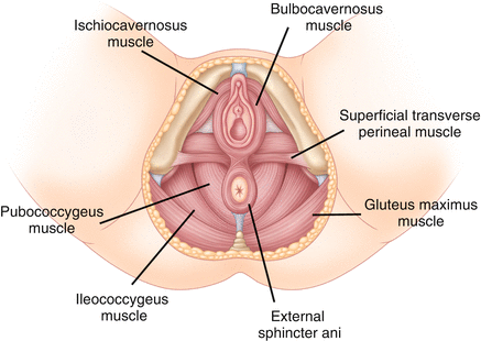
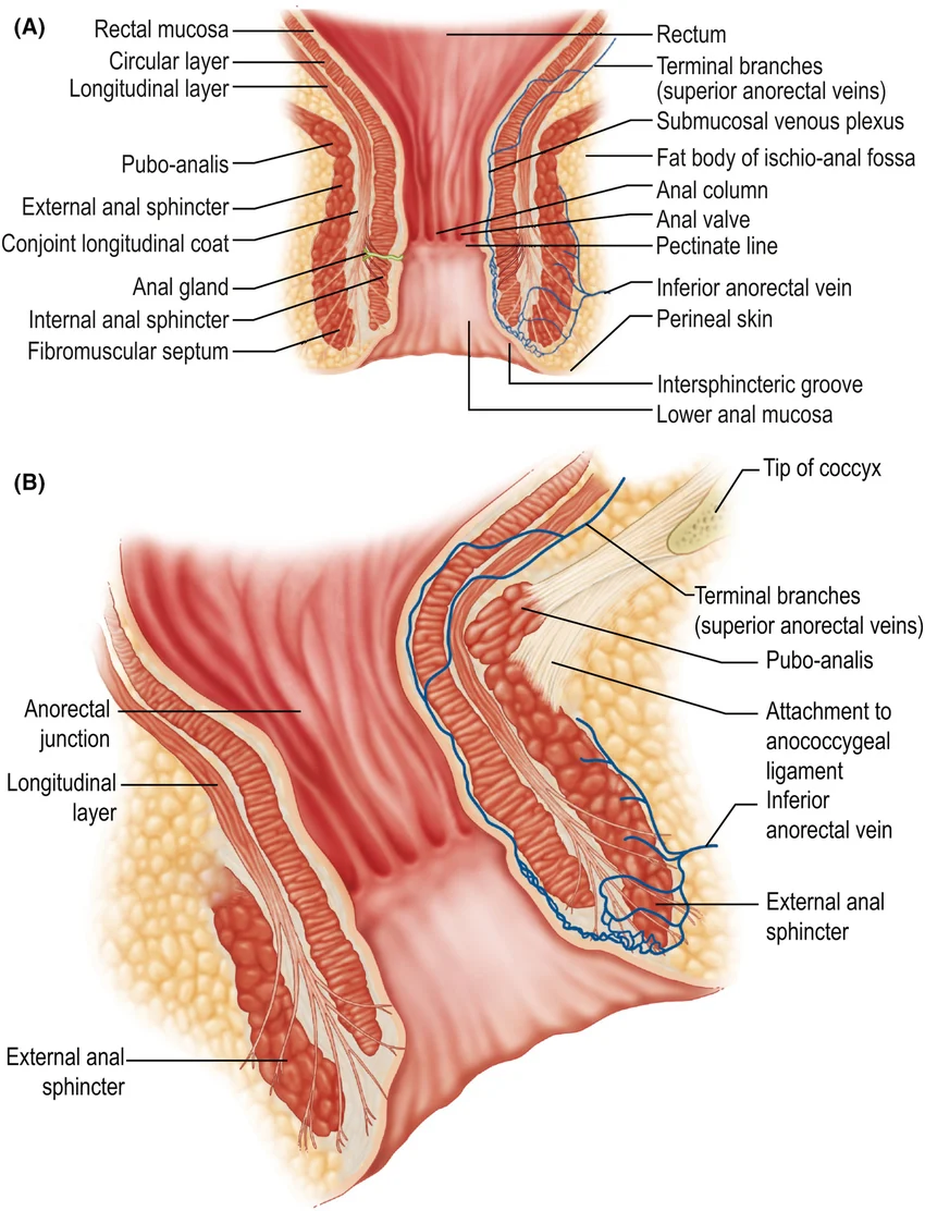
external anal sphincter
a short tube of skeletal muscle surrounding the inferior portion of the anal canal
largely under voluntary control by the somatic nervous system
origin → skin/fascia surrounding anal canal/anus
insertion → perineal body, anococcygeal ligament (encircles anus)
action → constricts anal canal, supports pelvic floor
innervation → inferior anal/rectal branch of pudendal nerve (S2-S4)
→ mnemonic: S2, S3, S4 keep the shit off the floor
blood supply → inferior rectal arteries
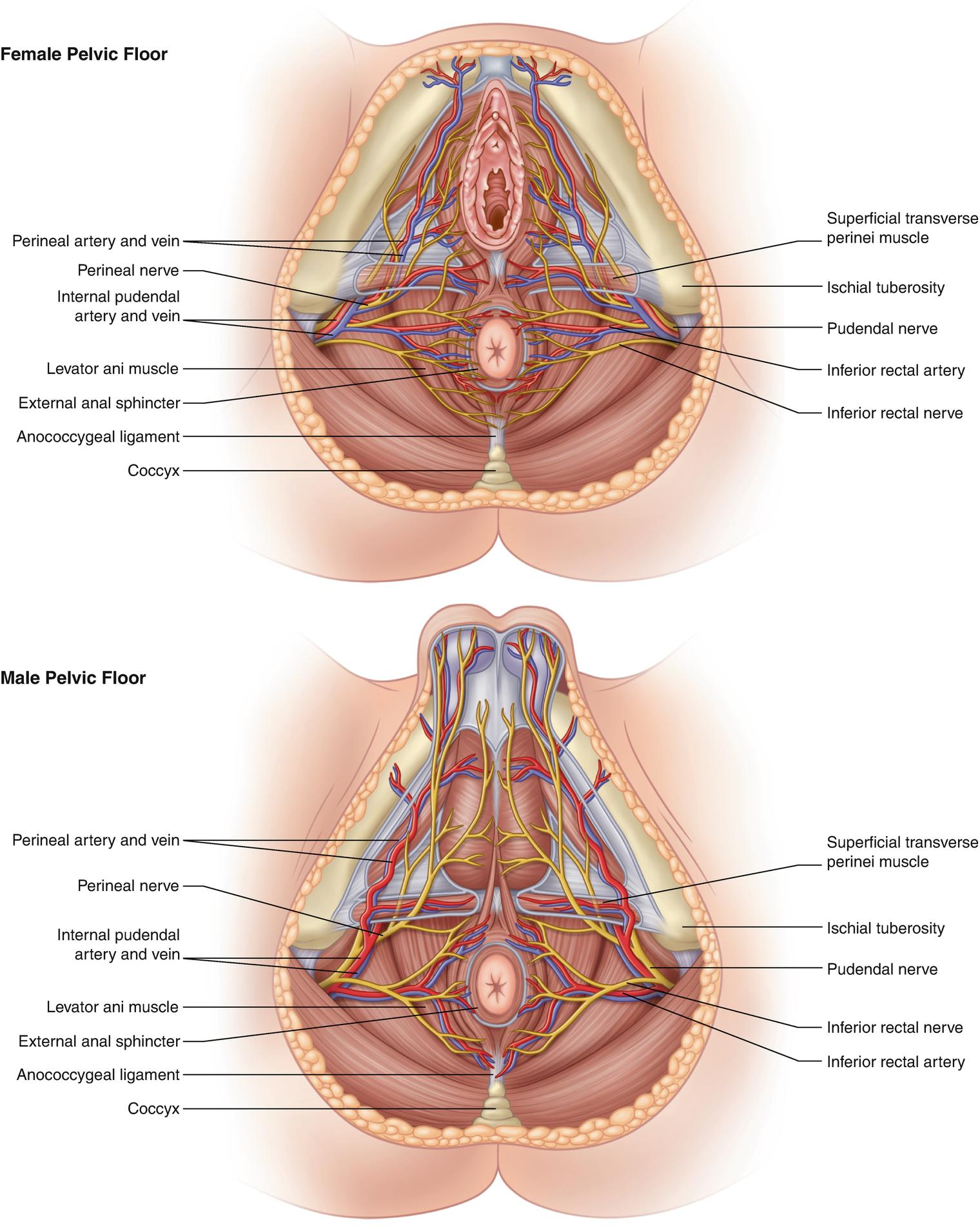
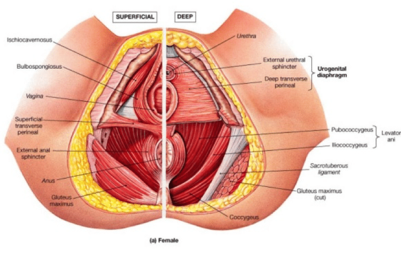
superficial transverse perineal muscle
a paired, narrow muscular slip in the superficial perineal pouch that helps stabilize the perineal body (the central tendon of the perineum)
origin → ischial tuberosity (medial surface) and the ramus of the ischium
insertion → the perineal body (the fibres of both muscles meet in the midline, interlace, and decussate/criss cross with the contralateral muscle, while also blending with the bulbospongiosus and external anal sphincter
action → bilateral contraction fixes and stabilizes the perineal body, providing structural support to the adjacent pelvic floor and perineal structures
innervation → perineal nerve (a branch of the pudendal nerve, S2-S4)
blood supply → perineal artery, a branch of the internal pudendal artery
deep transverse perineal muscle
a paired muscle in the deep perineal space
provides vital structural support to the pelvic floor and stabilizes the perineal body (central tendon)
plays a role in urinary continence, ejaculation, and supporting the vaginal wall
origin → inferior ramus of the ischium
insertion → the muscle fibres travel medially and interlace in the midline with the contralateral deep transverse perineal muscle. they envelope the perineal body and blend with the external anal sphincter and urethral sphincter
action → fixes and stabilizes the perineal body, supports the pelvic floor, aids in expulsion of semen (males), and the final drops of urine (both sexes)
innervation → deep muscular branch of the pudendal nerve (sacral plexus, spinal roots S2-S4)
blood supply → internal pudendal artery, perineal artery
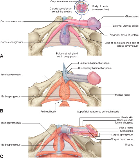
bulbospongiosus muscle
a paired muscle of the pelvic floor found in the superficial perineal space along with the ischiocavernosus and superficial transverse perineal muscles
sexually dimorphic: wrapped around the bulb of the penis and corpus spongiosum in males, encircles the vaginal orifice in females
compresses erectile tissues
facilitates urination, ejaculation, erection in males
contributes to erection of the clitoris and emptying the greater vestibular glands in females
supports the perineal body in both sexes
origin → male: perineal body, median penile raphe
→ female: perineal body
insertion → male: perineal membrane, dorsal aspect of corpus spongiosum and corpa cavernosa, fascia of bulb of penis
→ female: pubic arch, fascia of corpa cavernosa and clitoris
action → male: compresses the bulb of the penis during urination/ejaculation, assists in erection of the penis, supports perineal body
→ female: assists in erection of clitoris/bulb of vestibule, supports perineal body
innervation → deep branch of perineal nerve (of pudendal nerve) (S2-S4)
blood supply → perineal artery
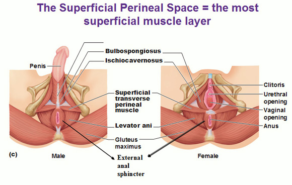
ischiocavernosus muscle
a bilateral perineal muscle located in the superficial perineal space of the urogenital triangle
extends between the ischium of the hip bone and the crura of the penis or clitoris
contraction helps to maintain penile/clitoral erections during sexual arousal and intercourse
origin → ischial tuberosity and ramus
insertion → crus of penis or clitoris
action → pushes blood from root of clitoris/penis to body (maintains erection of penis/clitoris)
innervation → deep branch of perineal nerve (of pudendal nerve (S2-S4)
blood supply → perineal artery
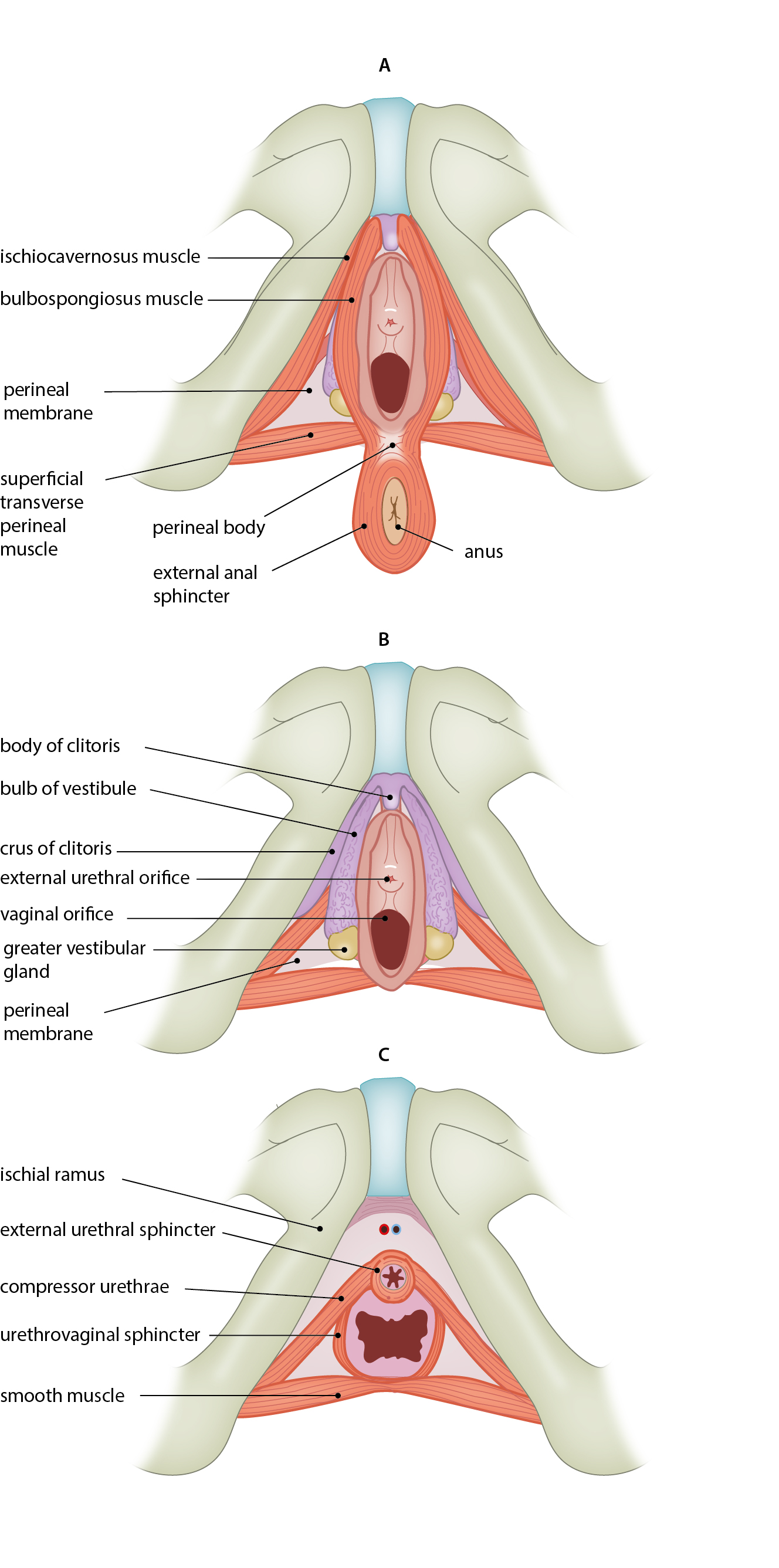
compressor urethrae
a female specific muscle
works as an accessory urinary sphincter by wrapping around and squeezing the urethra against the vagina to prevent involuntary leakage and maintain continence
origin → ischiopubic rami
insertion → its fibres cross anterior to the urethra and interlace with the muscle fibres of the opposite side
action → constricts and compresses the urethra, augmenting urinary continence
innervation → perineal branch of the pudendal nerve (S2-S4)
blood supply → internal pudendal artery
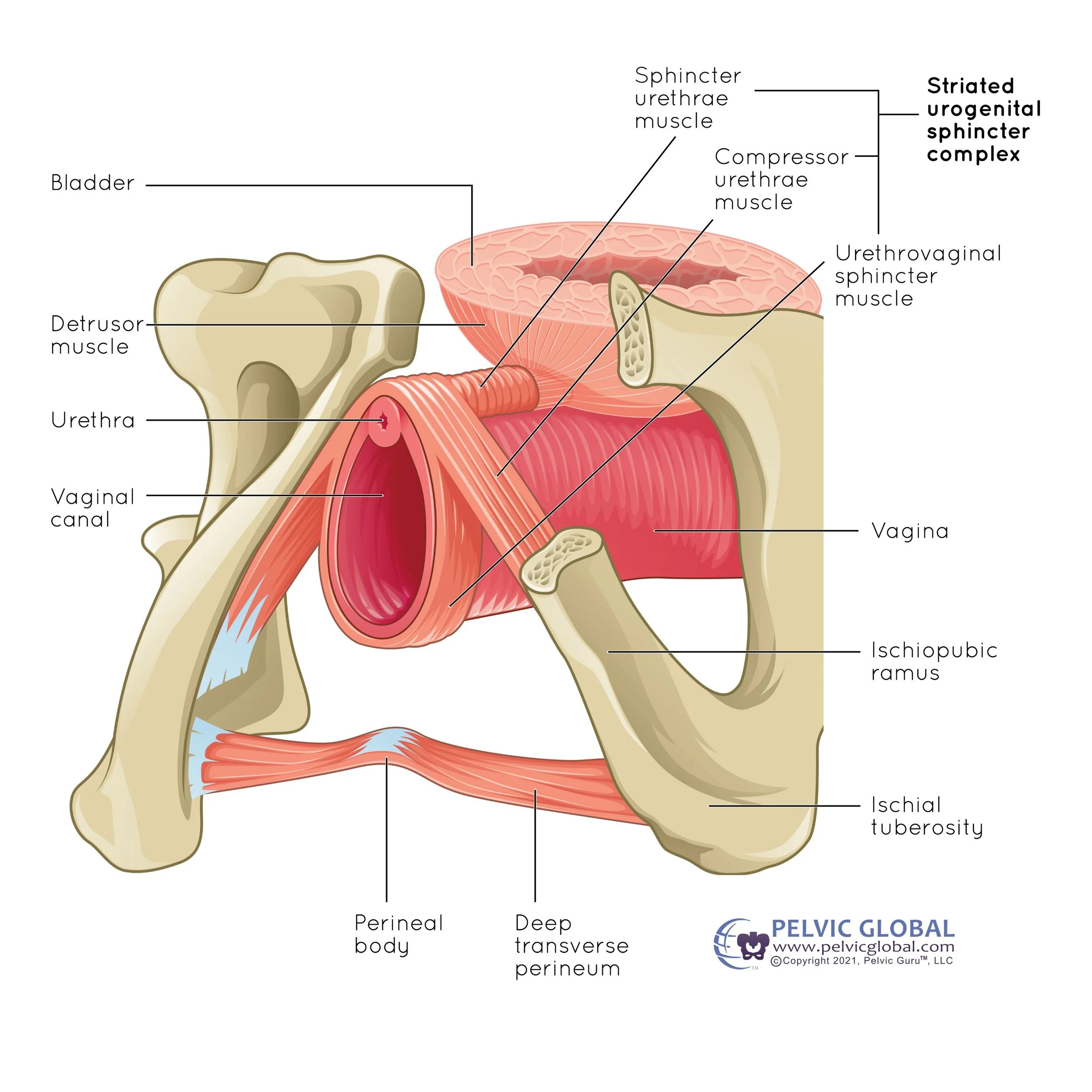
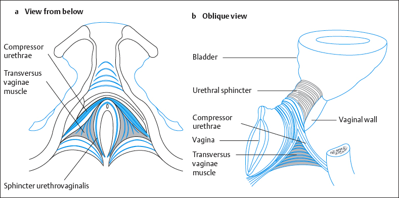
sphincter urethrovaginalis
specific to the female urogenital triangle
an accessory sphincter for the urinary tract and vagina
a key component in maintaining urinary continence and supporting pelvic floor function
origin → perineal body
insertion → the muscle fibres course anterolaterally to wrap around and encircle both the urethra and vagina, blending with fibres from the opposite side
action → constricts both the urethra and vagina to aid in urinary continence and regulate vaginal tone
innervation → somatic pudendal nerve, specifically perineal branches (S2-S4)
blood supply → perineal artery (branch of internal pudendal artery)
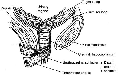
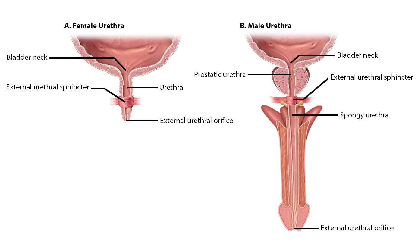
external urethral sphincter
a skeletal muscle that wraps around the urethra, critical for voluntary urinary continence
origin → ischiopubic ramus and neighbouring perineal fascia
insertion → fibrous raphe (median septum) on the posterior aspect of the urethra and intermeshing fibres with the opposite side
action → constricts the urethra to stop the flow of urine
innervation → somatic: perineal branch of pudendal nerve (s2, S3, S4)
blood supply → internal pudendal artery and its branches (perineal artery, bulbourethral artery)