Ch: 8 Gallbladder and Biliary System
 Call Kai
 Learn
Learn Practice Test
 Spaced Repetition
 Match
Match Flashcards
 Knowt Play
1/76

There's no tags or description
Looks like no tags are added yet.
Name | Mastery | Learn | Test | Matching | Spaced | Call with Kai | Chat |
|---|
No analytics yet
Send a link to your students to track their progress
77 Terms
Physiology: Functions of the Gallbladder
concentrates and stores bile, regulates biliary pressure, and contracts after eating
Physiology: what causes the gallbladder to contract
the secretion of CCK (cholecystokinin). could be after fatty food enters small intestine, or by IV CCK injections (for patients who are TPN (total parenteral nutrition))
Physiology: steps/process of bile do duodenum
food enters small intestine and CCK is secreted, the gb contracts and sphincter of Oddi relaxes. bile is released into the cystic duct, flows through the CBD, and enters the duodenum
Normal Variants/locations of the Gallbladder
mobile, ectopic (on the left, midline, transversely), in RLQ, partially or totally embedded in liver parenchyma, in abdominal wall or falciform ligament, in retroperitoneum, or above the liver
How common are Congenital Anomalies of the Gallbladder and when do they occur
They are rare and most occur between 4-12 gestational weeks
Congenital anomaly symptoms
the anomalies alone do not give rise to any characteristic symptoms but some defects do predispose to bile stasis (flow of bile is slowed or stopped) and attacks of cholecystitis
What is septate gallbladder and how is it found
Congenital anomaly. The gallbladder normally develops from cannulation of a blind sac (hepatic diverticulum), but septum forms if that is not complete.
it is often found incidentally
Septate gallbladder sonographic appearance
single septum appears as thin, linear echo
WHat can mimic septate gallbladder and how to differentiate
Simple junctional fold (gb is partially folded over itself) may mimic a septum. junctional fold will most likely shift when patient is rolled, unlike septate which will remain.
What is Biliary Atresia and where is it most commonly found
Congenital Anomaly. Destruction of extrahepatic biliary system and its the most common type of obstructive biliary disease in infants and young children
what percent of neonates have transient jaundice and when does it indicate serious abnormality, and where does it commonly come from
more that 50% have transient jaundice, but if a neonate has persistent or sudden onset jaundice after 1-2 weeks of life, that may indicate serious abnormality
Where does jaundice commonly come from in neonates
biliary atresia or neonatal hepatitis
What sex is biliary atresia commonly found
2x more common in males
What sex is neonatal hepatitis commonly found
4x more common in females
How can biliary atresia be repaired and its outcome
surgically with liver transplant or Kasai procedure (uses portion of small intestine as extrahepatic duct system).
outcome is better with early surgical intervention
Complications of untreated biliary atresia
cirrhosis, cholangitis (inflammation of intrahepatic ducts), portal HTN, failure of biliary drainage
2 sonographic indicators seem with biliary atresia
Portal HTN and small GB
What is/what causes a Choledochal Cyst
A congenital weakness of duct walls which results in cyst formation, or angulation of CBD causing partial obstruction leading to dilatation and cyst formation
Choledochal Cyst symptoms
Intermittent jaundice with colicky pain, failure to thrive, or palpable subhepatic mass displacing stomach or duodenum
What sex is Choledochal Cyst more common in
4x more common in females
Why is surgical management of Choledochal Cyst recommended
Due to increased incidence of malignant transformation
Choledochal Cyst sonographic appearance
localized cystic mass separate from GB in region of porta hepatis, and may be intrahepatic (adjacent to bile duct).
Must verify peristalsis to avoid mistaking fluid-filled bowel for a cyst (if there is movement, its most likely bowel appearing cystic)
What is biliary sludge
An acquired disease that represents precipitates (cholesterol depostis, calcium salts) formed in bile and settles in the gb.
Pathogenesis of biliary sludge
uncertain
Biliary sludge causes/associations and precursor for what
comes from stagnant bile, periods of prolonged fasting, total parenteral nutrition (TPN), pregnancy, or rapid weight loss.
May also be associated with other complications that can be a precursor for GB disease
Biliary sludge symptoms
Can produce pain bc gb is trying to contract with thick “mud like” material inside
Biliary sludge Sonographic appearance
homogeneous, non-shadowing echo pattern, often forms a fluid-fluid level that remains constant in sagittal and tranverse images, and a gb filled with sludge can be difficult to distinguish from adjacent liver parenchyma

What is fluid-fluid level
When sludge in gb has division of where anechoic fluid is and sludge area is by distinct line
What is Tumefactive sludge and why use color doppler
not layered sludge, has more mass appearance (sludge ball).
color doppler will help differentiate sludge ball from other pathologies/abnormalities bc sludge ball will be avascular

What is cholelithiasis and how common is it and what they contain
An acquired disease. gallstones, and they are very common. majority of calculi contain mixture of cholesterol, bilirubin, and calcium
Cholelithiasis size and quantity
size varies, could be small or large (small are more dangerous bc there easier to cause blockages)
they could be single or multiple
Cholelithiasis symptoms
Could be asymptomatic or experience RUQ pain, N/V, right shoulder or back pain
Cholelithiasis prevalence and risk factors
More prevalent in females.
most common risk factors are female, obesity, age, pregnancy, or diabetes but could also be from TPN, ethnicity, cirrhosis, or crohns disease
Cholelithiasis sonographic appearance
mobile, gravity dependent, echogenic with posterior enhancement, and small stone are typically multiple and described as gravel and mostly lay on posterior wall with shadowing

Non-mobile structures
polyps, impacted stones, or stones adherent to gb
Cholelithiasis: Wall-Echo-Shadow Triad (WES)
LOOK BACK AT SLIDES IDK

What is Acute Cholecystitis and what does it result from/in
An acquired disease with swelling/irritation of the gb.
up to 25% of cases result from impacted stones with gb neck or cystic duct.
may result in necrosis, ulceration, swelling, or edema.
Acute Cholecystitis symptoms
RUQ pain or palpable mass, positive murphy sign (pain with pressure), N/V, or abdominal distention, fever, and/or jaundice
Acute Cholecystitis lab values
can be non-specific. things to look for include leukocytosis (elevated WBC), hyperbilirubinemia, or elevated ALP
Acute Cholecystitis surgical intervention or what is done if surgery isn’t an option
leads to fewer complications. poor surgical candidates will receive IV antibiotics or gb drainage
What percent of Acute Cholecystitis cases resolve spontaneously
about 60%
Acute Cholecystitis Sonographic appearance
gb wall thickening, gallstones, pericholecystic fluid (fluid around gb), hydropic gb (distended gb filled with mucous, water, or fluid other than bile from prolonged blockage of cystic duct)
color doppler detects hyperemia

Complications of Cholecystitis
empyema (build up of pus in/around gb), gallbladder perforation (tear in gb wall and bile leaks into peritoneal cavity and cause biloma), or gangrenous (from necrosis or infection) or Emphysematous cholecystitis (gas buildup)
What is Acute Acalculous Cholecystitis and where is it commonly found
An acquired disease. its acute cholecystitis with no gallstones present. common in ICU patients
What is Acute Acalculous Cholecystitis a result of
stagnant bile and vascular changes. stagnant bile leads to overdistension of gb and vascular changes may lead to thrombosis of vessels supplying gb.
Acute Acalculous Cholecystitis causes
long term TPN, trauma, or surgery
Acute Acalculous Cholecystitis Symptoms
May be non specific but typically could experience RUQ pain, positive murphy sign, N/V, abdominal distention, or fever.
The difficulty diagnosing yields high morbidity and mortality
Acute Acalculous Cholecystitis sonographic appearance
GB distended, thickened wall, internal debris or sludge, pericholecystic fluid and/or edema

What is Chronic Cholecystitis and Associated Conditions and what to patients tend to have intolerance to
An acquired disease of recurrent inflammatory changes in GB secondary to infection, obstruction, or metabolic disorders. this is often associated with stones, and patients tend to have intolerance to fatty foods
Chronic inflammation of GB wall results in what
thickening and fibrosis (tough/hardened wall which is harder to contract)
Chronic Cholecystitis and Associated Conditions clinical clues
N/V, RUQ pain, or jaundice
Chronic Cholecystitis and Associated Conditions Sonographic clues
look for gallstones, evenly thickened walls, smaller GB, and decreased response to CCK

What is porcelain gallbladder and where is comes from and what it can cause
occurs when all parts of GB wall is calcified. It is a rare manifestation of chronic cholecystitis and is associated with high incidence of GB carcinoma
Porcelain gallbladder sonographic appearance
Single echogenic line representing calcified wall. All parts of the wall may be calcified and could have posterior shadowing that would obscure the GB

What measurement is considered thickening of Gallbladder wall
>3mm
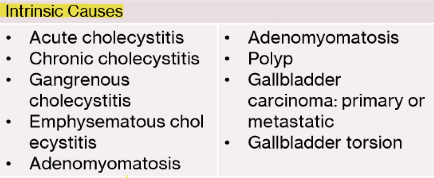
Intrinsic causes of gallbladder wall thickening
Something within the GB or a condition of the GB that causes thickened wall.
Acute cholecystitis, Chronic cholecystitis, Gangrenous cholecystitis, Emphysematous cholecystitis, Adenomyomatosis, Polyp, Gallbladder carcinoma: primary or metastatic, or Gallbladder torsion
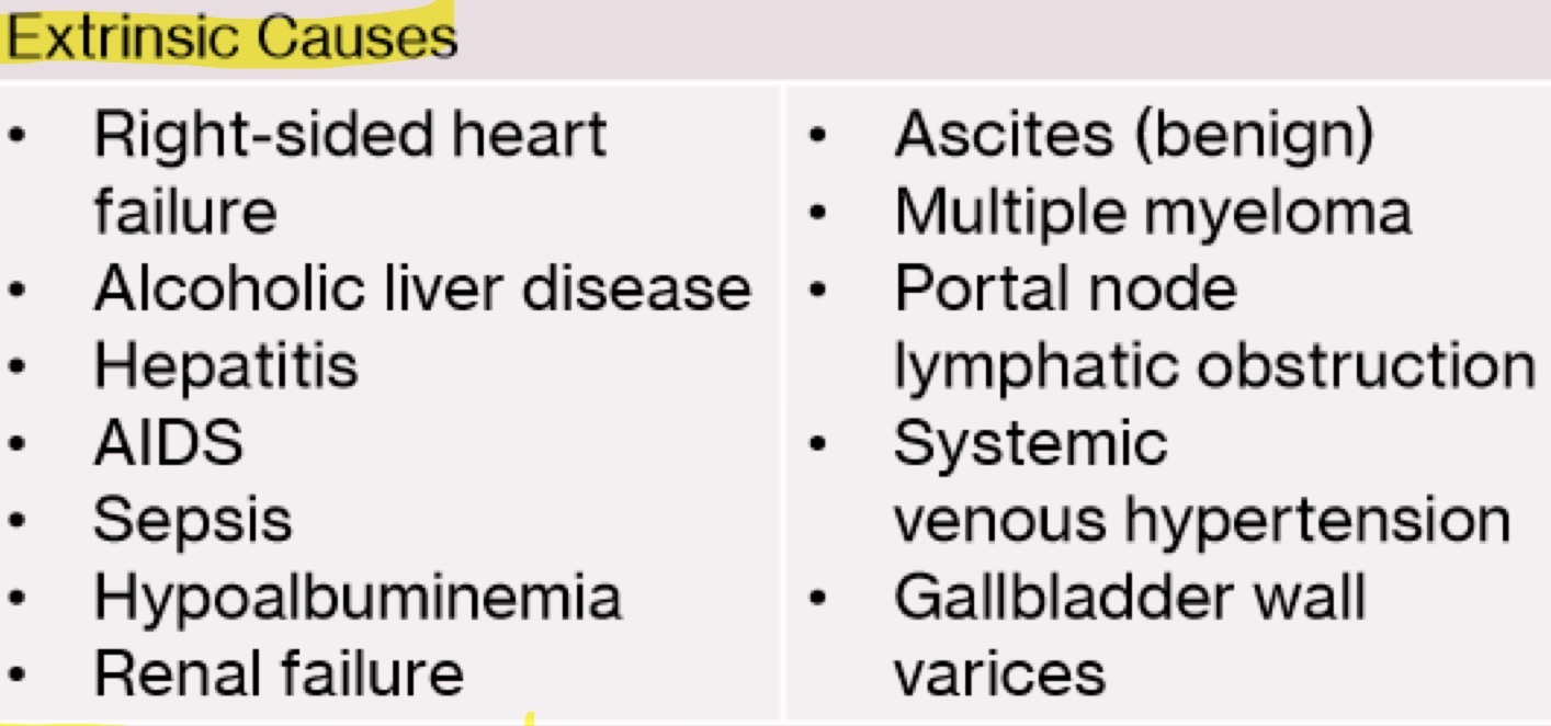
Extrinsic causes of gallbladder wall thickening
Non-GB related issue that may have side effects/complications that result in wall thickening.
Right-sided heart failure, Alcoholic liver disease, Hepatitis, AIDS, Sepsis, Hypoalbuminemia, Renal failure, Ascites (benign), Multiple myeloma, Portal node lymphatic obstruction, Systemic venous hypertension, or Gallbladder wall varices
Physiologic causes of gallbladder wall thickening
contracted gallbladder after eating
What is Cholestasis and Pregnancy relevance
Acquired disease where flow of bile from the liver is reduced or blocked. intrahepatic cholestasis occurs in 2nd+3rd trimester and resolves after delivery
What can the mother present with, with Cholestasis
pruritus (itchy skin), elevated ALP, serum transaminase (ALT), and bile acid
Severity/risks of cholestasis for mother and baby
for the mother, its a benign condition
the fetus is at risk for prematurity (delivered prematurely), dysrhythmia (irregular heartbeat), distress, or intrauterine death
What is a Gallbladder Polyp
Acquired benign neoplasm that is a fix, non-mobile mass extending from GB mucosa (inner lining)
What is gallbladder polyp caused by
chronic inflammation, hyperplasia of GB wall, or lipid deposits
What can present as a polyp
early stages of GB cancer
When should polyps be removed
When they are >1cm because they are suggestive of malignancy
Gallbladder polyp symptoms and associations
They are typically found incidentally and asymptomatic. there are no associations with age, gender, obesity, etc.
Gallbladder polyp sonographic appearance
Echogenic small masses along the GB wall

How common is Gallbladder Carcinoma and who is effects
it is an acquired malignant neoplasm and its rare and affects women >72 years old
Gallbladder carcinoma risk factors
Chronic cholecystitis, cholelithiasis (approx. 95% of cases), rapidly growing polyps, or porcelain gallbladder (approx. 25% of cases)
Survival rate of Gallbladder carcinoma
5 year survival rate is <5%
Early stages of gallbladder carcinoma detectability and signs/symptoms
difficult to detect and patients may be asymptomatic or present with symptoms similar to cholecystitis. no lab value variances to assist early diagnosis
Late stages of gallbladder carcinoma findings
jaundice, malaise, and weight loss. by this point, cancer has grown/spread.
Gallbladder carcinoma sonographic appearance
Look for irregular shaped mass that replaces GB (GB looks filled with mass like structure), irregular wall thickening, direct invasion into liver, or gallstones surrounded by echogenic mass.
color doppler will demonstrate internal vascularity

Most common tumor to metastasize to GB
Melanoma
Metastasis to Gallbladder prognosis and association
has a poor prognosis and is not associated with cholelithiasis, which differs from primary GB cancer
Metastasis of Gallbladder sonographic appearance
asymmetric wall thickening and solitary or multiple masses in GB
Metastasis of Gallbladder treatment
cholecystectomy