Anatomy Lecture Exam 3

0.0(0)
Studied by 4 people
call kaiCall Kai
Locked
learnLearn
examPractice Test
spaced repetitionSpaced Repetition
heart puzzleMatch
flashcardsFlashcards
GameKnowt Play
Card Sorting

1/52

flashcard set

Earn XP

Description and Tags

Anatomy Lecture Exam 3

Last updated 3:22 AM on 7/13/26
Name
Mastery
Learn
Test
Matching
Spaced
Call with Kai
Chat

No analytics yet

Send a link to your students to track their progress

53 Terms
1
New cards
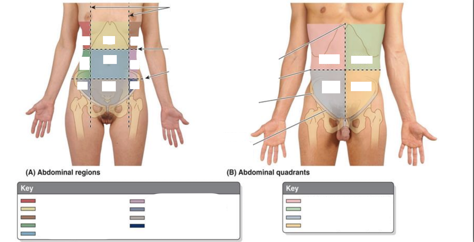
<p>Describe the regional and quadrant reference systems of the abdomen.</p>

Describe the regional and quadrant reference systems of the abdomen.
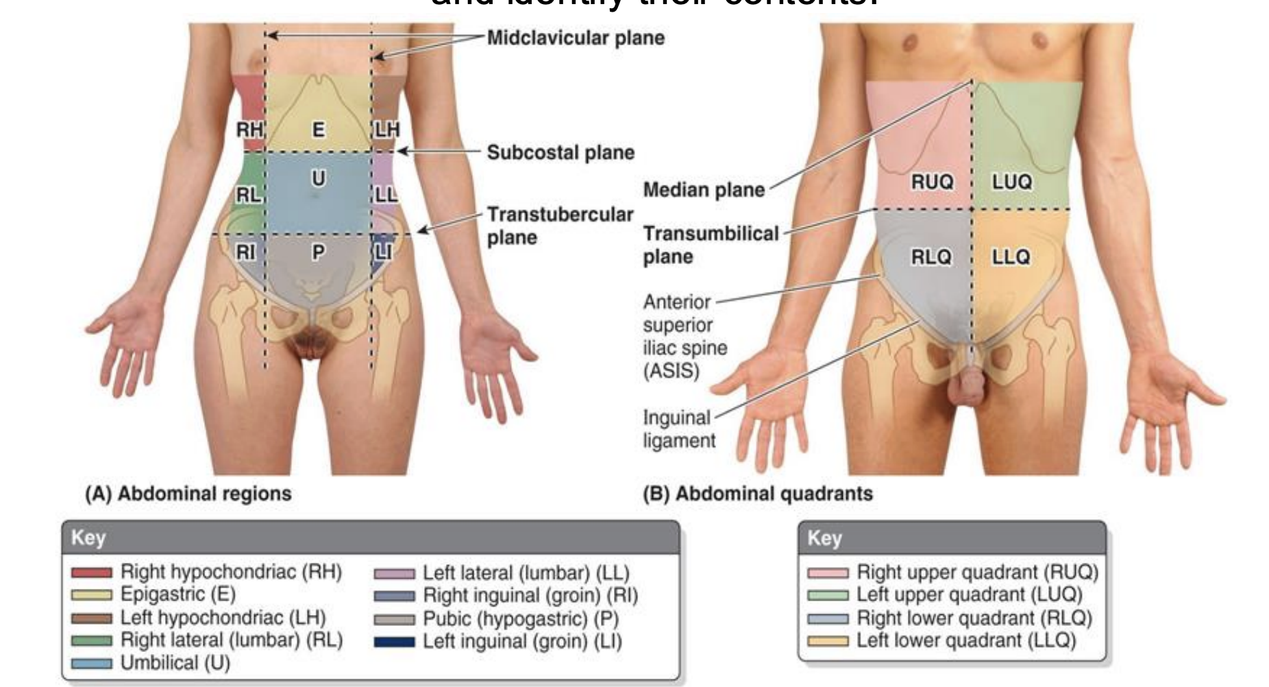
knowt flashcard image
2
New cards
<p>Identify the contents of the regional and quadrant reference systems of the abdomen.</p>

Identify the contents of the regional and quadrant reference systems of the abdomen.
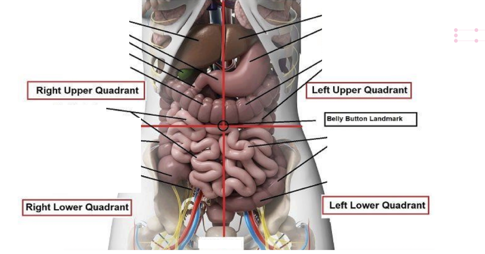
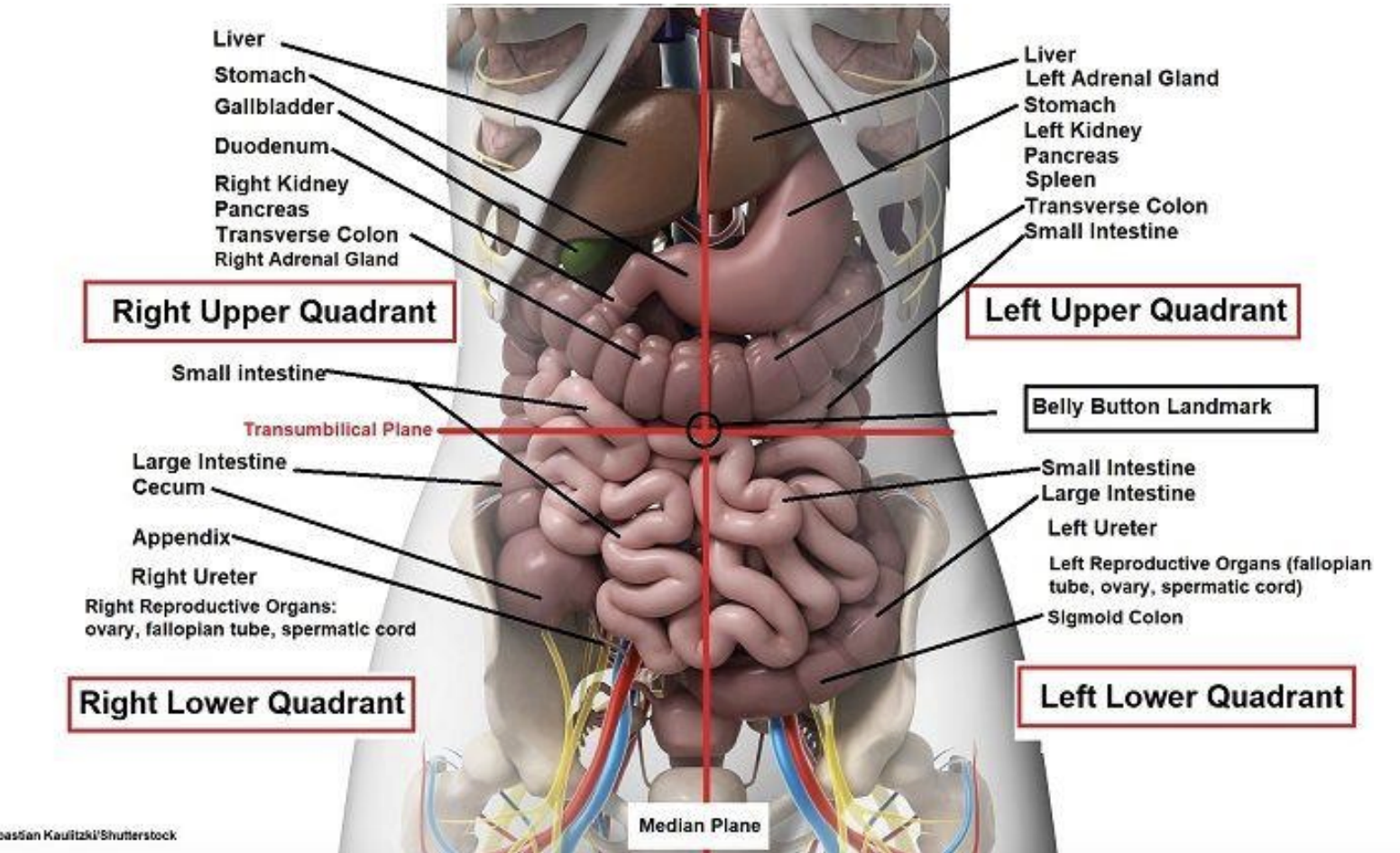
Right Upper Quadrant:

Liver, Stomach, Gallbladder, Duodenum, Right Kidney, Pancreas, Transverse Colon, Right Adrenal Gland, Small Intestine

Left Upper Quadrant:

Liver, Left Adrenal Gland, Stomach, Left Kidney, Pancreas, Spleen, Transverse Colon, Small Intestine

Right Lower Quadrant:

Small Intestine, Large Intestine, Cecum, Appendix, Right Ureter, Right Reproductive Organs: Ovary, Fallopian Tube, Spermatic Cord

Left Lower Quadrant:

Small Intestine, Large Intestine, Left Ureter, Left Reproductive Organs: Ovary, Fallopian Tube, Spermatic Cord, Sigmoid Colon
<p><strong>Right Upper Quadrant:</strong></p><p>Liver, Stomach, Gallbladder, Duodenum, Right Kidney, Pancreas, Transverse Colon, Right Adrenal Gland, Small Intestine</p><p></p><p><strong>Left Upper Quadrant:</strong></p><p>Liver, Left Adrenal Gland, Stomach, Left Kidney, Pancreas, Spleen, Transverse Colon, Small Intestine</p><p></p><p><strong>Right Lower Quadrant:</strong></p><p>Small Intestine, Large Intestine, Cecum, Appendix, Right Ureter, Right Reproductive Organs: Ovary, Fallopian Tube, Spermatic Cord</p><p></p><p><strong>Left Lower Quadrant:</strong></p><p>Small Intestine, Large Intestine, Left Ureter, Left Reproductive Organs: Ovary, Fallopian Tube, Spermatic Cord, Sigmoid Colon</p><p></p>
3
New cards
<p>Femoral Hernia</p>

Femoral Hernia

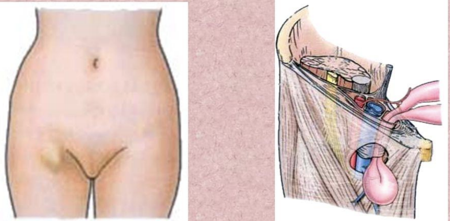
A femoral hernia is common in women than in men (possibly due to a wider pelvis and femoral canal). If a loop of intestine is forced into the femoral ring, it expands to form a swelling in the upper part of the thigh.
4
New cards
<p>Indirect Hernia</p>

Indirect Hernia

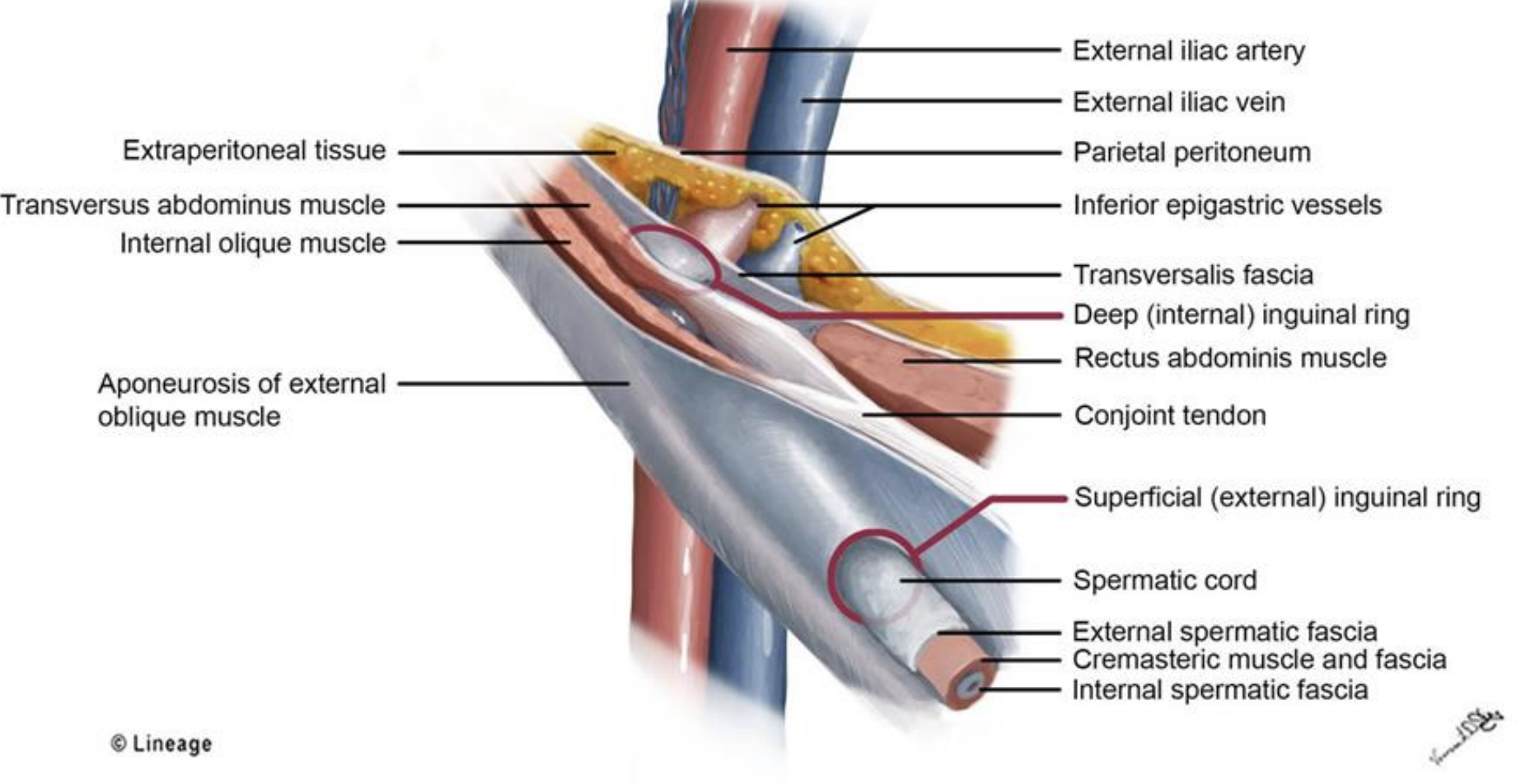
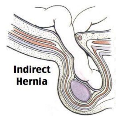
-Traverses inguinal canal

-Same course as spermatic cord

-Enter inguinal canal at the deep inguinal ring

-Lateral to the inferior epigastric vessels

-Can pass into the scrotum or labia majora

-Male > female

-Congenital, injury
5
New cards
<p>Direct Hernia</p>

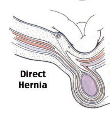
Direct Hernia

- Bulge through weakened fascia of abdominal wall

- Directly behind the superficial inguinal ring

- Medial to the inferior epigastric vessels

- Rarely enter the scrotum

- Common in elderly men with weak abdominal muscles
6
New cards
<p>Referred Pain</p>

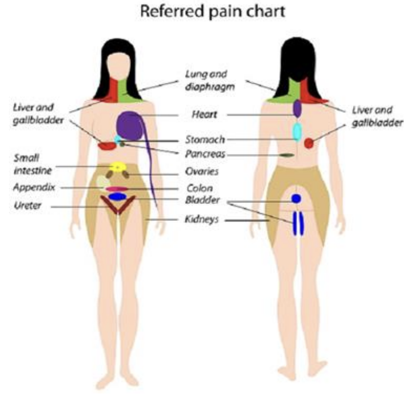
Referred Pain

For parietal peritoneum pain, the symptoms tend to be well localized as the sensory fibers travel directly with the corresponding spinal nerve in each dermatome.

In visceral peritoneal pain, the sensory fibers pass through the associated plexus before making their way back to the various spinal cord levels. This means visceral pain is poorly localized to the following regions;

● Foregut pain - experienced in the epigastric region

● Midgut pain - experienced in the umbilical region

● Hindgut pain - experienced in the pubic region
<p><strong>For parietal peritoneum pain, the symptoms tend to be well localized </strong>as the sensory fibers travel directly with the corresponding spinal nerve in each dermatome. </p><p></p><p>In visceral peritoneal pain, the sensory fibers pass through the associated plexus before making their way back to the various spinal cord levels. This means <strong>visceral pain is poorly localized </strong>to the following regions; </p><p>● Foregut pain - experienced in the epigastric region </p><p>● Midgut pain - experienced in the umbilical region </p><p>● Hindgut pain - experienced in the pubic region</p>
7
New cards

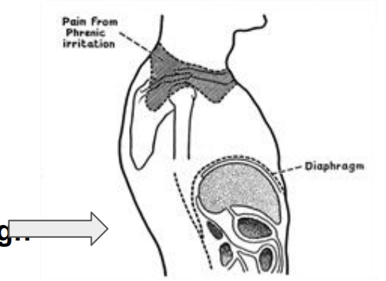
What is Kehr’s sign?

Parietal pain at the diaphragm relayed through the phrenic nerve - Kehr’s sign
<p>Parietal pain at the diaphragm relayed through the phrenic nerve - Kehr’s sign</p>
8
New cards
<p>Mesentery</p>

Mesentery

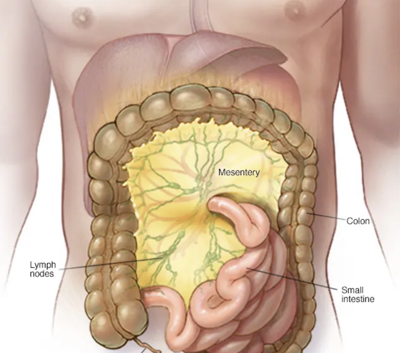
double layer of peritoneum covering neurovascular components and connective tissue, connecting intraperitoneal organs to the abdominal wall, such as with the small bowel, transverse colon and sigmoid colon
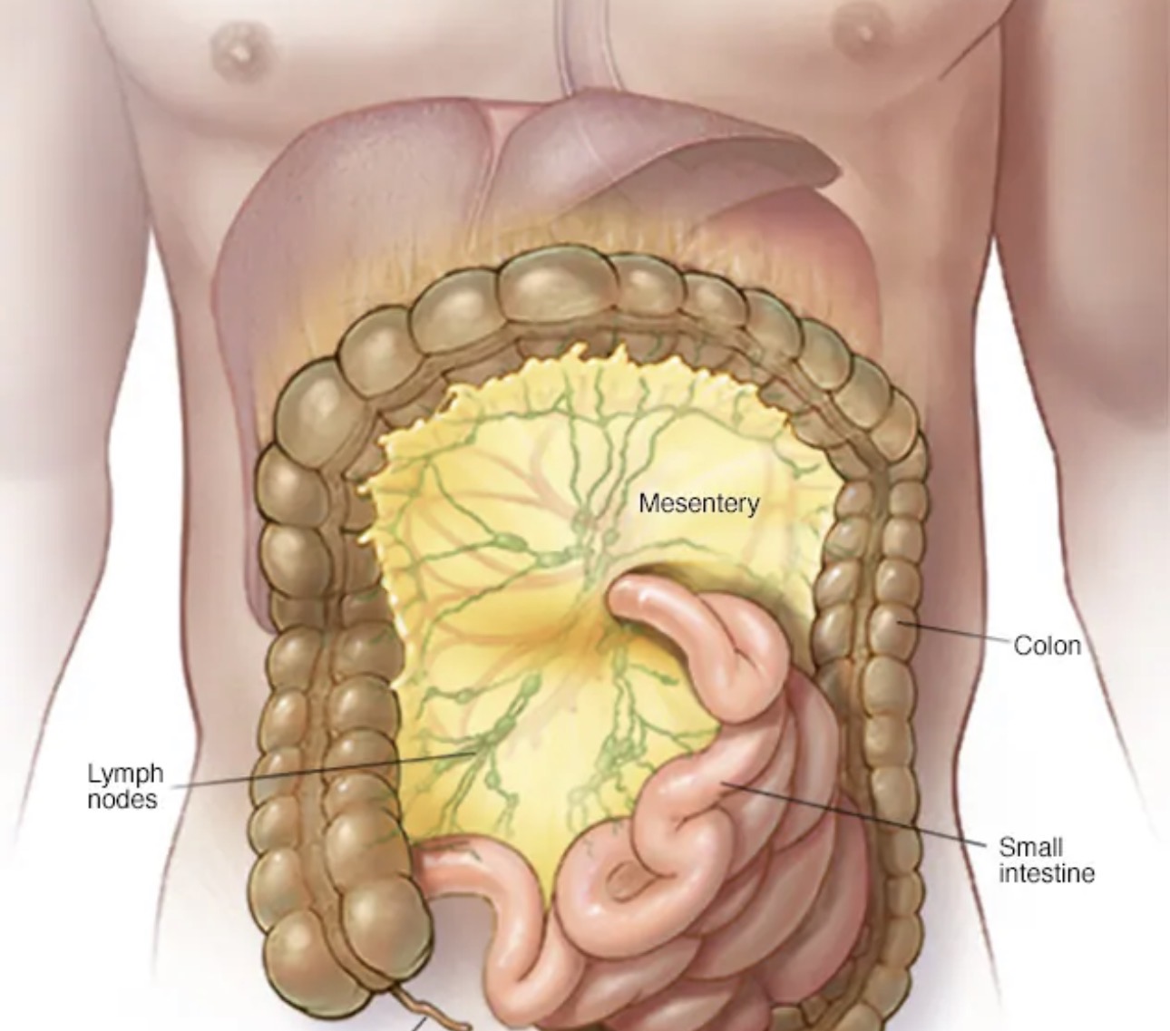
<p>double layer of peritoneum covering neurovascular components and connective tissue, connecting intraperitoneal organs to the abdominal wall, such as with the small bowel, transverse colon and sigmoid colon</p>
9
New cards
<p>Peritoneal Folds</p>

Peritoneal Folds

reflection of peritoneum that is raised from the abdominal wall by underlying structures (such as blood vessels, obliterated blood vessels, ligaments or ducts)
<p>reflection of peritoneum that is raised from the abdominal wall by underlying structures (such as blood vessels, obliterated blood vessels, ligaments or ducts)</p>
10
New cards
<p>Omentum</p>

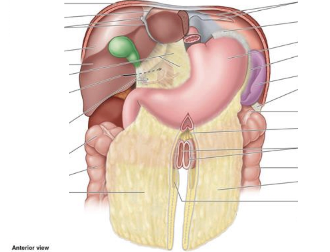
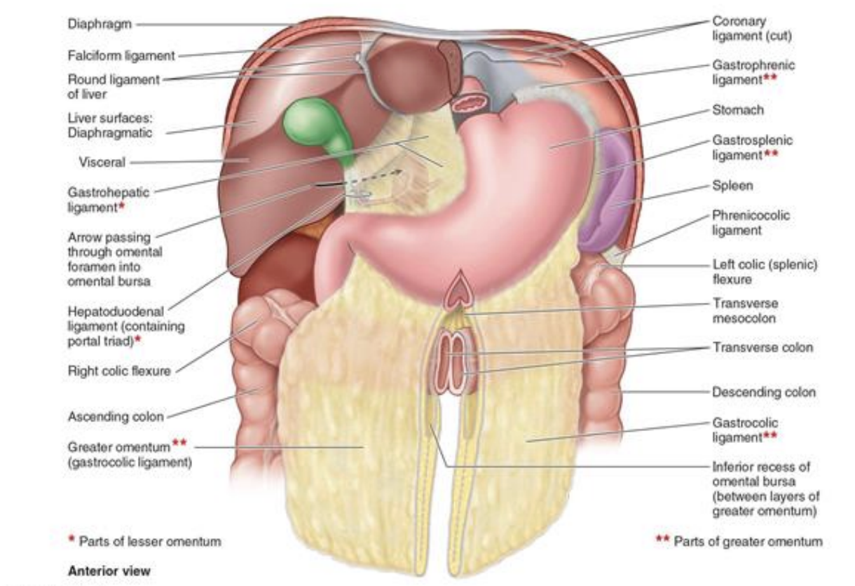
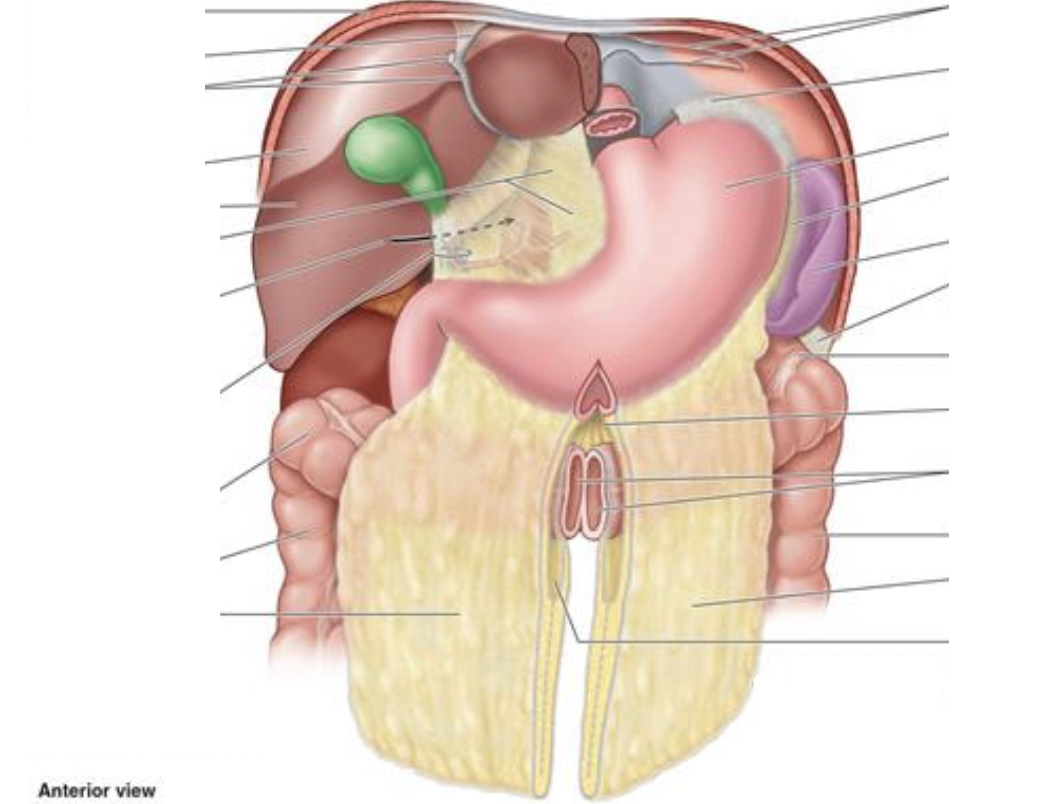
Omentum

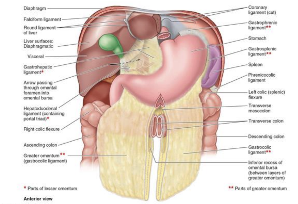
double layer fold or extension of peritoneum that attaches to the stomach and duodenum to adjacent structures.

Structure

Connects

Mesentery

Organ→abdominal wall

Peritoneal fold

Raised ridge over vessels/ligaments/ducts

Omentum

Organ→organ

<p>double layer fold or extension of peritoneum that attaches to the stomach and duodenum to adjacent structures.</p><p></p><table style="min-width: 50px;"><colgroup><col style="min-width: 25px;"><col style="min-width: 25px;"></colgroup><tbody><tr><td colspan="1" rowspan="1" style="width: 233.2px; border-style: solid; border-width: 1px; padding: 8px 12px;"><p style="text-align: center;"><strong>Structure</strong></p></td><td colspan="1" rowspan="1" style="width: 680.8px; border-style: solid; border-width: 1px; padding: 8px 12px;"><p style="text-align: center;"><strong>Connects</strong></p></td></tr><tr><td colspan="1" rowspan="1" style="width: 233.2px; border-style: solid; border-width: 1px; padding: 8px 12px;"><p><strong>Mesentery</strong></p></td><td colspan="1" rowspan="1" style="width: 680.8px; border-style: solid; border-width: 1px; padding: 8px 12px;"><p>Organ→abdominal wall</p></td></tr><tr><td colspan="1" rowspan="1" style="width: 233.2px; border-style: solid; border-width: 1px; padding: 8px 12px;"><p><strong>Peritoneal fold</strong></p></td><td colspan="1" rowspan="1" style="width: 680.8px; border-style: solid; border-width: 1px; padding: 8px 12px;"><p>Raised ridge over vessels/ligaments/ducts</p></td></tr><tr><td colspan="1" rowspan="1" style="width: 233.2px; border-style: solid; border-width: 1px; padding: 8px 12px;"><p><strong>Omentum</strong></p></td><td colspan="1" rowspan="1" style="width: 680.8px; border-style: solid; border-width: 1px; padding: 8px 12px;"><p>Organ→organ</p></td></tr></tbody></table><p></p>
11
New cards
<p>Muscles of the Anterior Abdominal Wall</p>

Muscles of the Anterior Abdominal Wall

External Oblique:

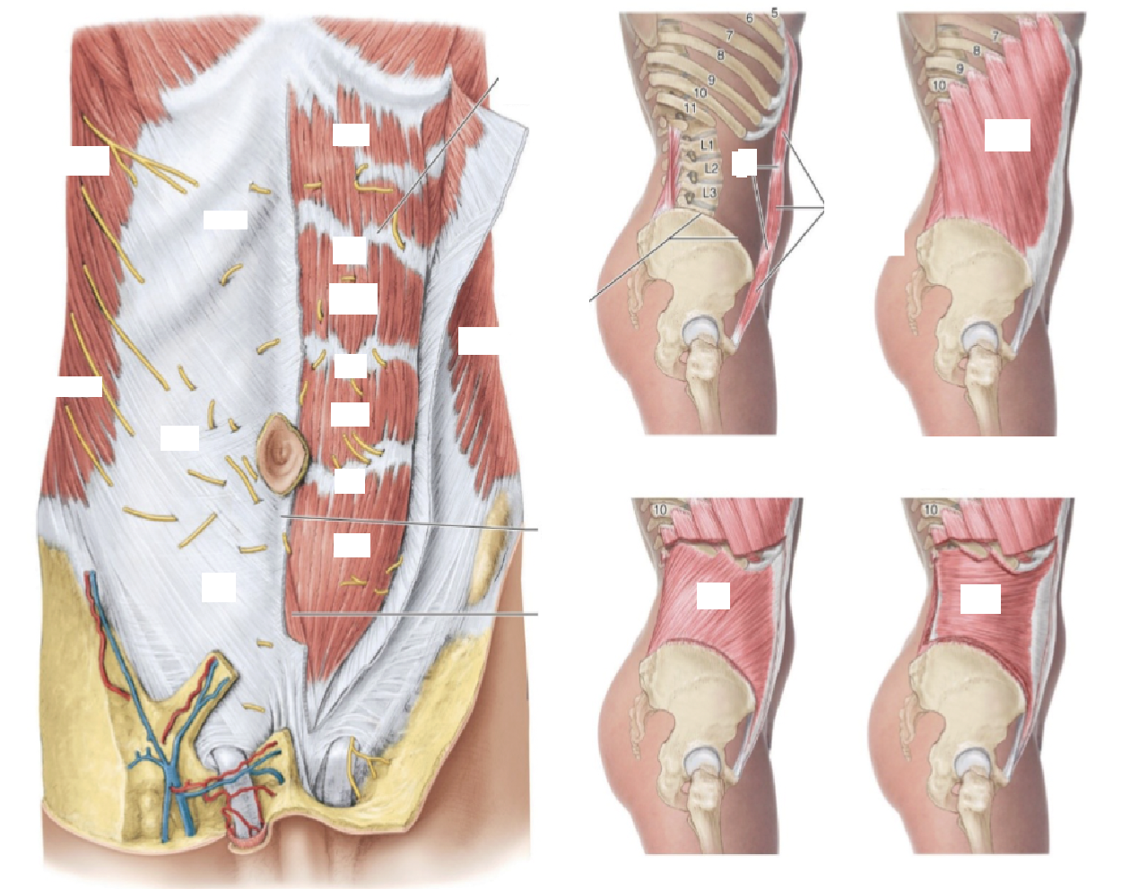
Compress and support abdomen viscera; flex and rotate trunk

Internal Oblique:

Compress and support abdomen viscera; flex and rotate trunk

Transversus abdominis:

Compress and support abdomen viscera

Rectus Abdominis:

Flex trunk; Compress abdominal viscera
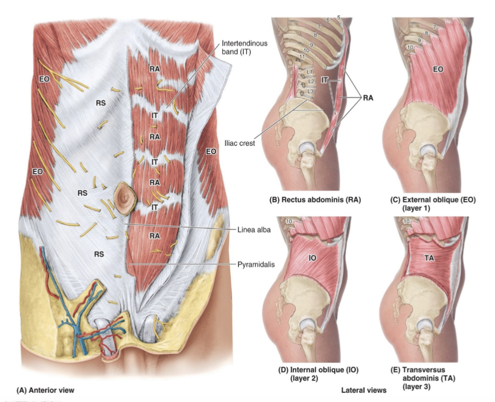
<p><strong>External Oblique:</strong></p><p>Compress and support abdomen viscera; flex and rotate trunk </p><p></p><p><strong>Internal Oblique:</strong></p><p>Compress and support abdomen viscera; flex and rotate trunk </p><p></p><p><strong>Transversus abdominis:</strong></p><p>Compress and support abdomen viscera </p><p></p><p><strong>Rectus Abdominis: </strong></p><p>Flex trunk; Compress abdominal viscera</p>
12
New cards
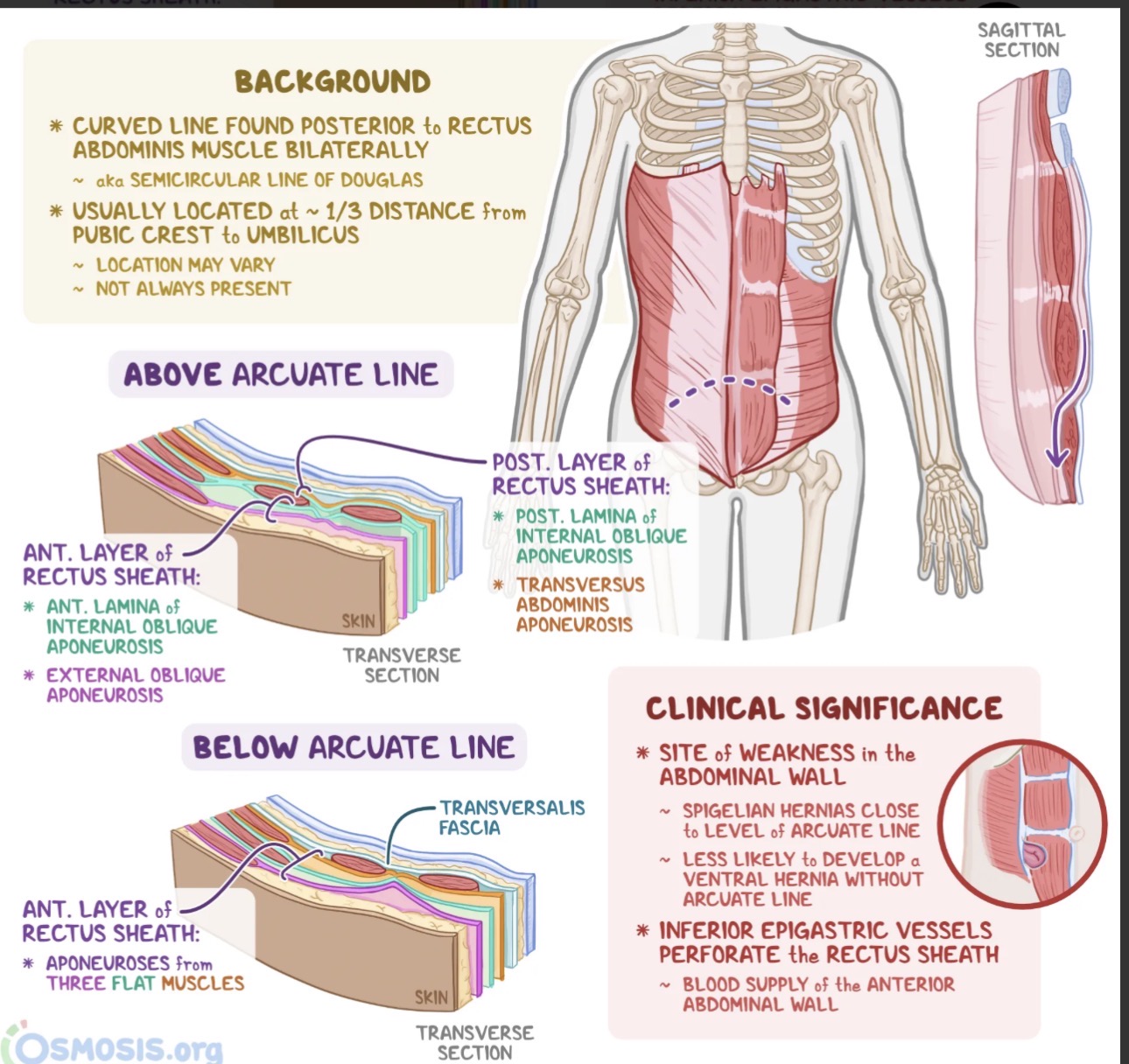
<p>Anterior Abdominal Wall (Rectus Sheath, Rectus Sheath Regions &amp; Aponeurosis layers)</p>

Anterior Abdominal Wall (Rectus Sheath, Rectus Sheath Regions & Aponeurosis layers)

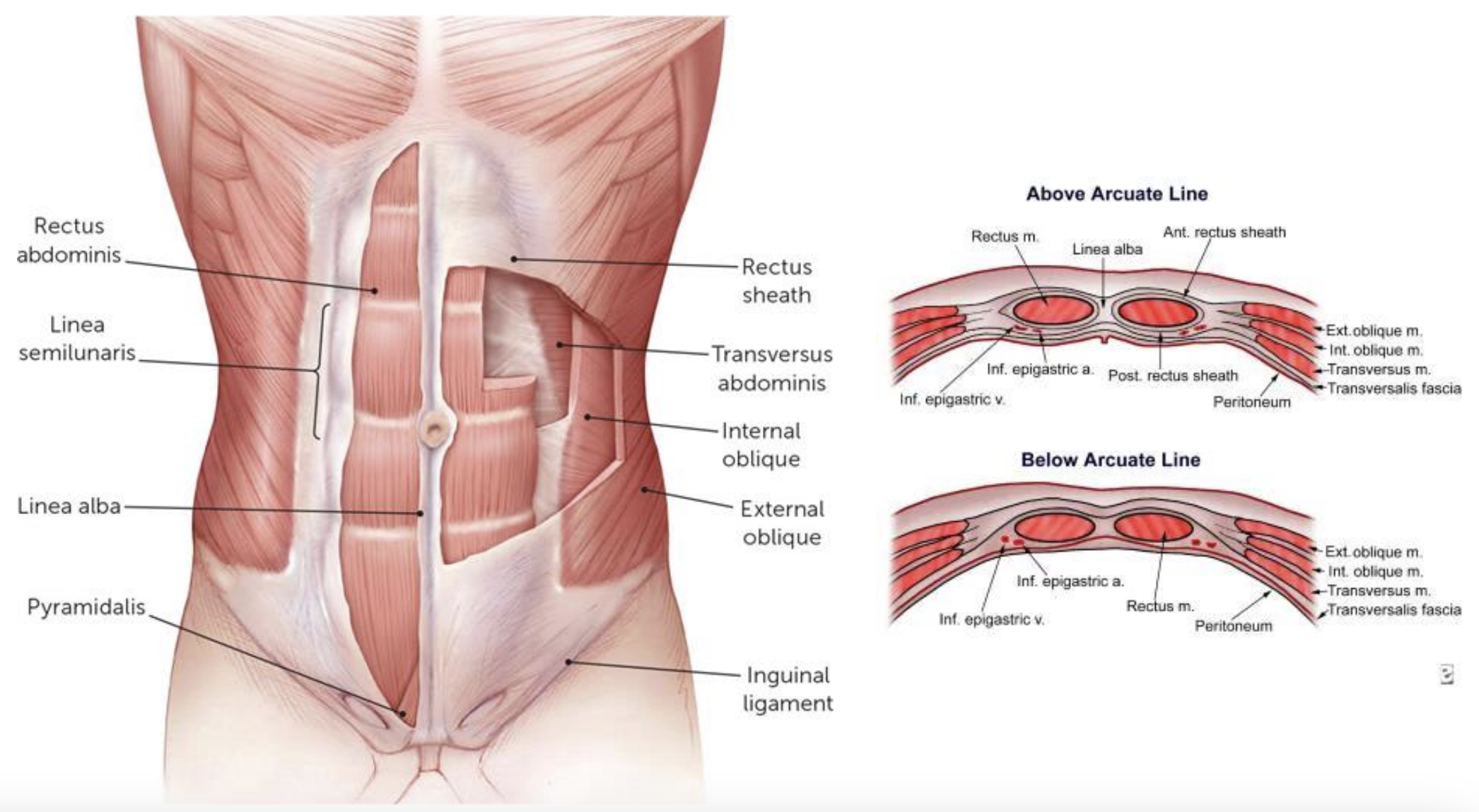
*The rectus sheath covers the rectus abdominis anteriorly and posteriorly. The sheath is made from the aponeurotic fibers of the obliques and transversus abdominis muscles.

Rectus Sheath Regions

1. Anterior rectus sheath layers (above arcuate line)

Aponeurosis layers: Fused aponeurosis of external and internal oblique muscles

2. Posterior rectus sheath layers (above arcuate line)

Aponeurosis layers: Fused aponeurosis of internal oblique and transversus abdominis muscles

3. Anterior rectus sheath layers (below arcuate line)

Aponeurosis layers: Fused aponeurosis of external oblique, internal oblique and transversus abdominis muscles

4. Posterior rectus sheath layers (below arcuate line)

Aponeurosis layers: Transversalis fascia only (no aponeurotic layers)

*The arcuate line is the name given to the horizontal line across the anterior abdomen that marks the end of the posterior portion of the rectus sheath
<p>*The <strong>rectus sheath</strong> covers the rectus abdominis anteriorly and posteriorly. The sheath is made from the aponeurotic fibers of the obliques and transversus abdominis muscles.</p><p></p><p>Rectus Sheath Regions</p><p><strong>1. Anterior rectus sheath layers (above arcuate line)</strong></p><p>Aponeurosis layers: Fused aponeurosis of external and internal oblique muscles</p><p></p><p><strong>2. Posterior rectus sheath layers (above arcuate line)</strong></p><p>Aponeurosis layers: Fused aponeurosis of internal oblique and transversus abdominis muscles</p><p></p><p><strong>3. Anterior rectus sheath layers (below arcuate line)</strong></p><p>Aponeurosis layers: Fused aponeurosis of external oblique, internal oblique and transversus abdominis muscles</p><p></p><p><strong>4. Posterior rectus sheath layers (below arcuate line)</strong></p><p>Aponeurosis layers: Transversalis fascia only (no aponeurotic layers)</p><p></p><p>*The arcuate line is the name given to the horizontal line across the anterior abdomen that marks the end of the posterior portion of the rectus sheath</p>
13
New cards
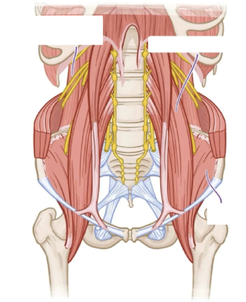
<p>Muscles of the Posterior Abdominal Wall</p>

Muscles of the Posterior Abdominal Wall

Psoas major

Innervation: L1-L4 ventral rami

Function: Flex hip (w/Iliacus); Flex vertebral column laterally; balance during sitting; Flex trunk (w/ iliacus)

Iliacus

Innervation: Femoral nerve

Function: Flex hip, stabilize hip joint; Works in conjunction with psoas major

Quadratus lumborum

Innervation: T12-L4 ventral rami

Function: Extend and laterally flex vertebral column; Fixes 12th rib during inspiration

Diaphragm

Innervation: Phrenic nerve

Function: Inspiration (major respiratory muscle)
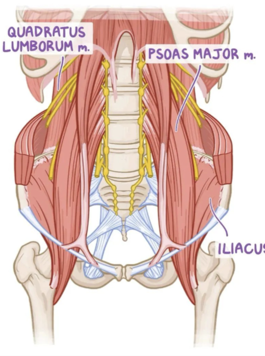
<p><strong>Psoas major</strong></p><p>Innervation: L1-L4 ventral rami</p><p>Function: Flex hip (w/Iliacus); Flex vertebral column laterally; balance during sitting; Flex trunk (w/ iliacus)</p><p></p><p><strong>Iliacus</strong></p><p>Innervation: Femoral nerve</p><p>Function: Flex hip, stabilize hip joint; Works in conjunction with psoas major</p><p></p><p><strong>Quadratus lumborum</strong></p><p>Innervation: T12-L4 ventral rami</p><p>Function: Extend and laterally flex vertebral column; Fixes 12th rib during inspiration</p><p></p><p><strong>Diaphragm</strong></p><p>Innervation: Phrenic nerve</p><p>Function: Inspiration (major respiratory muscle)</p>
14
New cards
<p>Inguinal Ligament and Canal</p>

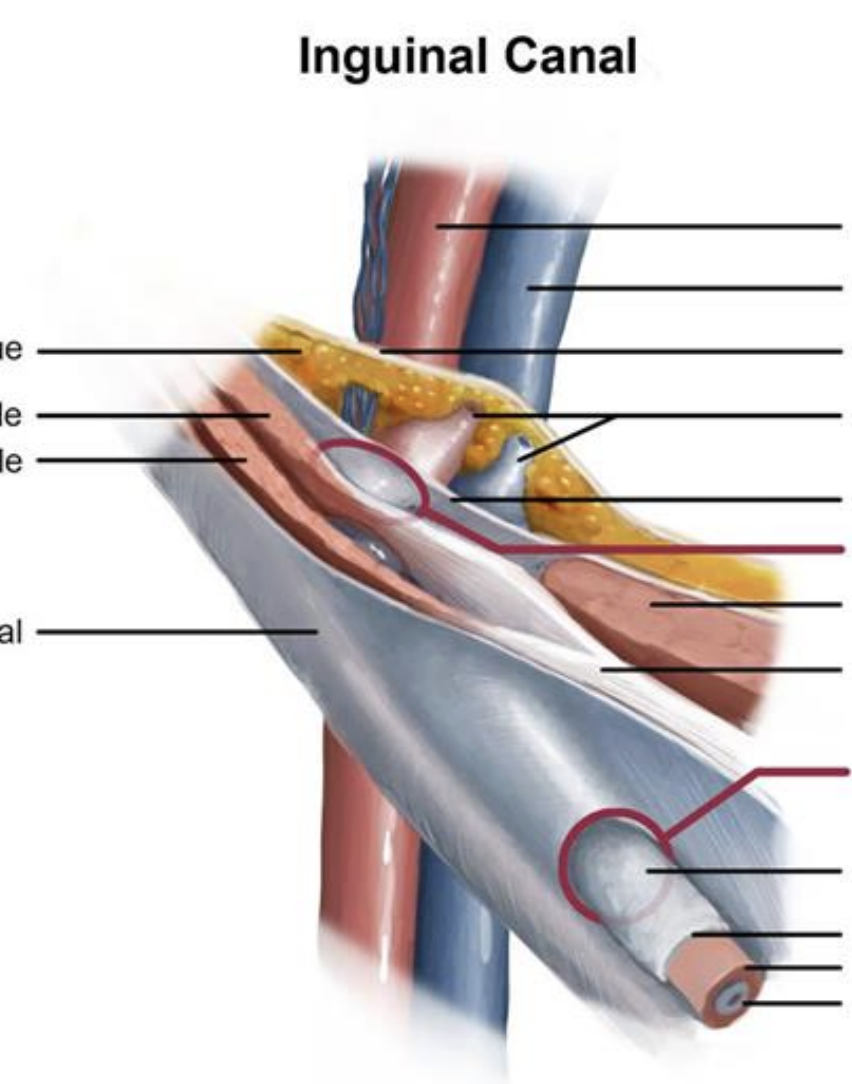
Inguinal Ligament and Canal

●The inguinal ligament is a fibrous band connecting the anterior superior iliac spine to the pubic tubercle of the pelvis. It marks the junction between pelvis and lower extremity

● The inguinal canal is formed by fibrosis structures and sits on the medial half of the inguinal ligament

Contents of Inguinal Canal

● Spermatic cord (biological males)

● Round ligament (biological females)

● Ilioinguinal nerve

● Genital branch of genitofemoral nerve

<p>●The inguinal ligament is a fibrous band connecting the anterior superior iliac spine to the pubic tubercle of the pelvis. It marks the junction between pelvis and lower extremity </p><p>● The inguinal canal is formed by fibrosis structures and sits on the medial half of the inguinal ligament</p><p></p><p><strong>Contents of Inguinal Canal</strong></p><p><strong>● Spermatic cord (biological males)</strong></p><p><strong>● Round ligament (biological females)</strong></p><p><strong>● Ilioinguinal nerve</strong></p><p><strong>● Genital branch of genitofemoral nerve</strong></p>
15
New cards
<p>Lumbar Plexus: Ventral Rami of T12-L4</p>

Lumbar Plexus: Ventral Rami of T12-L4

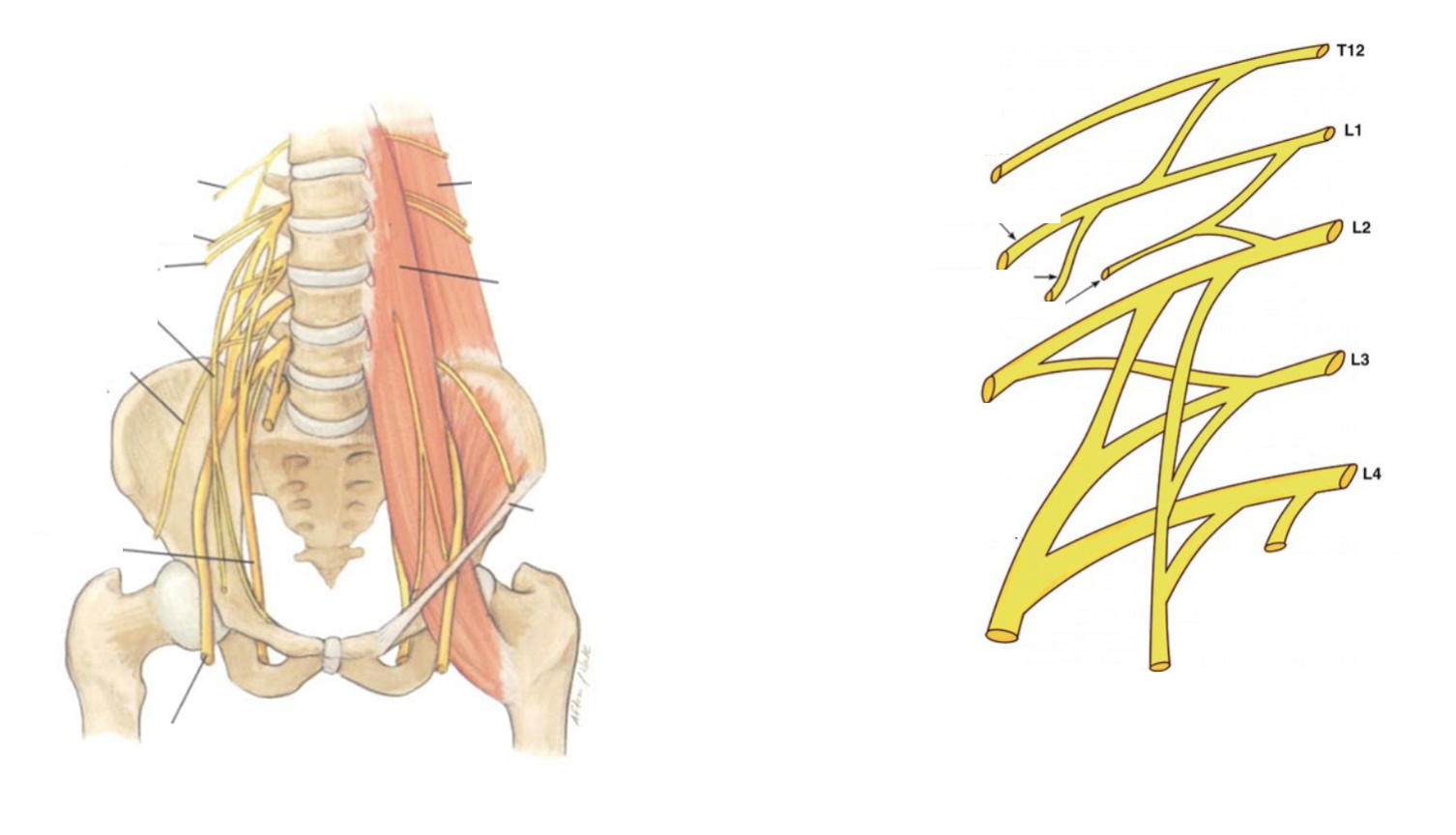
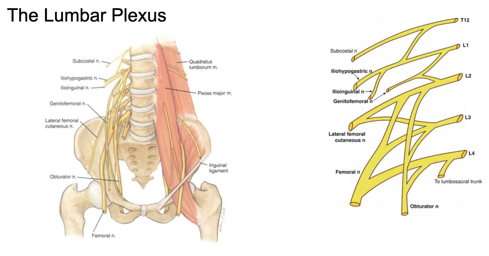
mnemonic: Strong Intellectuals In Gym Lift For Optimalism

Subcostal

Arises from: T12

Supplies: Sensory and Motor to 12th intercostal space

Iliohypogastric

Arises from: L1

Supplies: Sensory to inguinal region; some Motor to internal oblique and transversus abdominis

Ilioinguinal

Arises from: L1

Supplies: Sensory to inguinal region and external genitalia; some Motor to internal oblique and transversus abdominis

Genitofemoral

Arises from: L1 & L2

Supplies: Sensory to anteromedial thigh; Motor to cremaster muscle in males

Lateral femoral cutaneous

Arises from: L2 & L3

Supplies: Sensory to lateral thigh

Femoral

Arises from: L2 - L4

Supplies: Sensory to anterior thigh, anteromedial leg and ankle; Motor to anterior compartment of thigh

Obturator

Arises from: L2 - L4

Supplies:Sensory to medial thigh; Motor to medial compartment thigh muscles

<p>mnemonic: <strong>Strong Intellectuals In Gym Lift For Optimalism</strong></p><p><strong>Subcostal</strong></p><p>Arises from: T12</p><p>Supplies: Sensory and Motor to 12th intercostal space</p><p></p><p><strong>Iliohypogastric</strong></p><p>Arises from: L1</p><p>Supplies: Sensory to inguinal region; some Motor to internal oblique and transversus abdominis</p><p></p><p><strong>Ilioinguinal</strong></p><p>Arises from: L1</p><p>Supplies: Sensory to inguinal region and external genitalia; some Motor to internal oblique and transversus abdominis</p><p></p><p><strong>Genitofemoral</strong></p><p>Arises from: L1 &amp; L2</p><p>Supplies: Sensory to anteromedial thigh; Motor to cremaster muscle in males</p><p></p><p><strong>Lateral femoral cutaneous</strong></p><p>Arises from: L2 &amp; L3</p><p>Supplies: Sensory to lateral thigh</p><p></p><p><strong>Femoral</strong></p><p>Arises from: L2 - L4</p><p>Supplies: Sensory to anterior thigh, anteromedial leg and ankle; Motor to anterior compartment of thigh</p><p></p><p><strong>Obturator</strong></p><p>Arises from: L2 - L4</p><p>Supplies:Sensory to medial thigh; Motor to medial compartment thigh muscles</p>
16
New cards

Sympathetic Nerve Fibers

Liver/Gallbladder

Ganglion giving postsynaptic fibers: Celiac ganglion

Spleen

Ganglion giving postsynaptic fibers: Celiac ganglion

Pancreas

Ganglion giving postsynaptic fibers: Celiac ganglion

Small bowel

Ganglion giving postsynaptic fibers: Superior mesenteric ganglion

Proximal Colon

Ganglion giving postsynaptic fibers: Superior mesenteric ganglion

Distal Colon

Ganglion giving postsynaptic fibers: Inferior mesenteric ganglion

Kidney

Ganglion giving postsynaptic fibers: Inferior mesenteric ganglion

Adrenal Gland

Ganglion giving postsynaptic fibers: Celiac ganglion

17
New cards

Preganglionic Parasympathetic Fibers

Vagus Nerve

● Liver

● Gallbladder

● Spleen

● Stomach

● Pancreas

● Small bowel

● Right colon

Pelvic Splanchnic Nerves (S2 - S4)

● Left Colon

● Kidney

● Bladder

● Gonads

18
New cards
<p>Arteries of the Abdomen (Foregut)</p>

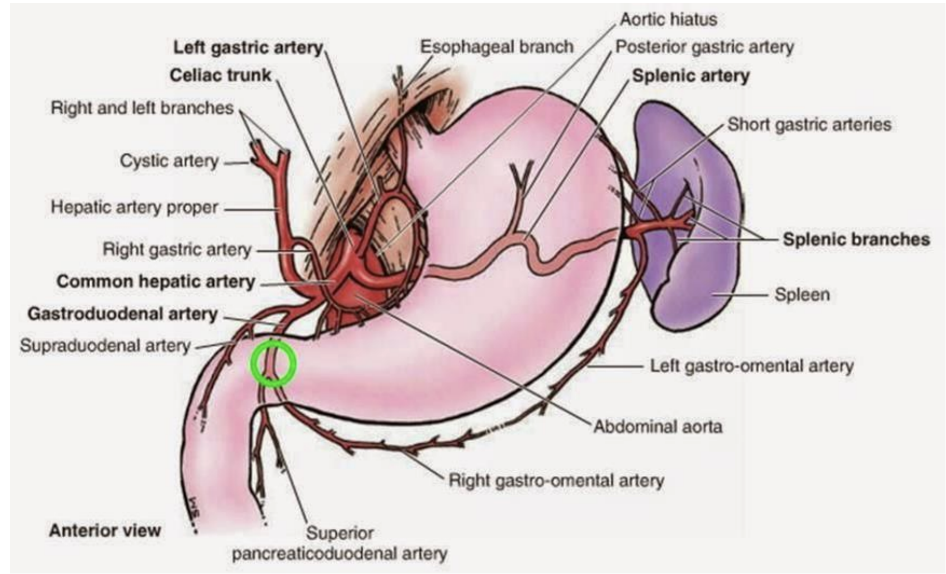
Arteries of the Abdomen (Foregut)

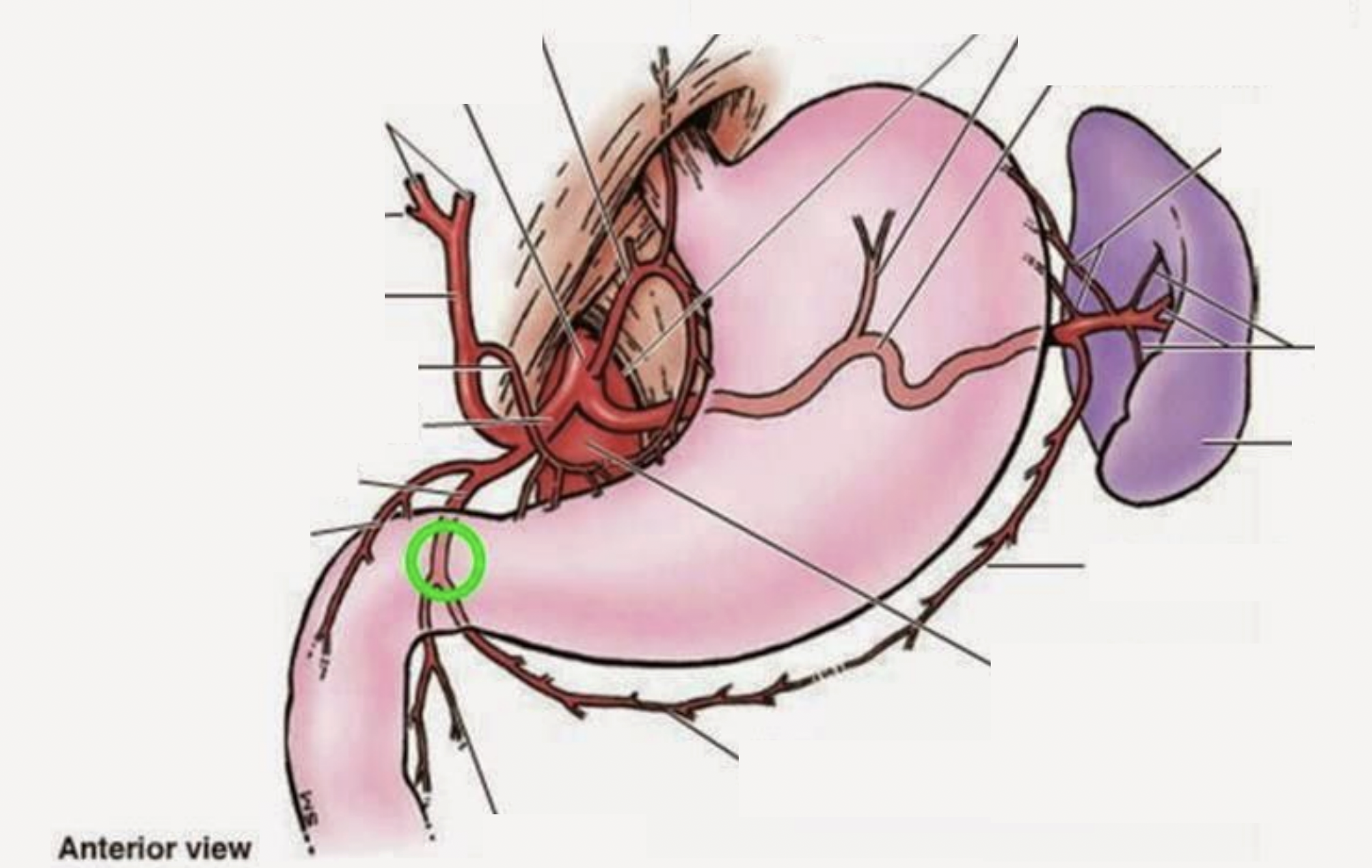
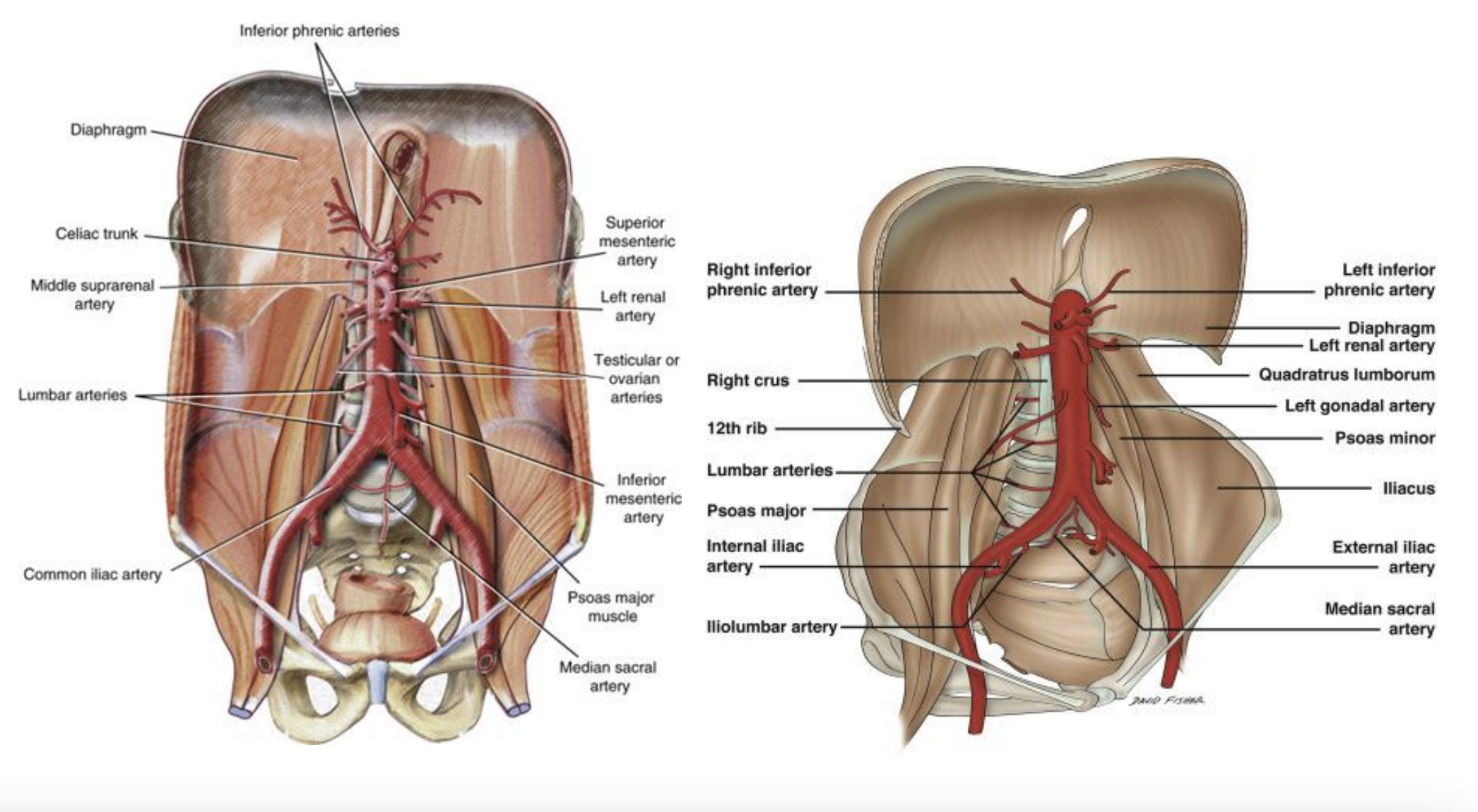
Descending (Abdominal) Aorta

Arises from: Descending (Thoracic) Aorta

Celiac Trunk:

  1. Left Gastric Arises from: Celiac Trunk

  2. Splenic Arises from: Celiac Trunk

  3. Common Hepatic Arises from: Celiac Trunk, Supplies: Liver (divides into left and right), gallbladder via *Cystic artery

<p><strong>Descending (Abdominal) Aorta</strong></p><p>Arises from: Descending (Thoracic) Aorta</p><p></p><p><strong>Celiac Trunk:</strong></p><ol><li><p><strong>Left Gastric </strong>Arises from: Celiac Trunk</p></li><li><p><strong>Splenic </strong>Arises from: Celiac Trunk</p></li><li><p><strong>Common Hepatic </strong>Arises from: Celiac Trunk, Supplies: Liver (divides into left and right), gallbladder via *<em>Cystic artery</em></p></li></ol><p></p>
19
New cards
<p>Arteries of the Abdomen (Midgut)</p>

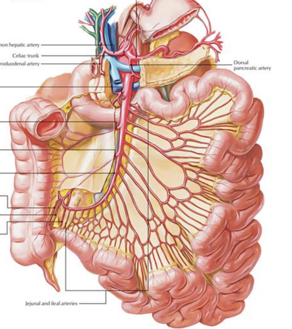
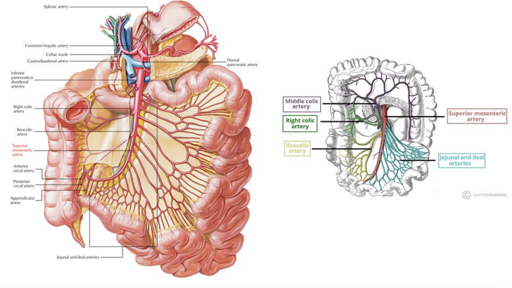
Arteries of the Abdomen (Midgut)

Descending (Abdominal) Aorta

Arises from: Descending (Thoracic) Aorta

Superior Mesenteric Artery (SMA)

Arises from: Descending (Abdominal) Aorta

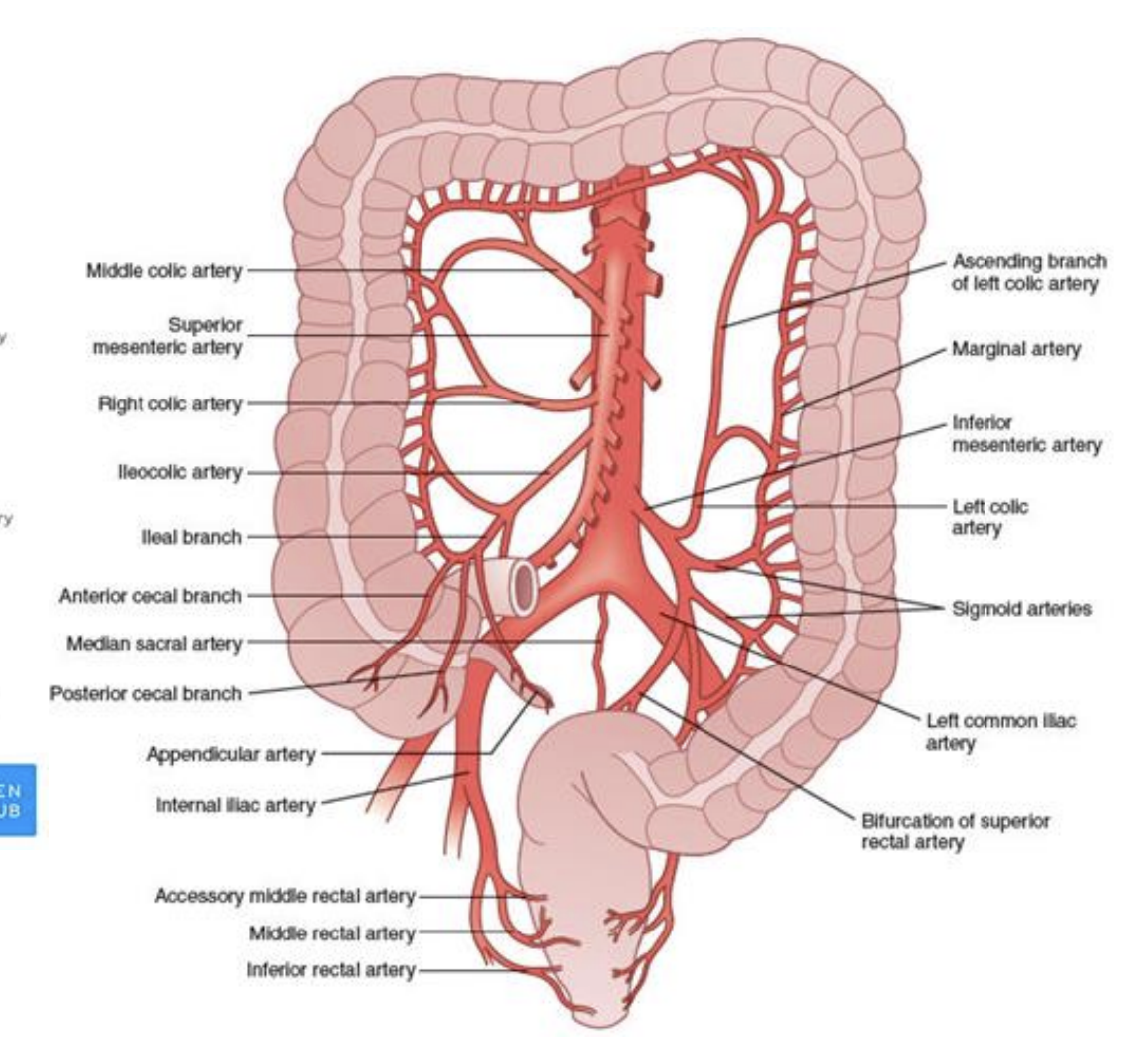
*A majority of the small intestine and part of the large intestine make up the hindgut, which receive their blood from the superior mesenteric artery

<p><strong>Descending (Abdominal) Aorta</strong></p><p>Arises from: Descending (Thoracic) Aorta</p><p></p><p><strong>Superior Mesenteric Artery (SMA)</strong></p><p>Arises from: Descending (Abdominal) Aorta</p><p>*A majority of the small intestine and part of the large intestine make up the hindgut, which receive their blood from the superior mesenteric artery</p>
20
New cards
<p>Arteries of the Abdomen (Hindgut)</p>

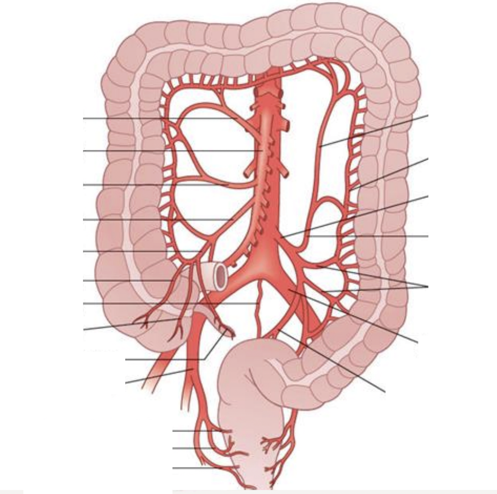
Arteries of the Abdomen (Hindgut)

Descending (Abdominal) Aorta

Arises from: descending thoracic aorta

Inferior Mesenteric Artery (IMA)

Arises from: Descending (Abdominal) Aorta

<p><strong>Descending (Abdominal) Aorta</strong></p><p>Arises from: descending thoracic aorta </p><p></p><p><strong>Inferior Mesenteric Artery (IMA)</strong></p><p>Arises from: Descending (Abdominal) Aorta</p>
21
New cards

Arteries of the Abdomen (Paired Arteries)

Descending (Abdominal) Aorta

Arises from: descending thoracic aorta

Renal Arteries

Arises from: Descending Aorta (laterally)

Common Iliac Arteries

Arises from: Descending Aorta (laterally)

<p><strong>Descending (Abdominal) Aorta</strong></p><p>Arises from: descending thoracic aorta</p><p></p><p><strong>Renal Arteries</strong></p><p>Arises from: Descending Aorta (laterally)</p><p></p><p><strong>Common Iliac Arteries</strong></p><p>Arises from: Descending Aorta (laterally)</p>
22
New cards

Veins of the Abdomen (Caval System)

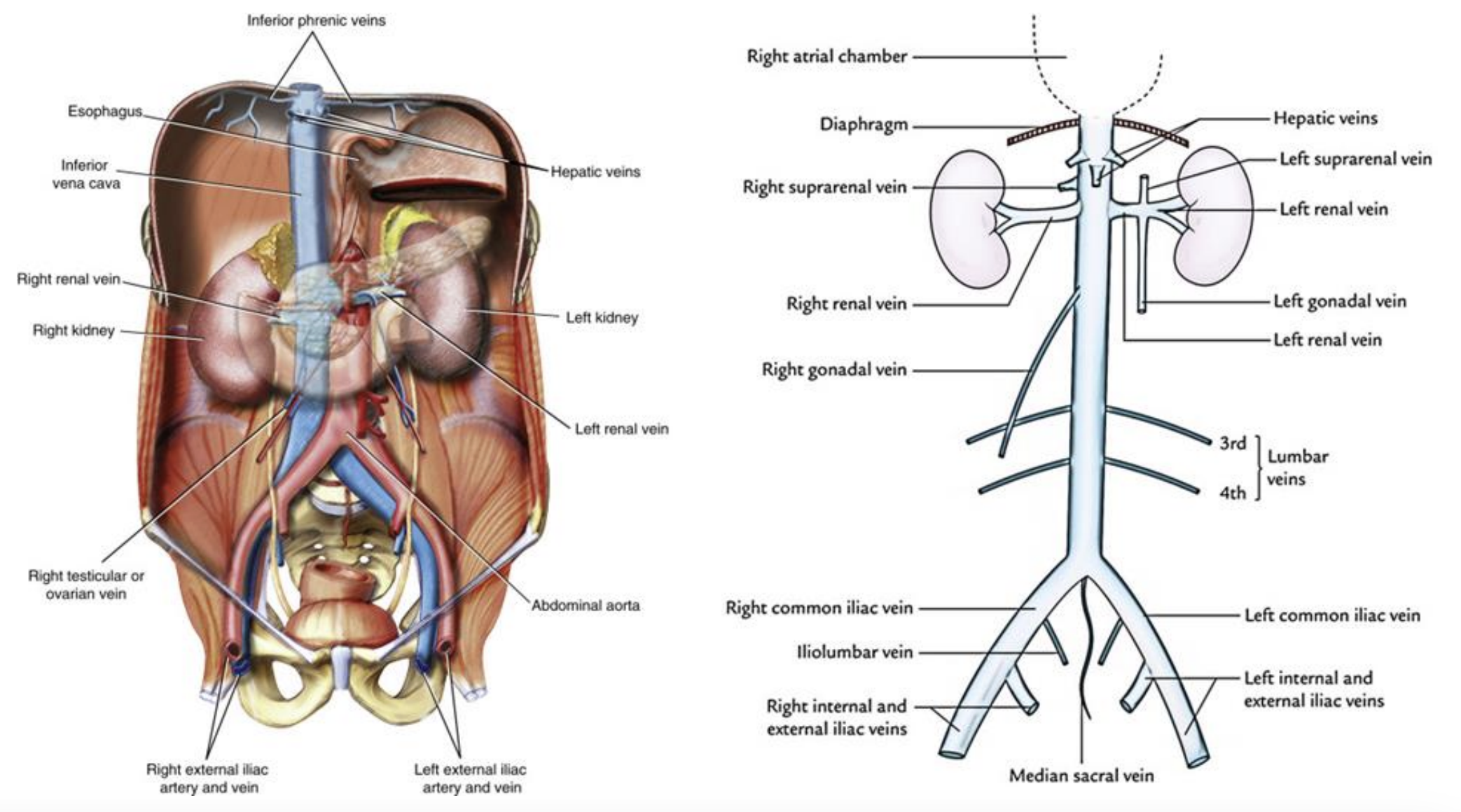
Inferior Vena Cava (IVC)

Runs to: Right atrium of the heart

Hepatic Veins

Runs to: IVC

Renal Veins

Runs to: IVC

Common Iliac Veins

Runs to: IVC (Converge to create IVC)

<p><strong>Inferior Vena Cava (IVC) </strong></p><p>Runs to: Right atrium of the heart</p><p></p><p><strong>Hepatic Veins </strong></p><p>Runs to: IVC</p><p></p><p><strong>Renal Veins </strong></p><p>Runs to: IVC</p><p></p><p><strong>Common Iliac Veins</strong></p><p>Runs to: IVC (Converge to create IVC)</p>
23
New cards

Veins of the Abdomen (Portal System)

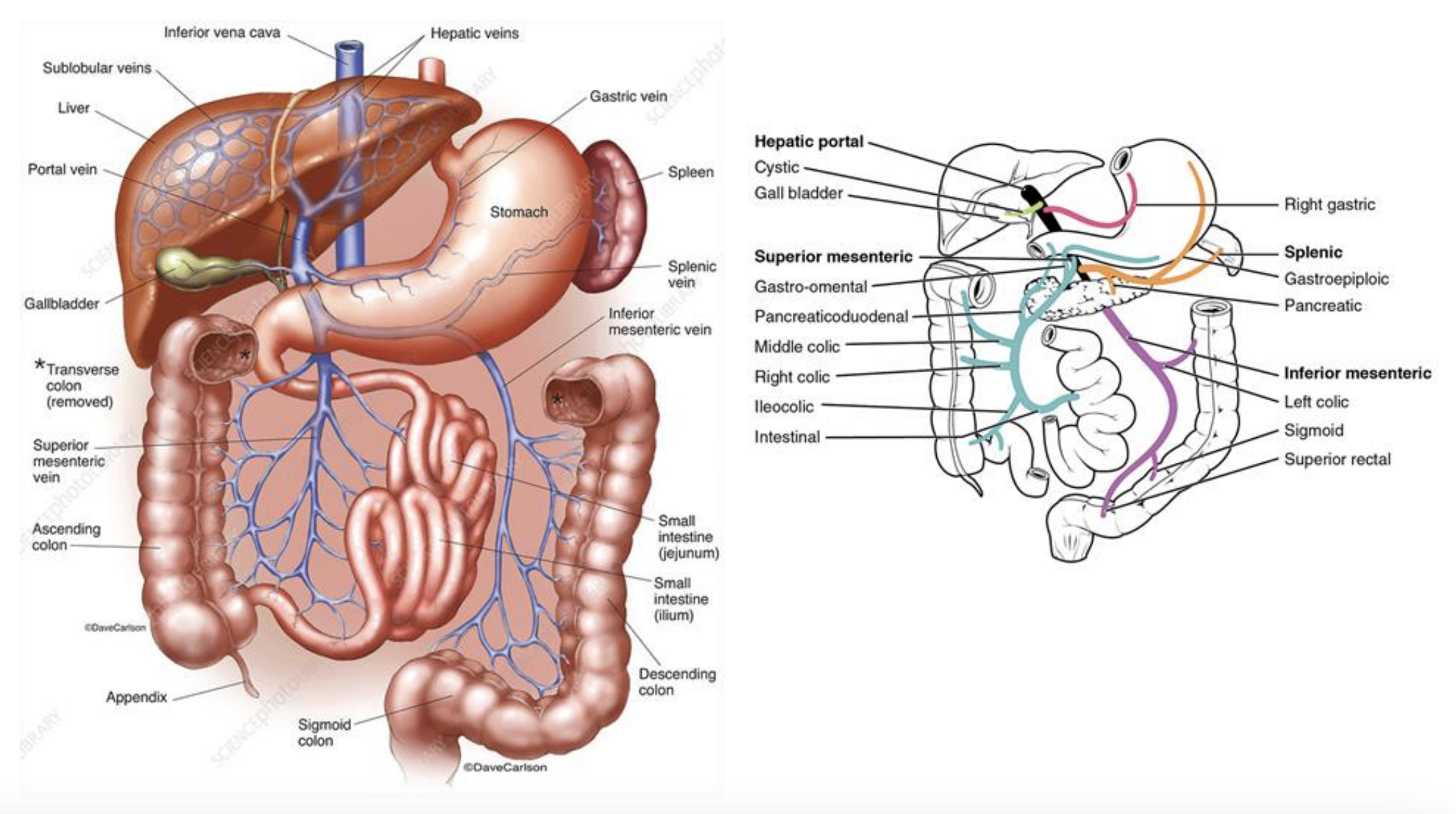
Hepatic Portal Vein

Runs to: Hilum of the Liver

Gastric Veins

Runs to: Hepatic portal vein

Splenic Vein

Runs to: Hepatic portal vein (Converges with SMV to form hepatic portal vein)

Inferior Mesenteric Vein (IMV)

Runs to: Splenic Vein


Hepatic Portal Vein

Runs to: Hilum of the Liver

Superior Mesenteric Vein (SMV)

Runs to: Hepatic portal vein (Converges with splenic vein to form hepatic portal vein)

<p><strong>Hepatic Portal Vein </strong></p><p>Runs to: Hilum of the Liver </p><p></p><p><strong>Gastric Veins</strong> </p><p>Runs to: Hepatic portal vein </p><p></p><p><strong>Splenic Vein </strong></p><p>Runs to: Hepatic portal vein (Converges with SMV to form hepatic portal vein) </p><p></p><p><strong>Inferior Mesenteric Vein (IMV) </strong></p><p>Runs to: Splenic Vein</p><p><br><strong>Hepatic Portal Vein </strong></p><p>Runs to: Hilum of the Liver</p><p></p><p><strong>Superior Mesenteric Vein (SMV) </strong></p><p>Runs to: Hepatic portal vein (Converges with splenic vein to form hepatic portal vein)</p>
24
New cards
<p>Liver</p>

Liver

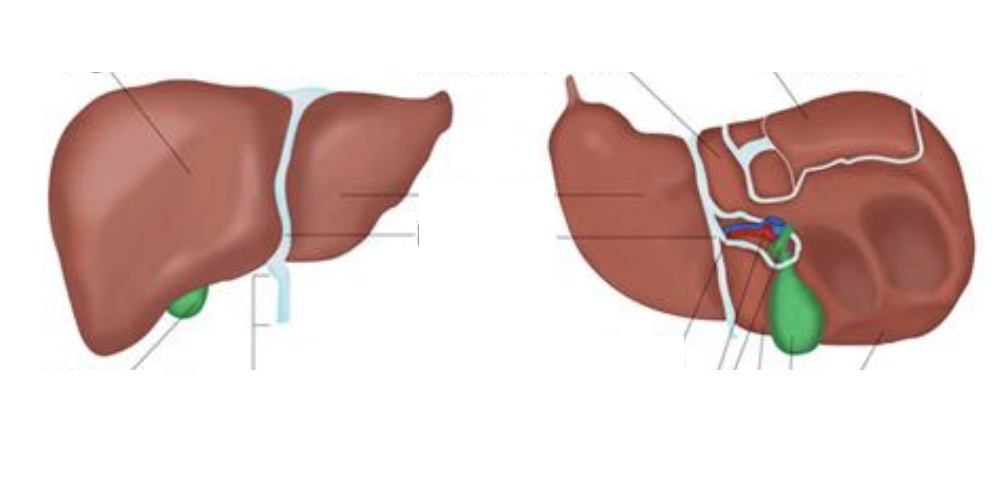
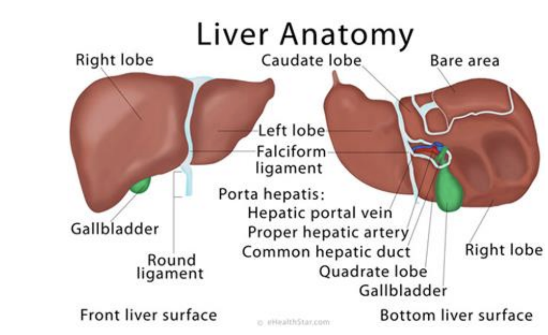
● Final destination of the portal system before returning blood to the inferior vena cava

● Gallbladder located on inferior right lobe

● Located on right side of body just inferior to diaphragm (secured to diaphragm with coronary ligament)

● Round ligament of liver (ligamentum teres) connects to umbilicus

● Lesser omentum is comprised of hepatoduodenal and hepatogastric ligaments, attaching the liver to the duodenum and lesser curvature of the stomach respectively

<p>● Final destination of the portal system before returning blood to the inferior vena cava </p><p>● Gallbladder located on inferior right lobe </p><p>● Located on right side of body just inferior to diaphragm (secured to diaphragm with coronary ligament) </p><p>● Round ligament of liver (ligamentum teres) connects to umbilicus </p><p>● Lesser omentum is comprised of hepatoduodenal and hepatogastric ligaments, attaching the liver to the duodenum and lesser curvature of the stomach respectively</p>
25
New cards
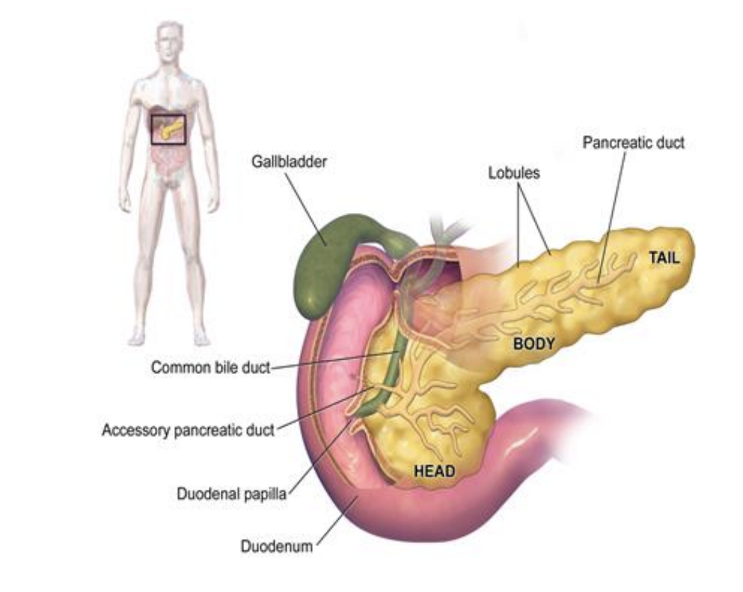
<p>Anatomy of the Biliary System</p>

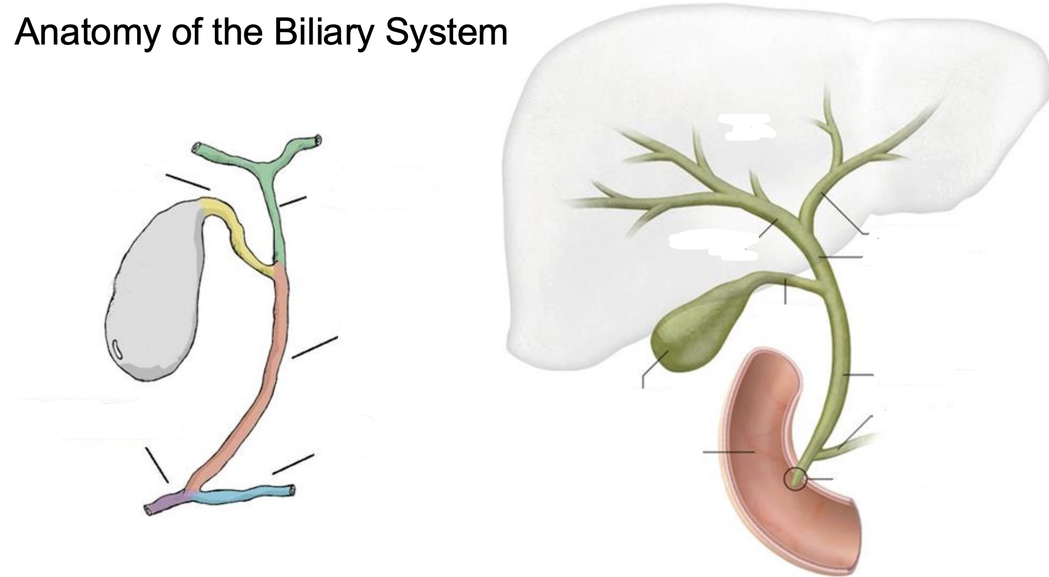
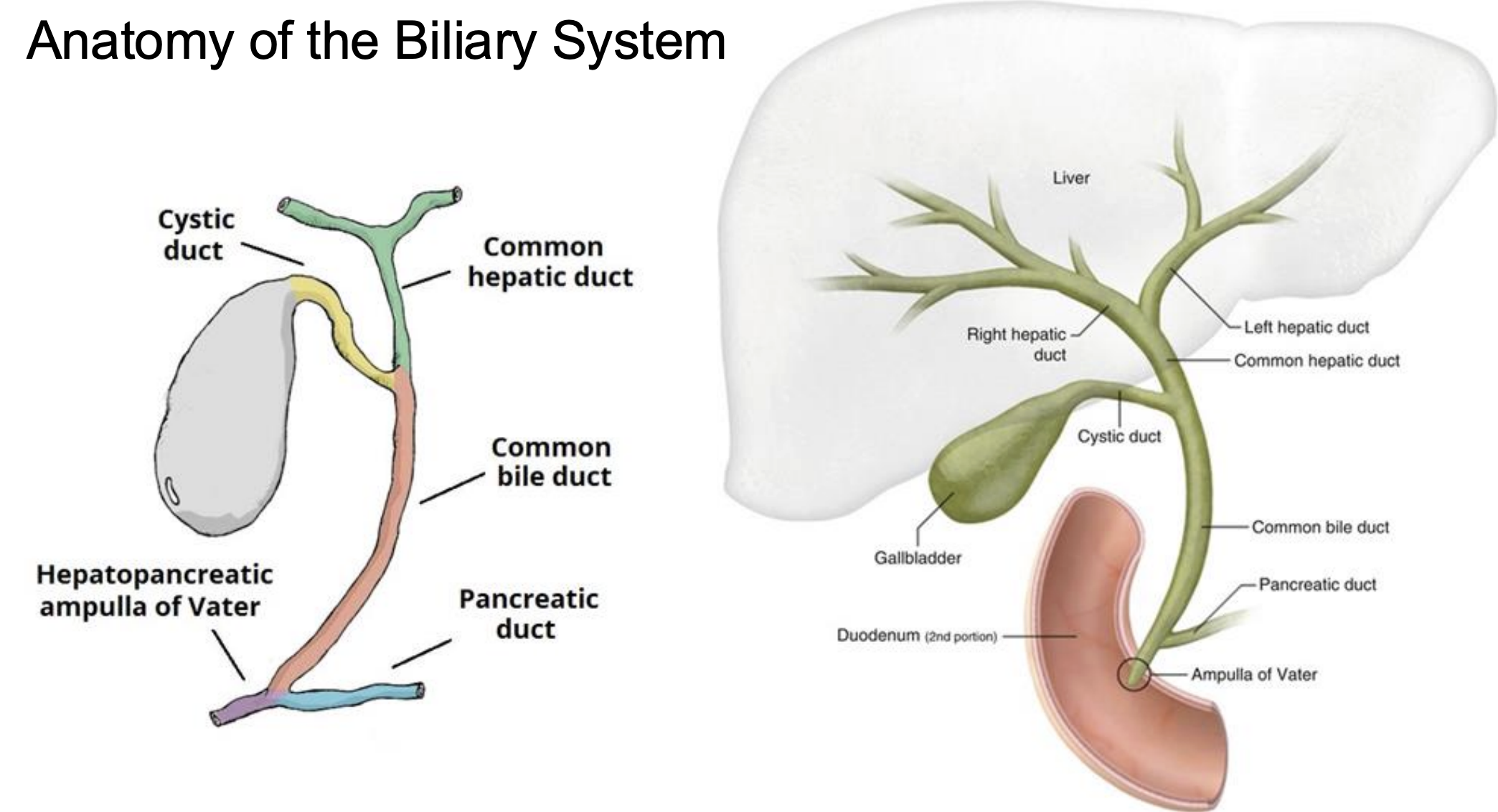
Anatomy of the Biliary System

knowt flashcard image
26
New cards
<p>Stomach</p>

Stomach

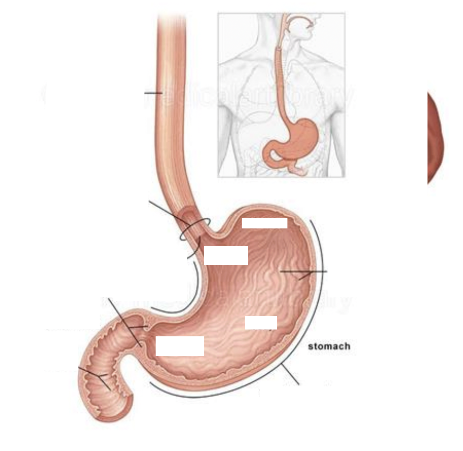
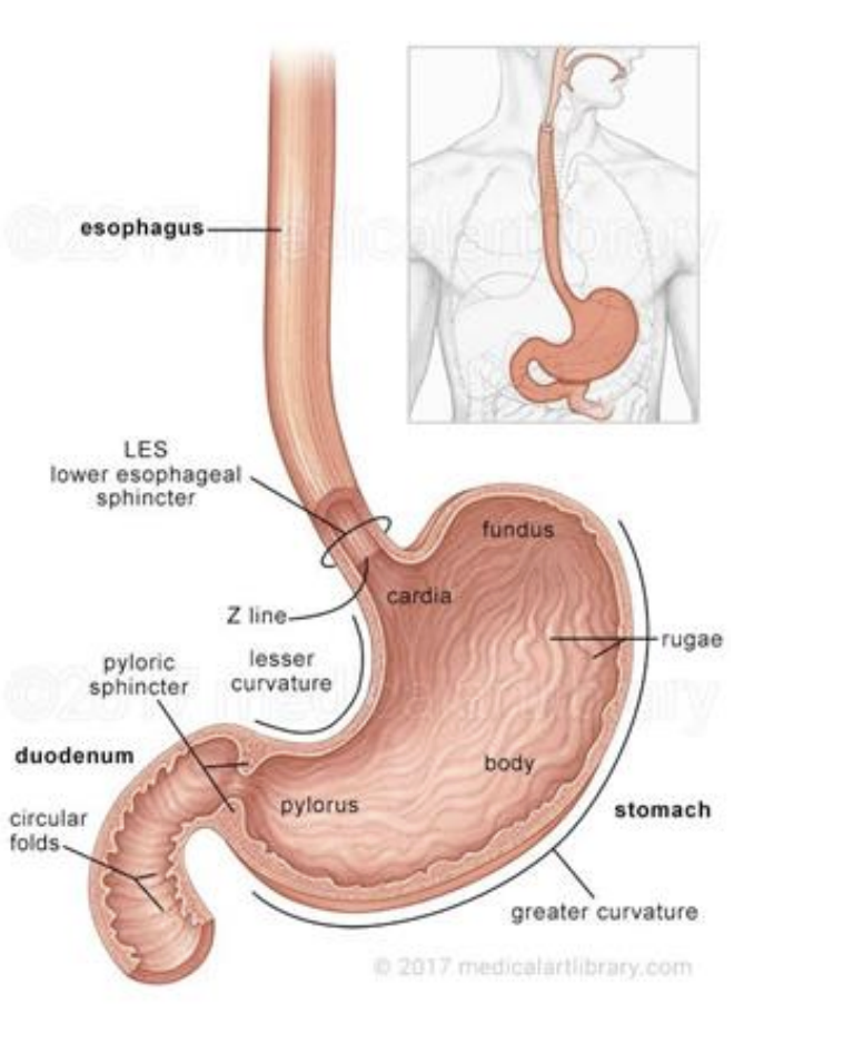
● The stomach has four parts: the cardia, the fundus, the body and the pyloric part.

● Esophagus enters at the cardia

● Lesser curvature contains lesser omentum, attaching stomach to liver

● Greater curvature has greater omentum, a fatty structure lying on anterior abdominal structures and connecting with transverse colon

● Passes food into duodenum

<p>● The stomach has four parts: the cardia, the fundus, the body and the pyloric part. </p><p>● Esophagus enters at the cardia </p><p>● Lesser curvature contains lesser omentum, attaching stomach to liver </p><p>● Greater curvature has greater omentum, a fatty structure lying on anterior abdominal structures and connecting with transverse colon </p><p>● Passes food into duodenum</p>
27
New cards
<p>Spleen</p>

Spleen

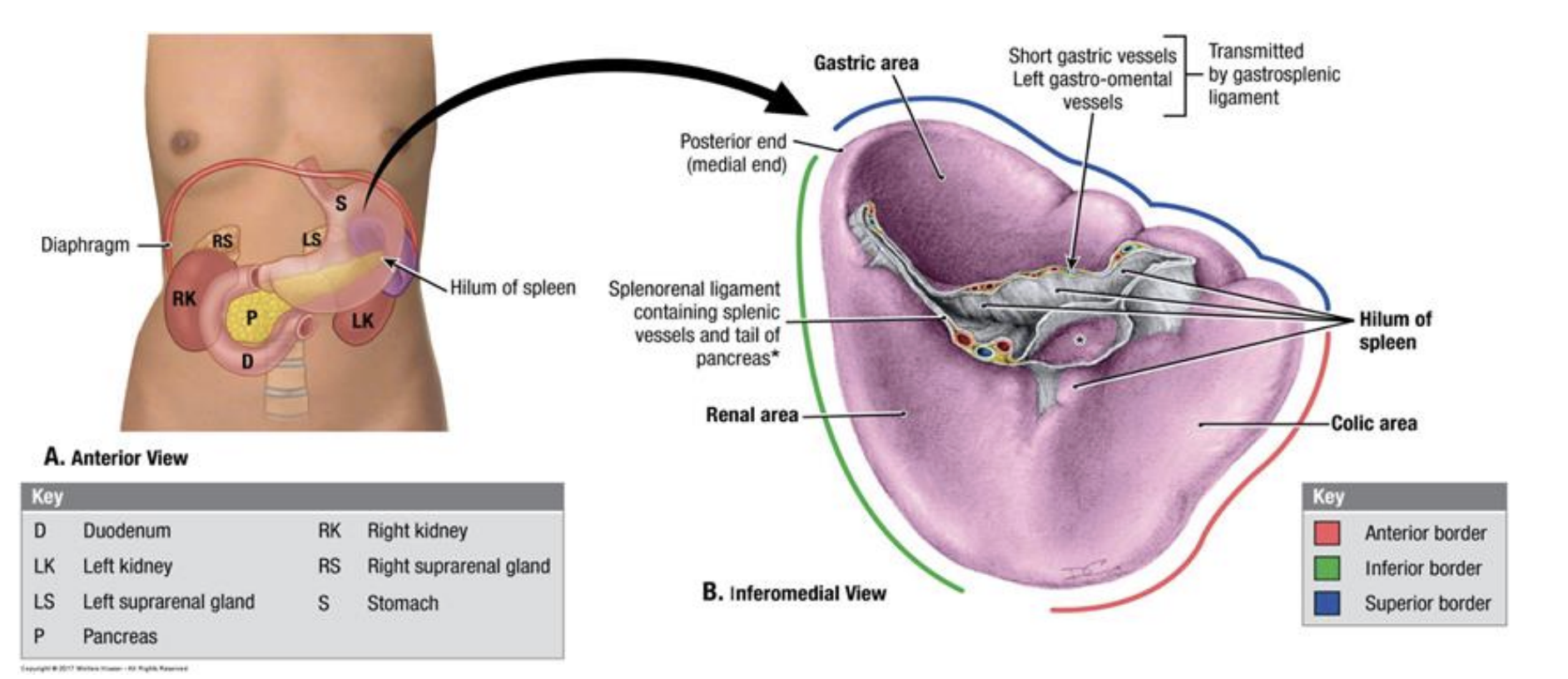
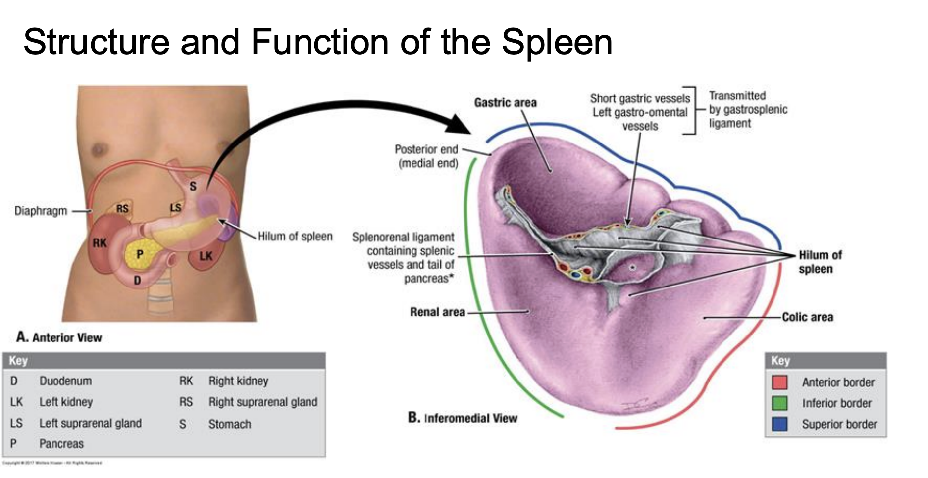
• located inferior to the diaphragm on the left side of the body (upper left quadrant)

• is a lymphatic organ, the largest lymphatic organ and plays an important part in the immune system

<p>• located inferior to the diaphragm on the left side of the body (upper left quadrant) </p><p>• is a lymphatic organ, the largest lymphatic organ and plays an important part in the immune system</p>
28
New cards
<p>Pancreas</p>

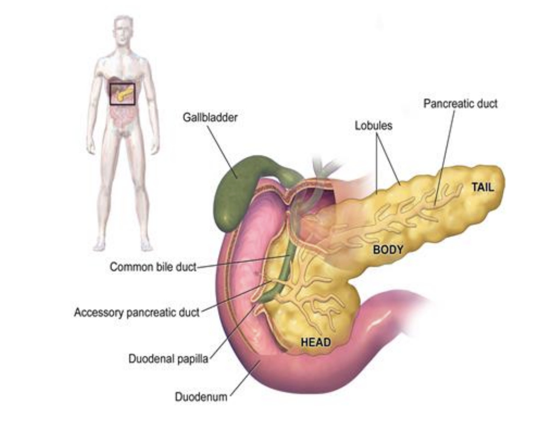
Pancreas

● Adjacent to pylorus of stomach inferiorly and to the duodenal bulb medially

● Receives bile from the liver via the common bile duct

● Main pancreatic duct meets with common bile duct near the ampulla of Vater, feeding digestive juices into duodenal bulb

● Sits on superior mesenteric artery and vein anteriorly

**The pancreas lacks any significant capsule other than the visceral peritoneum around it. This, in addition to being adjacent to the superior mesenteric artery and vein, makes pancreatic cancers particular deadly

Exocrine function: Pancreatic juices are secreted into the duodenum via the main pancreatic duct

Endocrine function: Insulin and glucagon secrete directly into the bloodstream to regulate blood sugar metabolism

<p>● Adjacent to pylorus of stomach inferiorly and to the duodenal bulb medially </p><p>● Receives bile from the liver via the common bile duct </p><p>● Main pancreatic duct meets with common bile duct near the ampulla of Vater, feeding digestive juices into duodenal bulb </p><p>● Sits on superior mesenteric artery and vein anteriorly </p><p></p><p><strong>**</strong>The <u>pancreas lacks any significant capsule</u> other than the visceral peritoneum around it. This, in addition to being adjacent to the superior mesenteric artery and vein, <u>makes pancreatic cancers particular deadly</u></p><p></p><p><strong>Exocrine function:</strong> Pancreatic juices are secreted into the duodenum via the main pancreatic duct </p><p><strong>Endocrine function:</strong> Insulin and glucagon secrete directly into the bloodstream to regulate blood sugar metabolism</p>
29
New cards
<p>Small Intestine</p>

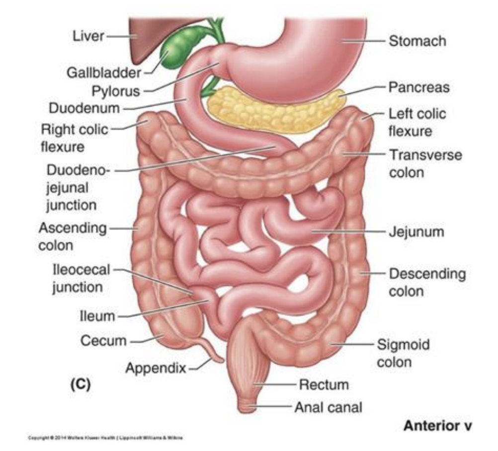
Small Intestine

● Three parts; duodenum, jejunum and ileum (proximal to distal)

● Intraperitoneal (suspending within peritoneal cavity), connected to posterior peritoneal cavity via mesentery

● Mesentery contains neurovascular supply

● Functions to absorb nutrients, electrolytes and water from the recently digested food

<p>● Three parts; duodenum, jejunum and ileum (proximal to distal) </p><p>● Intraperitoneal (suspending within peritoneal cavity), connected to posterior peritoneal cavity via mesentery </p><p>● Mesentery contains neurovascular supply </p><p>● Functions to absorb nutrients, electrolytes and water from the recently digested food</p>
30
New cards
<p>Large Intestine or Colon</p>

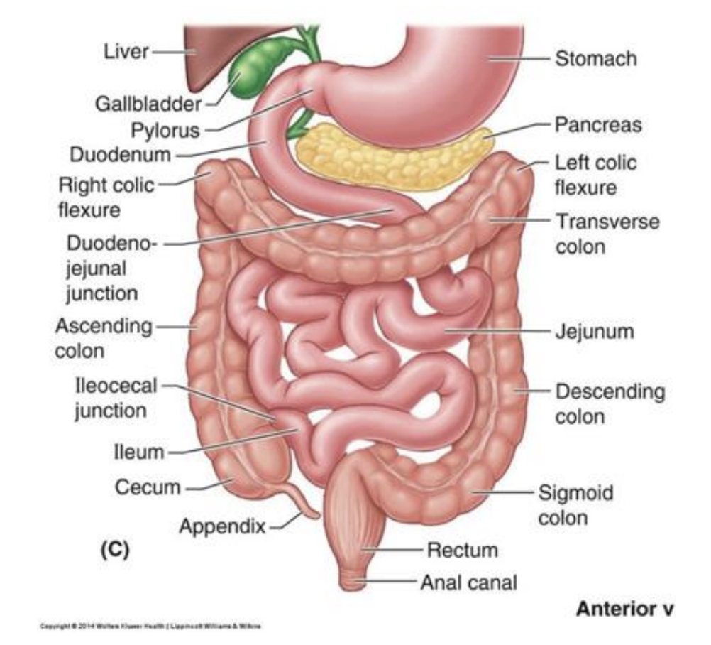
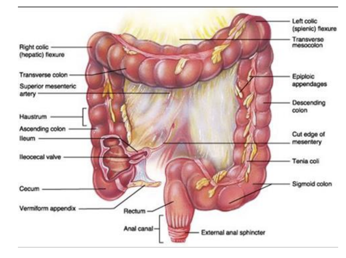
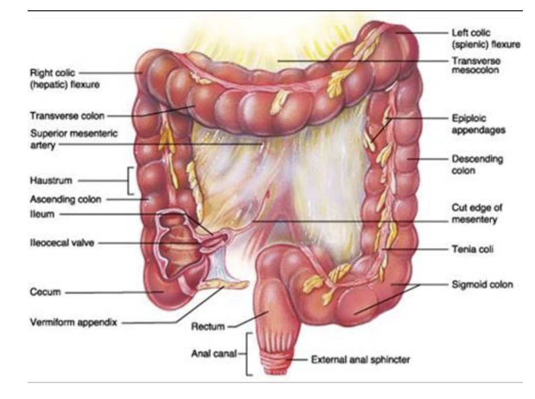
Large Intestine or Colon

● Starts at junction of ileum and cecum

● Cecum, transverse and sigmoid colon are intraperitoneal

● Ascending, descending colon and rectum are retroperitoneal (attached directly to posterior wall)

● Primarily serves to reabsorb water and electrolytes from and store feces, the final product after digestion and nutrient absorption

<p>● Starts at junction of ileum and cecum </p><p>● Cecum, transverse and sigmoid colon are intraperitoneal </p><p>● Ascending, descending colon and rectum are retroperitoneal (attached directly to posterior wall) </p><p>● Primarily serves to reabsorb water and electrolytes from and store feces, the final product after digestion and nutrient absorption</p>
31
New cards
<p>Kidney</p>

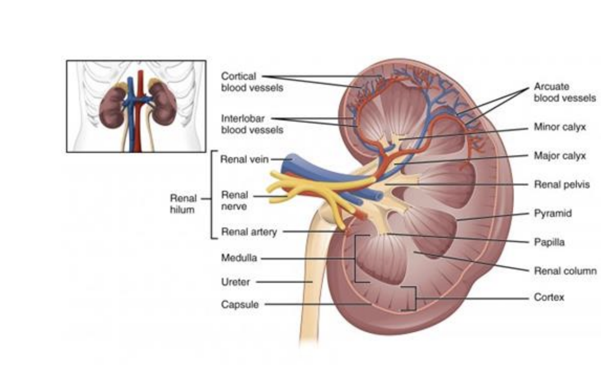
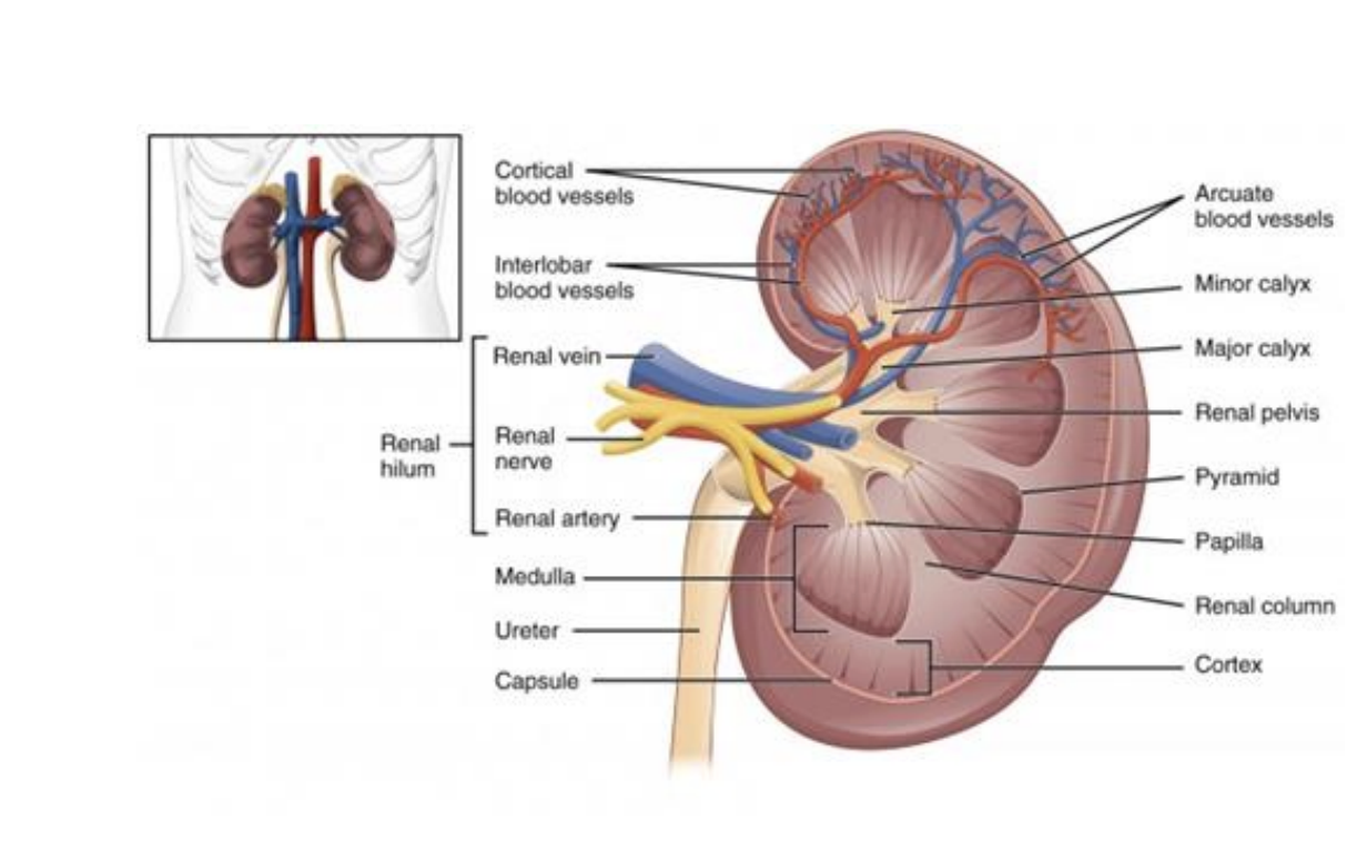
Kidney

● Collection of urine from renal pyramids into minor calyces

● Venous blood returns directly to caval system

● Adrenal glands sit on superior pole of kidney, contribute to endocrine functions

<p>● Collection of urine from renal pyramids into minor calyces </p><p>● Venous blood returns directly to caval system </p><p>● Adrenal glands sit on superior pole of kidney, contribute to endocrine functions</p>
32
New cards
<p><span style="font-family: &quot;Helvetica Neue&quot;;">Anatomic Landmarks of the Thorax</span></p>

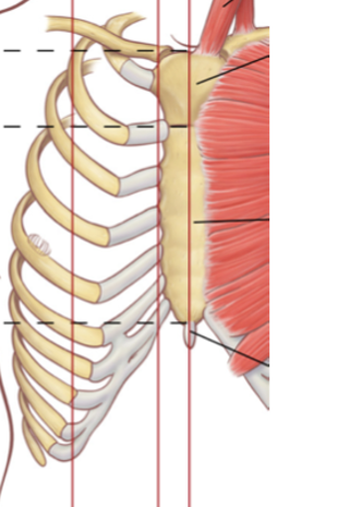
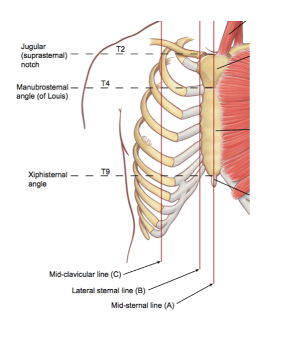
Anatomic Landmarks of the Thorax

•Sternal notch/Jugular notch

T2 vertebra

Rib 1

Sternal angle

T4 vertebra (T4-T5 intervertebral disk)

Rib 2

Aortic arch

Carina (bifurcation of trachea)

<p><span>•Sternal notch/Jugular notch</span></p><p><span style="font-family: &quot;Helvetica Neue&quot;;">•</span><span>T2 vertebra</span></p><p><span style="font-family: &quot;Helvetica Neue&quot;;">•</span><span>Rib 1</span></p><p><span style="font-family: &quot;Helvetica Neue&quot;;">•</span><span>Sternal angle</span></p><p><span style="font-family: &quot;Helvetica Neue&quot;;">•</span><span>T4 vertebra (T4-T5 intervertebral disk)</span></p><p><span style="font-family: &quot;Helvetica Neue&quot;;">•</span><span>Rib 2</span></p><p><span style="font-family: &quot;Helvetica Neue&quot;;">•</span><span>Aortic arch</span></p><p><span style="font-family: &quot;Helvetica Neue&quot;;">•</span><span>Carina (bifurcation of trachea)</span></p>
33
New cards
<p><span>Rib Anatomy</span></p>

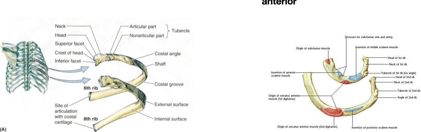
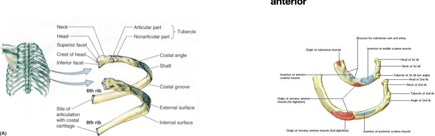
Rib Anatomy

Typical Ribs

Ribs 3 through 9

Head is wedged

2 articulation sites

***Articulates with superior costal facet on same numbered vertebrae and inferior costal facet on vertebrae superior to that

Neck has tubercle for articulation with vertebrae transverse process

Shaft has costal groove where neurovascular bundle travels

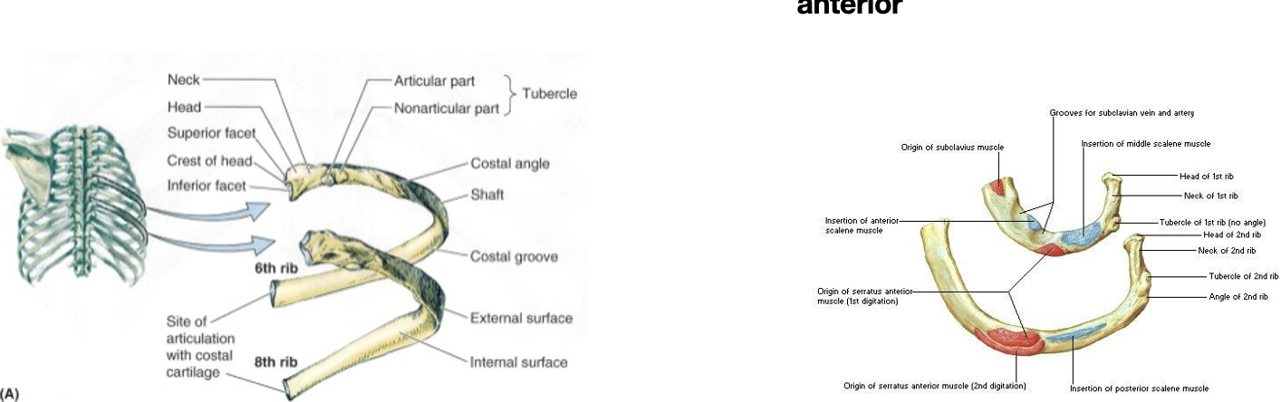
Atypical Ribs

First rib

Single facet at head (articulates with T1 only)

Widest rib

Tubercle for scalene

Grooves for subclavian artery and vein

Second rib

Two facets at head (articulates with T1 and T2)

Tubercle for scalene

Tuberosity for serratus anterior

<p><span style="font-family: &quot;Helvetica Neue&quot;;">❖</span><span><strong>Typical Ribs</strong></span></p><p><span style="font-family: &quot;Helvetica Neue&quot;;">➢</span><span><strong>Ribs 3 through 9</strong></span></p><p><span style="font-family: &quot;Helvetica Neue&quot;;">➢<strong><u>Head </u>is wedged</strong></span></p><p><span style="font-family: &quot;Helvetica Neue&quot;;">■</span><span><strong>2 articulation sites</strong></span></p><p style="text-align: left;"><span style="font-family: &quot;Helvetica Neue&quot;;"><strong>***Articulates with superior costal facet on same numbered vertebrae and inferior costal facet on vertebrae superior to that</strong></span></p><p style="text-align: left;"></p><p><span style="font-family: &quot;Helvetica Neue&quot;;">➢<strong><u>Neck </u>has <u>tubercle </u>for articulation with vertebrae transverse process</strong></span></p><p><span style="font-family: &quot;Helvetica Neue&quot;;">➢<strong><u>Shaft </u>has <u>costal groove </u>where neurovascular bundle travels</strong></span></p><p></p><p><span style="font-family: &quot;Helvetica Neue&quot;;">❖</span><span><strong>Atypical Ribs</strong></span></p><p><span style="font-family: &quot;Helvetica Neue&quot;;">➢</span><span><strong>First rib</strong></span></p><p><span style="font-family: &quot;Helvetica Neue&quot;;">■</span><span><strong>Single facet at head (articulates with T1 only)</strong></span></p><p><span style="font-family: &quot;Helvetica Neue&quot;;">■</span><span><strong>Widest rib</strong></span></p><p><span style="font-family: &quot;Helvetica Neue&quot;;">■</span><span><strong>Tubercle for scalene</strong></span></p><p><span style="font-family: &quot;Helvetica Neue&quot;;">■</span><span><strong>Grooves for subclavian artery and vein</strong></span></p><p><span style="font-family: &quot;Helvetica Neue&quot;;">➢</span><span><strong>Second rib</strong></span></p><p><span style="font-family: &quot;Helvetica Neue&quot;;">■</span><span><strong>Two facets at head (articulates with T1 and T2)</strong></span></p><p><span style="font-family: &quot;Helvetica Neue&quot;;">■</span><span><strong>Tubercle for scalene</strong></span></p><p><span style="font-family: &quot;Helvetica Neue&quot;;">■</span><span><strong>Tuberosity for serratus anterior</strong></span></p>
34
New cards

Joints of the Thorax

Costovertebral

Type: Synovial (mobile)

Costotransverse

Type: Synovial (mobile)

Costochondral

Type: Synchondrosis (immobile)

Sternocostal

Type: Synovial (mobile)

<p><span><strong>Costovertebral</strong></span></p><p><span>Type: Synovial (mobile)</span></p><p></p><p><span><strong>Costotransverse</strong></span></p><p>Type: <span>Synovial (mobile)</span></p><p></p><p><span><strong>Costochondral</strong></span></p><p>Type: <span>Synchondrosis (immobile)</span></p><p></p><p><span><strong>Sternocostal</strong></span></p><p>Type: <span>Synovial (mobile)</span></p>
35
New cards
<p>Respiration</p>

Respiration

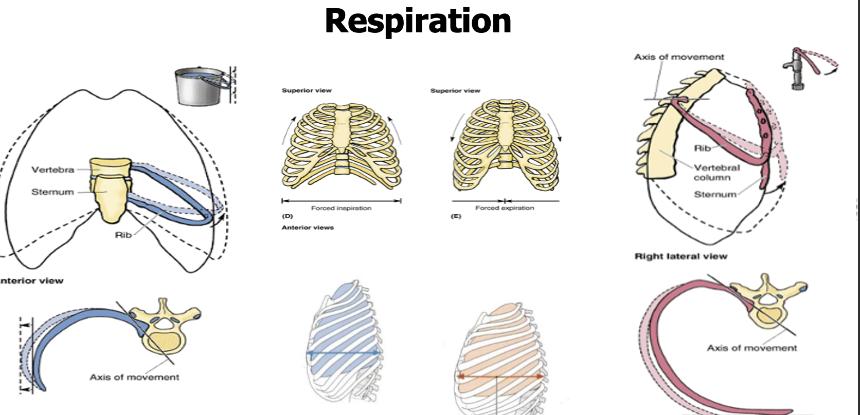
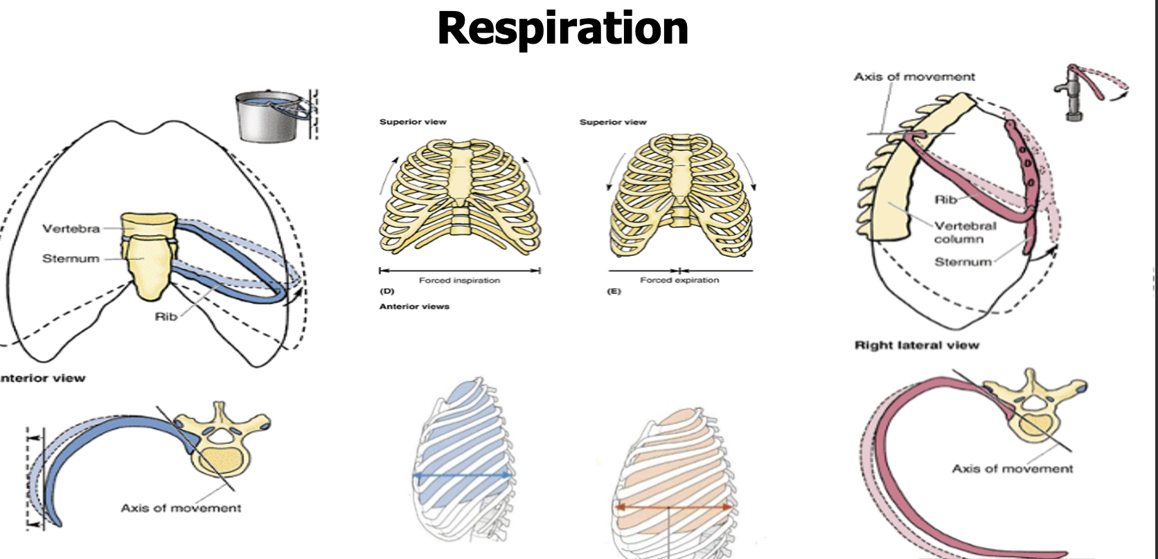
★Bucket-handle motion

○Costovertebral and sternocostal joints

○Rib moves laterally-superiorly

○Increases the transverse diameter

★Pump-handle motion

○Costotransverse joints

○Ribs and sternum moves anterior-superior

○Increases the Anterior- posterior diameter

<p><span><strong>★Bucket-handle motion</strong></span></p><p><span>○Costovertebral and sternocostal joints</span></p><p><span>○Rib moves laterally-superiorly</span></p><p><span>○Increases the transverse diameter</span></p><p></p><p><span><strong>★Pump-handle motion</strong></span></p><p><span>○Costotransverse joints</span></p><p><span>○Ribs and sternum moves anterior-superior</span></p><p><span>○Increases the Anterior- posterior diameter</span></p>
36
New cards

Muscles of the Thorax

Muscle

Innervation

Function

Location

Diaphragm

Phrenic nerve

Inspiration (major respiratory muscle)
Inferior boundary of thoracic cavity.  Inserts on central tendon

External Intercostals

Intercostal

Intercostal spaces; Fibers run anterior-caudally; Becomes membrane anteriorly

Internal Intercostals

Intercostal

Intercostal spaces; Fibers run posterior-caudally; Becomes membrane posteriorly.  Superficial to intercostal neurovascular

Innermost Intercostals

Intercostal

Intercostal spaces; Fibers run posterior-caudally; Becomes membrane posteriorly.  Deep to intercostal neurovascular

Subcostalis

Intercostal

Transversus thoracis

Intercostal

37
New cards

Nerves of the Thorax

Nerve name

Arises from

Supplies

Intercostal (T3 - T11)

T3 - T11 (ventral rami)

Sensory to parietal pleura and cutaneous region adjacent to ribs.  Motor to intercostal muscles

Subcostal (T12)

T12

Sensory and Motor to 12th intercostal space

Phrenic

C3 - C5

Sensory to diaphragm, pericardium and mediastinal parietal pleura region; some Motor to diaphragm

Vagus (CN X)

Parasympathetic to pleural and cardiac plexus

Sympathetic fibers

Sympathetic chain

Sympathetic to pleural and cardiac plexus

38
New cards

Autonomic Innervation of the Heart and Lungs

Sympathetic (Via Sympathetic trunk and plexuses)

Parasympathetic (via Vagus nerve and plexuses)

   HEART

Increase Heart Rate

Vasodilation of Coronaries

Decrease Heart Rate

Vasoconstriction of Coronaries

   LUNGS

Bronchodilation

•Vasoconstriction (includes pulmonary artery)

Bronchoconstriction

•Vasodilation (includes pulmonary artery)

39
New cards

Thoracic Wall Arteries (Posterior Thoracic Wall)

Vessel Name

Arises from;

Subclavian

Aortic Arch/Brachiocephalic (right)

Costocervical

Subclavian

Thoracic aorta

Aortic Arch

Posterior intercostals

Thoracic aorta

Subcostal

Thoracic aorta

40
New cards

Thoracic Wall Arteries (Anterior Thoracic Wall)

Vessel Name

Arises from;

Subclavian

Aortic Arch/Brachiocephalic (right)

Internal thoracic

Subclavian

Anterior intercostals (ribs 1 to 6)

Internal thoracic

Musculophrenic

Internal thoracic

Superior epigastric

Internal thoracic

Anterior intercostals (ribs 7 to 9)

Musculophrenic

41
New cards

Other Arteries of the Thorax

Pericardiacophrenic

Bronchial

Inferior phrenic
42
New cards
<p><span>Veins of the Thorax</span></p>

Veins of the Thorax

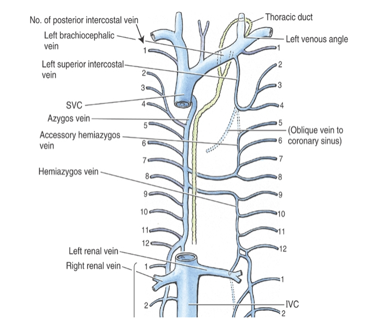
Vessel Name

Runs into;

Inferior Vena Cava (IVC)

Right atrium of the heart

Superior Vena Cava (SVC)

Right atrium of the heart

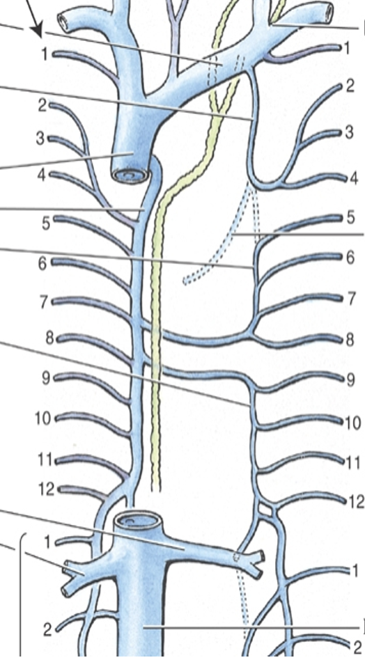
Azygos

Superior vena cava

Hemiazygos

Azygos

Accessory hemiazygos

Azygos

Posterior intercostals/Subcostal

Azygos veins

<table style="min-width: 50px;"><colgroup><col style="min-width: 25px;"><col style="min-width: 25px;"></colgroup><tbody><tr><td colspan="1" rowspan="1" style="height: 55.27pt; width: 314pt;"><p style="text-align: left;"><span><strong><u>Vessel Name</u></strong></span></p></td><td colspan="1" rowspan="1" style="width: 314pt;"><p style="text-align: left;"><span><strong><u>Runs into;</u></strong></span></p></td></tr><tr><td colspan="1" rowspan="1" style="height: 103.67pt; width: 314pt;"><p style="text-align: left;"><span>Inferior Vena Cava (IVC)</span></p></td><td colspan="1" rowspan="1" style="width: 314pt;"><p style="text-align: left;"><span>Right atrium of the heart</span></p></td></tr><tr><td colspan="1" rowspan="1" style="height: 52.41pt; width: 314pt;"><p style="text-align: left;"><span>Superior Vena Cava (SVC)</span></p></td><td colspan="1" rowspan="1" style="width: 314pt;"><p style="text-align: left;"><span>Right atrium of the heart</span></p></td></tr><tr><td colspan="1" rowspan="1" style="height: 50.84pt; width: 314pt;"><p style="text-align: left;"><span>Azygos</span></p></td><td colspan="1" rowspan="1" style="width: 314pt;"><p style="text-align: left;"><span>Superior vena cava</span></p></td></tr><tr><td colspan="1" rowspan="1" style="height: 48.82pt; width: 314pt;"><p style="text-align: left;"><span>Hemiazygos</span></p></td><td colspan="1" rowspan="1" style="width: 314pt;"><p style="text-align: left;"><span>Azygos</span></p></td></tr><tr><td colspan="1" rowspan="1" style="height: 61.43pt; width: 314pt;"><p style="text-align: left;"><span>Accessory hemiazygos</span></p></td><td colspan="1" rowspan="1" style="width: 314pt;"><p style="text-align: left;"><span>Azygos</span></p></td></tr><tr><td colspan="1" rowspan="1" style="height: 61.43pt; width: 314pt;"><p style="text-align: left;"><span>Posterior intercostals/Subcostal</span></p></td><td colspan="1" rowspan="1" style="width: 314pt;"><p style="text-align: left;"><span>Azygos veins</span></p></td></tr></tbody></table><p></p>
43
New cards
<p><span><strong>Mediastinum</strong></span></p>

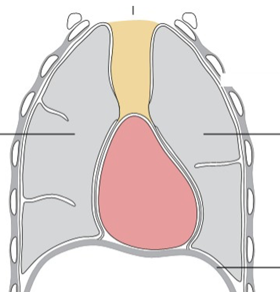
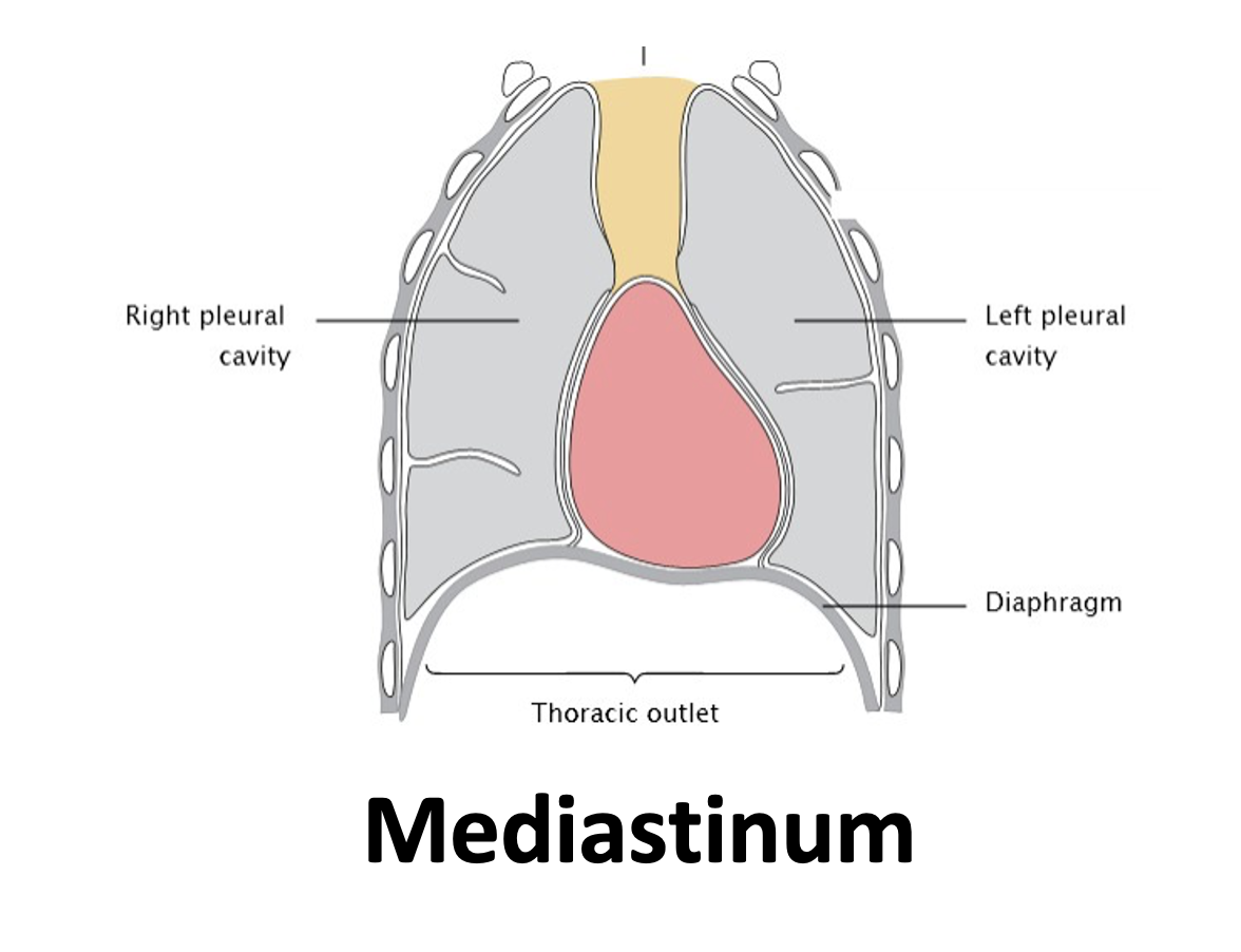
Mediastinum

Mediastinum Region

Contents

Superior

Thymus, trachea, esophagus; Aortic arch and its branches; Superior vena cava, brachiocephalic veins, thoracic duct; Vagus, phrenic nerve, cardiac plexus, left recurrent laryngeal

Inferior: Anterior

Thymus; Internal thoracic artery/vein and branches

Inferior: Middle

Heart, pericardium; Ascending aorta, pulmonary trunk, pericardiacophrenic arteries/veins; Pulmonary veins, superior vena cava; Phrenic nerve

Inferior: Posterior

Esophagus; Thoracic aorta (and branches); Azygos, hemiazygos v. thoracic duct; Vagus nerve, sympathetic trunk

<table style="min-width: 50px;"><colgroup><col style="min-width: 25px;"><col style="min-width: 25px;"></colgroup><tbody><tr><td colspan="1" rowspan="1" style="height: 85.58pt; width: 219pt;"><p style="text-align: left;"><span><strong><u>Mediastinum Region</u></strong></span></p></td><td colspan="1" rowspan="1" style="width: 674pt;"><p style="text-align: left;"><span><strong><u>Contents</u></strong></span></p></td></tr><tr><td colspan="1" rowspan="1" style="height: 113pt; width: 219pt;"><p style="text-align: left;"><span>Superior</span></p></td><td colspan="1" rowspan="1" style="width: 674pt;"><p style="text-align: left;"><span>Thymus, trachea, esophagus; <em>Aortic arch and its branches; </em>Superior vena cava, brachiocephalic veins, thoracic duct; <em>Vagus, phrenic nerve, cardiac plexus, left recurrent laryngeal</em></span></p></td></tr><tr><td colspan="1" rowspan="1" style="height: 63.29pt; width: 219pt;"><p style="text-align: left;"><span>Inferior: Anterior</span></p></td><td colspan="1" rowspan="1" style="width: 674pt;"><p style="text-align: left;"><span>Thymus; <em>Internal thoracic artery/vein and branches</em></span></p></td></tr><tr><td colspan="1" rowspan="1" style="height: 85.58pt; width: 219pt;"><p style="text-align: left;"><span>Inferior: Middle</span></p><p style="text-align: left;"></p></td><td colspan="1" rowspan="1" style="width: 674pt;"><p style="text-align: left;"><span>Heart, pericardium; <em>Ascending aorta, pulmonary trunk, pericardiacophrenic arteries</em>/veins; Pulmonary veins, superior vena cava; <em>Phrenic nerve</em></span></p></td></tr><tr><td colspan="1" rowspan="1" style="height: 113pt; width: 219pt;"><p style="text-align: left;"><span>Inferior: Posterior</span></p><p style="text-align: left;"></p></td><td colspan="1" rowspan="1" style="width: 674pt;"><p style="text-align: left;"><span>Esophagus; <em>Thoracic aorta (and branches)</em>; Azygos, hemiazygos v. thoracic duct; <em>Vagus nerve, sympathetic trunk</em></span></p></td></tr></tbody></table><p></p>
44
New cards

Pleura and Pericardium (Thorax Viscera)

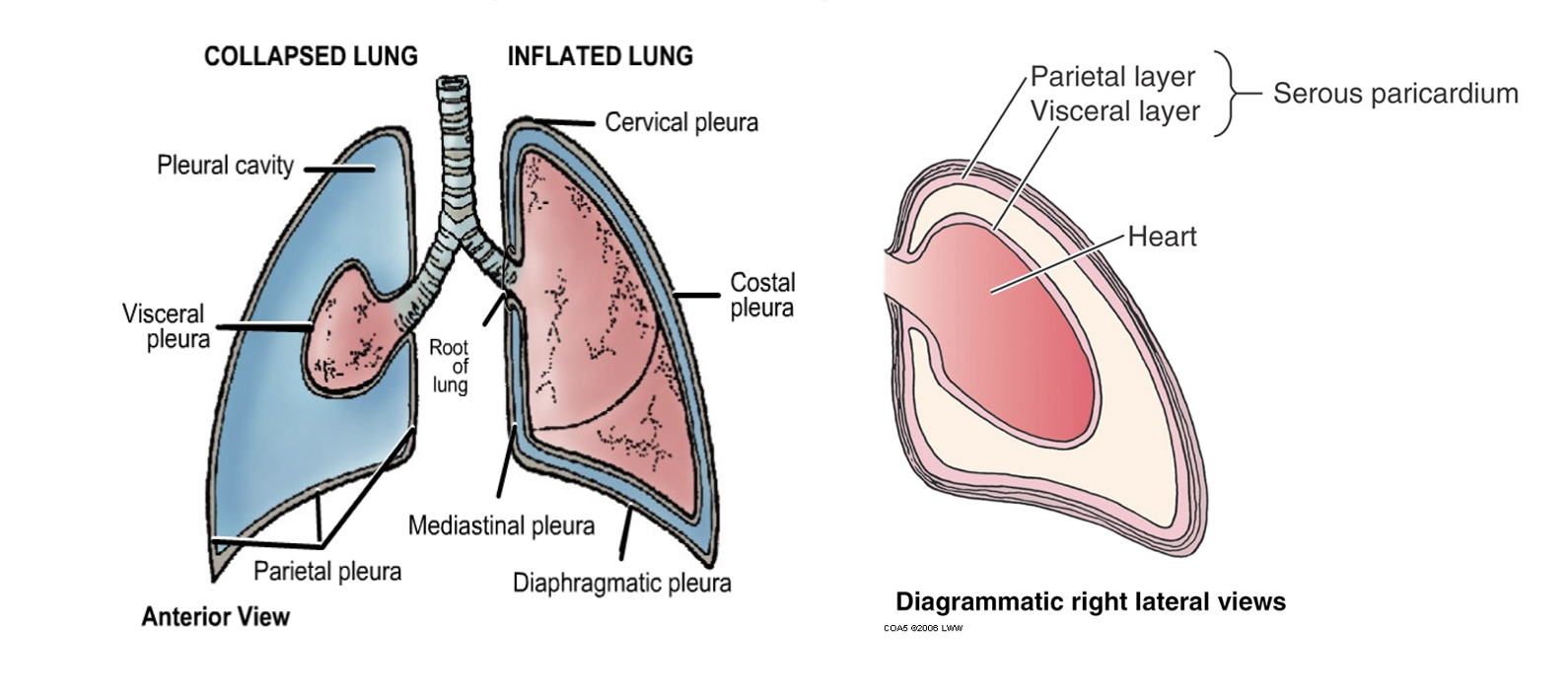
●Two continuous membranes (at the hilum)

○Visceral

-Invests on surfaces of organ

○Parietal

-Invests on outer cavity lining

○Pleural cavity & pericardial cavity

-Cavity formed between visceral layers and parietal layers

-Contains serous fluid (lubricates surfaces)
<p style="text-align: left;"></p><p><span>●Two continuous membranes (at the hilum)</span></p><p></p><p><span>○Visceral</span></p><p><span>-Invests on surfaces of organ</span></p><p></p><p><span>○Parietal</span></p><p><span>-Invests on outer cavity lining</span></p><p></p><p><span>○Pleural cavity &amp; pericardial cavity</span></p><p><span>-Cavity formed between visceral layers and parietal layers</span></p><p><span>-Contains serous fluid (lubricates surfaces)</span></p>
45
New cards
<p><span>Lungs</span></p>

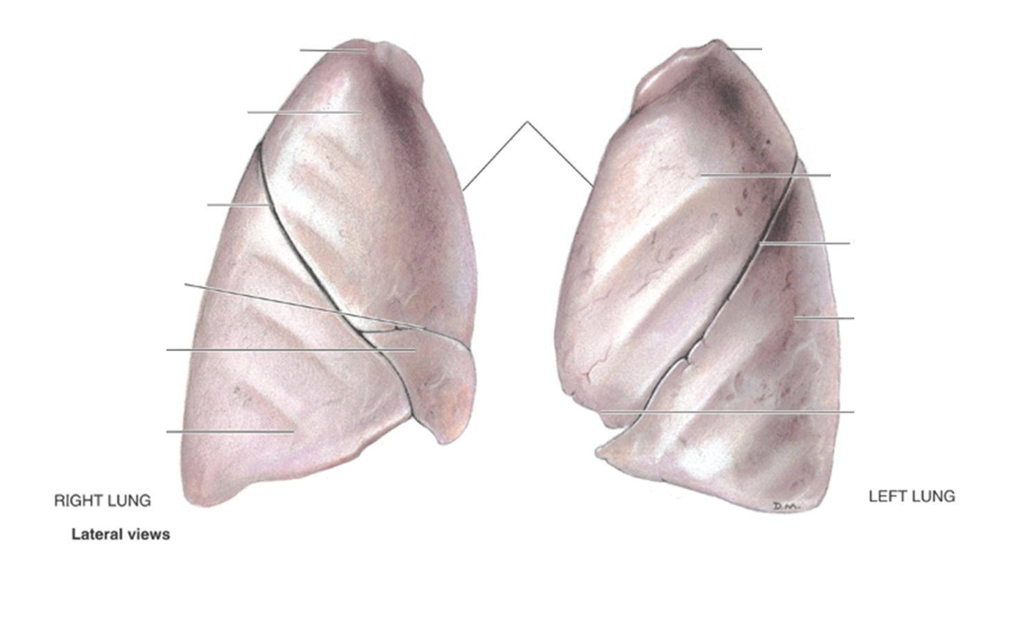
Lungs

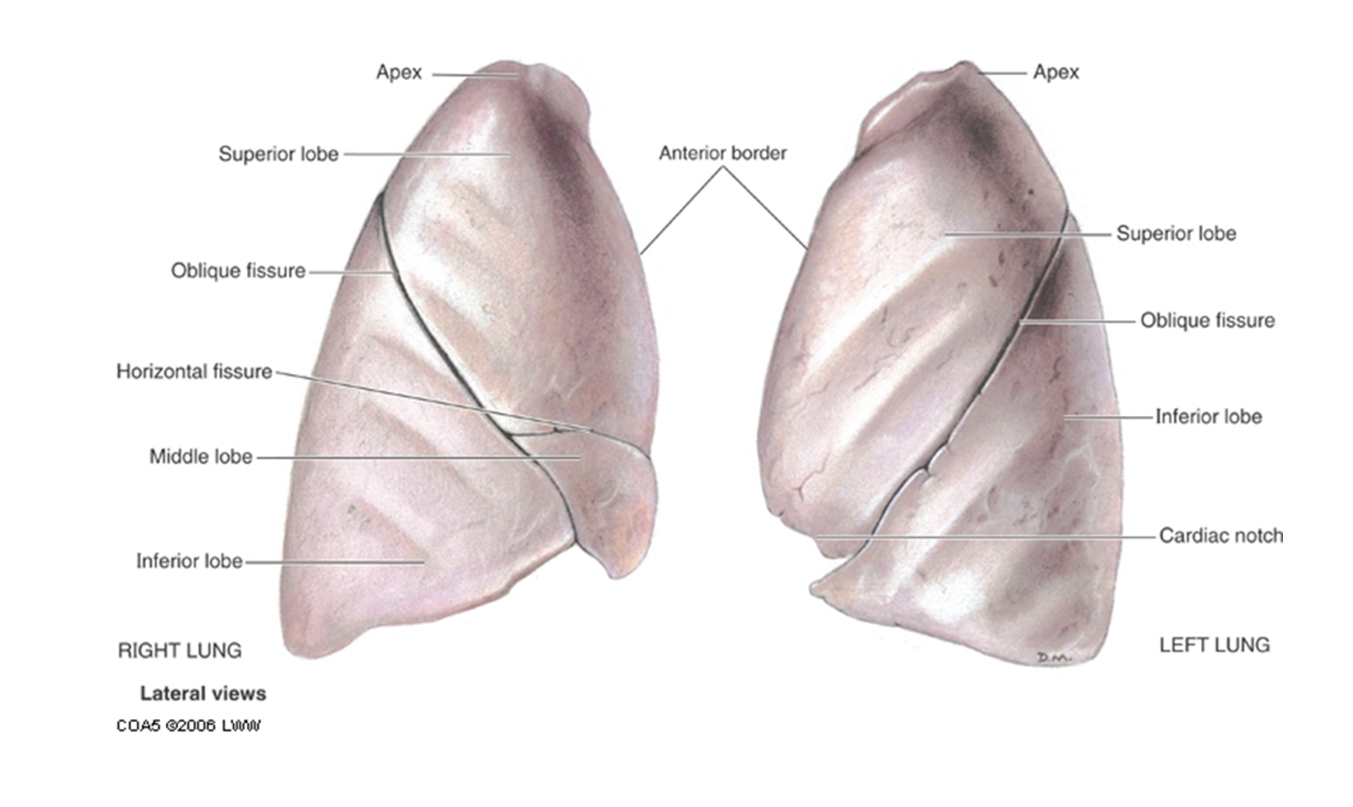
Right Lung

●Three lobed

-Superior

-Middle

-Inferior

●Fissures

-Horizontal fissure

-Oblique fissure

●Primary bronchi more vertical than left

●Wider than left

●Pulmonary artery is anterior to bronchus at hilum

Left lung

•Two lobed

–Superior

–Inferior

•Oblique fissure

•Prominent impression from aorta and heart

•Lingula on inferior aspect of Superior lobe

•Pulmonary artery is superior to bronchus at hilum

RALS: Pulmonary artery to Bronchus → Right anterior, left superior
<p><span><strong>Right Lung</strong></span></p><p><span>●Three lobed</span></p><p><span>-Superior</span></p><p><span>-Middle</span></p><p><span>-Inferior</span></p><p></p><p><span>●Fissures</span></p><p><span>-Horizontal fissure</span></p><p><span>-Oblique fissure</span></p><p></p><p><span>●Primary bronchi more vertical than left</span></p><p><span>●Wider than left</span></p><p><span>●Pulmonary artery is anterior to bronchus at hilum</span></p><p></p><p><span><strong>Left lung</strong></span></p><p><span>•Two lobed</span></p><p><span>–Superior</span></p><p><span>–Inferior</span></p><p></p><p><span>•Oblique fissure</span></p><p><span>•Prominent impression from aorta and heart</span></p><p><span>•Lingula on inferior aspect of Superior lobe</span></p><p><span>•Pulmonary artery is superior to bronchus at hilum</span></p><p></p><p><span><em>RALS:  Pulmonary artery to Bronchus → Right anterior, left superior</em></span></p>
46
New cards
<p><span>Tracheobronchial Tree of the Lungs</span></p>

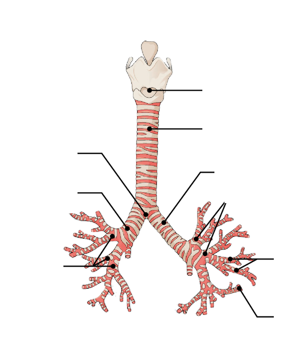
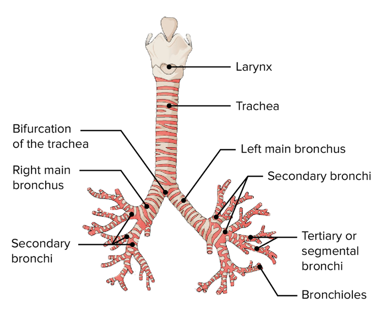
Tracheobronchial Tree of the Lungs

Tracheobronchial Tree

Primary bronchi branches after carina of trachea

Right bronchus is wider, shorter, more vertical

Secondary (lobar) bronchi branches to each separate lobe of lung (5 total, 3 right, 2 left)

Tertiary (segmental) bronchi  branch to bronchopulmonary segments (~10 segments in each lung)

Pulmonary arteries follow this segmenting pattern
<p><span style="font-family: &quot;Helvetica Neue&quot;;"><strong>Tracheobronchial Tree</strong></span></p><p><span style="font-family: &quot;Helvetica Neue&quot;;">•</span><span><strong>Primary bronchi</strong></span><span style="font-family: &quot;Helvetica Neue&quot;;"> branches after carina of trachea</span></p><p><span style="font-family: &quot;Helvetica Neue&quot;;">–</span><span><strong>Right bronchus is wider, shorter, more vertical</strong></span></p><p><span style="font-family: &quot;Helvetica Neue&quot;;">•</span><span><strong>Secondary (lobar) bronchi branches to each separate lobe of lung </strong>(5 total, 3 right, 2 left)</span></p><p><span style="font-family: &quot;Helvetica Neue&quot;;">•</span><span><strong>Tertiary (segmental) bronchi </strong></span><span style="font-family: &quot;Helvetica Neue&quot;;">&nbsp;branch to bronchopulmonary segments<strong> </strong>(~10 segments in each lung)</span></p><p style="text-align: left;"></p><p><span>❖</span><span style="font-family: &quot;Helvetica Neue&quot;;">Pulmonary arteries follow this segmenting pattern</span></p>
47
New cards
<p><span>Heart - Blood Flow</span></p>

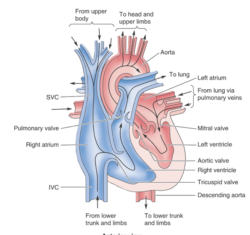
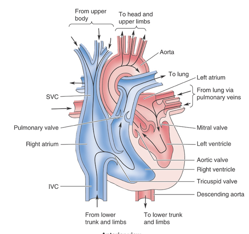
Heart - Blood Flow

Four chambers

Right and Left atrium receive blood from veins

Right and Left Ventricle receive blood from atria

  1. Right Atrium

    • Receives all venous drainage via;

      • Inferior vena cava

      • Superior vena cava

      • Coronary sinus

  2. Right Ventricle

    • Receives blood from atrium then pumps into the pulmonary trunk

  3. Pulmonary Trunk

    • Pulmonary circulation for oxygenation

  4. Left Atrium

    • Receives oxygenated blood from the pulmonary veins

  5. Left Ventricle

    • Pumps all oxygenated blood to the blood via the aortic arch (and the coronary ostia in the valves)
<p><span>•<strong>Four chambers</strong></span></p><p><span>–<strong><u>Right and Left atrium</u> </strong>– <strong>receive blood from veins</strong></span></p><p><span>–<strong><u>Right and Left Ventricle</u> </strong>– <strong>receive blood from atria</strong></span></p><p></p><ol><li><p><strong>Right Atrium</strong></p><ul><li><p>Receives all venous drainage via;</p><ul><li><p>Inferior vena cava</p></li><li><p>Superior vena cava</p></li><li><p>Coronary sinus</p></li></ul></li></ul></li><li><p><strong>Right Ventricle</strong></p><ul><li><p>Receives blood from atrium then pumps into the pulmonary trunk</p></li></ul></li><li><p><strong>Pulmonary Trunk</strong></p><ul><li><p>Pulmonary circulation for oxygenation</p></li></ul></li><li><p><strong>Left Atrium</strong></p><ul><li><p>Receives oxygenated blood from the pulmonary veins</p></li></ul></li><li><p><strong>Left Ventricle</strong></p><ul><li><p>Pumps all oxygenated blood to the blood via the aortic arch (and the coronary ostia in the valves)</p></li></ul></li></ol><p></p>
48
New cards

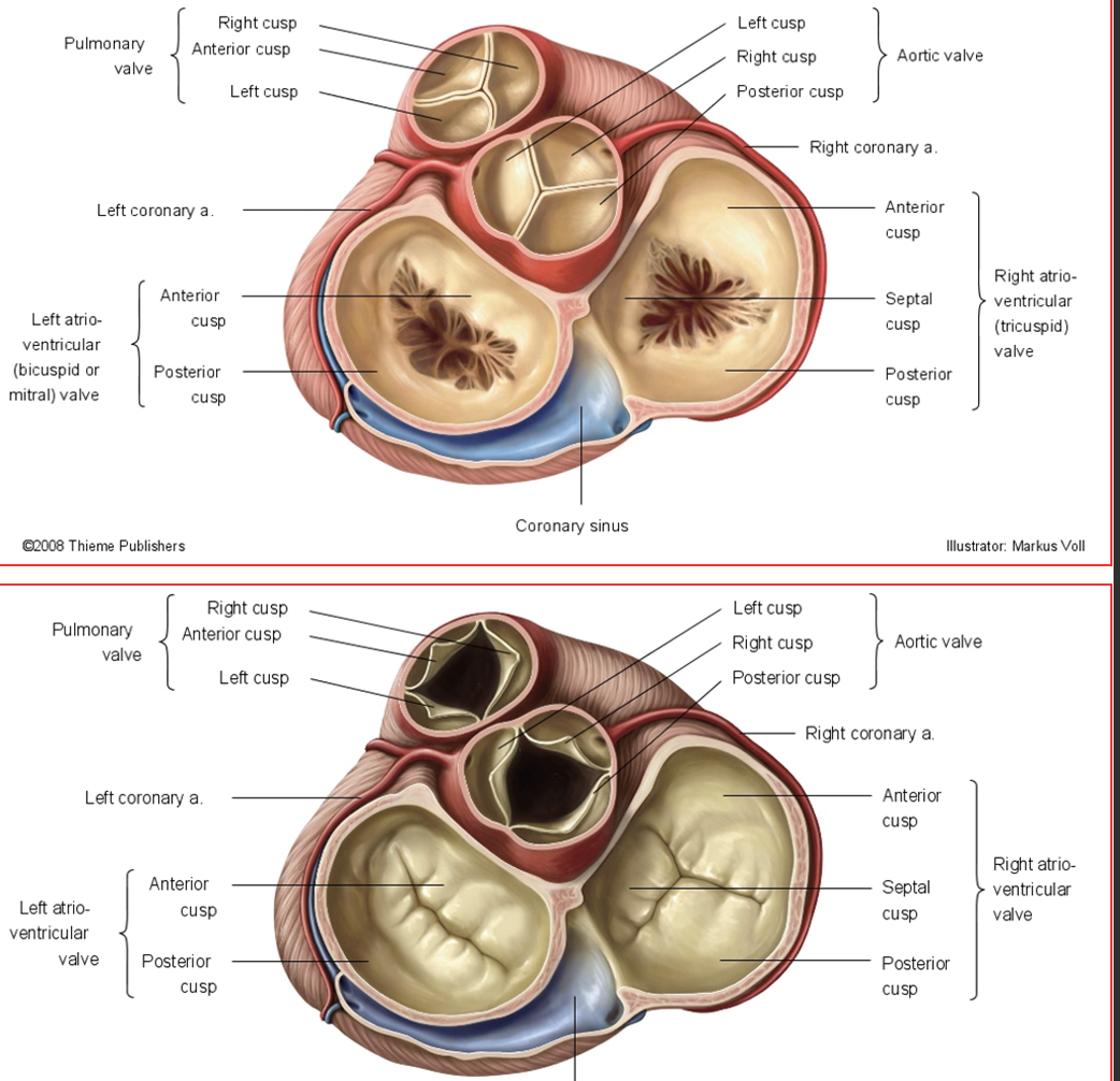
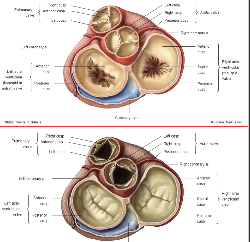
Valves of the Heart

Atrioventricular (AV) Valves

Tricuspid: Between right atrium and ventricle

Bicuspid/Mitral: Between left atrium and ventricle

Have an anterior AND posterior leaflet/cusp; Tricuspid has a septal leaflet/cusp

**AV valves are held closed during systole by contracted papillary muscles and the attached chordae tendineae

Semilunar Valves

Pulmonic: Between right ventricle and pulmonary trunk

Aortic: Between left ventricle and aorta

★Have a left AND right cusp; either a posterior OR anterior cusp

**Semilunar valves passively fill with blood during diastole to close off the ventricles from blood return
49
New cards
<p>Cardiac Cycle of the Heart</p>

Cardiac Cycle of the Heart

Diastole

Ventricle filling

AV valves open

Semilunar valves closed

Systole

Ventricle squeeze

AV valves closed

Semilunar valves open

<p><span style="font-family: &quot;Helvetica Neue&quot;;"><strong><u>Diastole</u></strong></span></p><p><span style="font-family: &quot;Helvetica Neue&quot;;">➔</span><span><strong>Ventricle filling</strong></span></p><p><span style="font-family: &quot;Helvetica Neue&quot;;">➔</span><span><strong>AV valves open</strong></span></p><p><span style="font-family: &quot;Helvetica Neue&quot;;">➔</span><span><strong>Semilunar valves closed</strong></span></p><p></p><p><span style="font-family: &quot;Helvetica Neue&quot;;"><strong><u>Systole</u></strong></span></p><p><span style="font-family: &quot;Helvetica Neue&quot;;">➔</span><span><strong>Ventricle squeeze</strong></span></p><p><span style="font-family: &quot;Helvetica Neue&quot;;">➔</span><span><strong>AV valves closed</strong></span></p><p><span style="font-family: &quot;Helvetica Neue&quot;;">➔</span><span><strong>Semilunar valves open</strong></span></p>
50
New cards

Heart Chamber contents

Heart Chamber

Structures found

Right Atrium

Pectinate muscles, sinus venarum, fossa ovalis, coronary sinus, crista terminalis, right auricle

Right Ventricle

Trabeculae carneae, conus arteriosus, moderator band (septomarginal trabeculae), papillary muscles, chordae tendineae

Left Atrium

Pectinate muscles in left auricle only

Left Ventricle

Trabeculae carneae, papillary muscles, chordae tendineae

51
New cards
<p><span style="font-family: &quot;Helvetica Neue&quot;;">Fetal Heart Circulation</span></p>

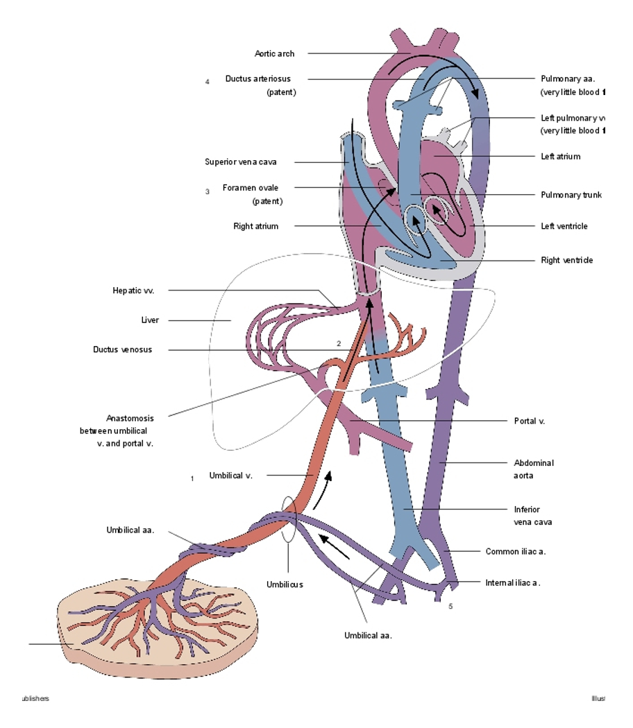
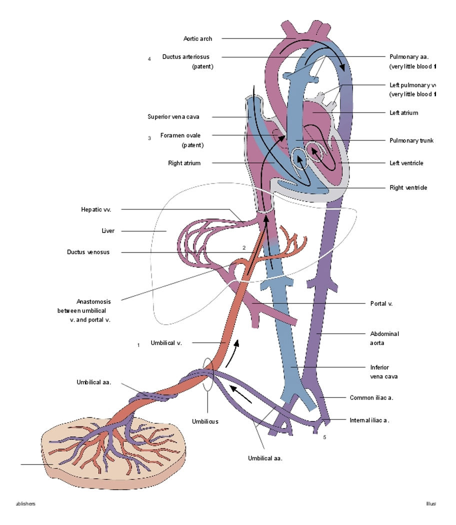
Fetal Heart Circulation

Bypasses pulmonary circulation

Foramen Ovale

Shunts blood from right atrium to left atrium

Closes at birth to become the Fossa Ovalis

Ductus Arteriosus,

Shunts blood from pulmonary trunk to aorta

Shrinks at birth to become the Ligamentum Arteriosum
<p><span><em>Bypasses pulmonary circulation</em></span></p><p></p><p><span style="font-family: &quot;Helvetica Neue&quot;;">•</span><span><strong>Foramen </strong></span><span style="font-family: &quot;Helvetica Neue&quot;;"><strong>Ovale</strong></span></p><p><span style="font-family: &quot;Helvetica Neue&quot;;">•</span><span>Shunts blood from right atrium to left atrium</span></p><p><span>•</span><span style="font-family: &quot;Helvetica Neue&quot;;">Closes at birth to become the <strong>Fossa Ovalis</strong></span></p><p style="text-align: left;"></p><p><span style="font-family: &quot;Helvetica Neue&quot;;">•</span><span><strong>Ductus Arteriosus</strong></span><span style="font-family: &quot;Helvetica Neue&quot;;"><strong>,</strong></span></p><p><span style="font-family: &quot;Helvetica Neue&quot;;">•</span><span>Shunts blood from pulmonary trunk to aorta</span></p><p><span>•</span><span style="font-family: &quot;Helvetica Neue&quot;;">Shrinks at birth to become the <strong>Ligamentum Arteriosum</strong></span></p>
52
New cards
<p>Blood Vessels of the Heart</p>

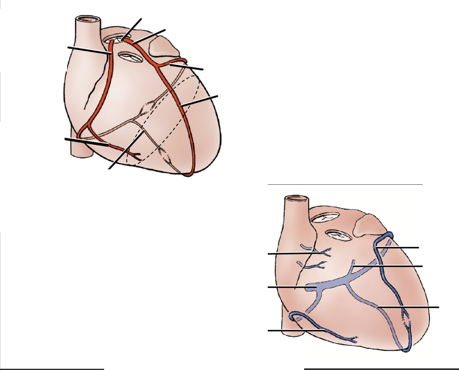
Blood Vessels of the Heart

Vessel

Arises from

Supplies

Pulmonary arteries

Pulmonary trunk exiting the right ventricle

Carries deoxygenated blood to the lungs

Pulmonary veins

Left and right lungs

Brings oxygenated blood back to the heart at the left atrium

Right coronary

Right marginal

Right coronary

Posterior interventricular branch

Right coronary (usually)

Left coronary

Anterior interventricular branch (left anterior descending, LAD)

Left coronary

Circumflex

Left coronary

Left marginal

Circumflex

Great cardiac vein

Anterior interventricular groove

Middle cardiac vein

Posterior interventricular groove

Merges with cardiac veins before becoming the coronary sinus

Small cardiac vein

Right side of heart near right marginal artery

Drains into coronary sinus right before it enters right atrium

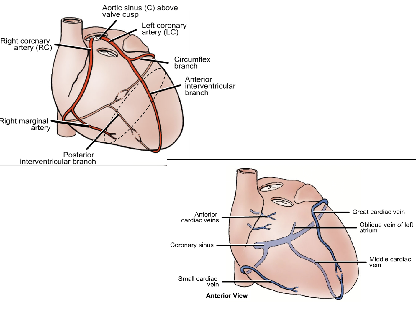
<table style="min-width: 75px;"><colgroup><col style="min-width: 25px;"><col style="min-width: 25px;"><col style="min-width: 25px;"></colgroup><tbody><tr><td colspan="1" rowspan="1" style="height: 28.07pt; width: 283pt;"><p style="text-align: left;"><span><strong><u>Vessel</u></strong></span></p></td><td colspan="1" rowspan="1" style="width: 212pt;"><p style="text-align: left;"><span><strong><u>Arises from</u></strong></span></p></td><td colspan="1" rowspan="1" style="width: 434pt;"><p style="text-align: left;"><span><strong><u>Supplies</u></strong></span></p></td></tr><tr><td colspan="1" rowspan="1" style="height: 48.74pt; width: 283pt;"><p style="text-align: left;"><span>Pulmonary arteries</span></p></td><td colspan="1" rowspan="1" style="width: 212pt;"><p style="text-align: left;"><span>Pulmonary trunk exiting the <u>right ventricle</u></span></p></td><td colspan="1" rowspan="1" style="width: 434pt;"><p style="text-align: left;"><span>Carries deoxygenated blood to the lungs</span></p></td></tr><tr><td colspan="1" rowspan="1" style="height: 59.81pt; width: 283pt;"><p style="text-align: left;"><span>Pulmonary veins</span></p></td><td colspan="1" rowspan="1" style="width: 212pt;"><p style="text-align: left;"><span>Left and right lungs</span></p></td><td colspan="1" rowspan="1" style="width: 434pt;"><p style="text-align: left;"><span>Brings oxygenated blood back to the heart at the l<u>eft atrium</u></span></p></td></tr><tr><td colspan="1" rowspan="1" style="height: 48.74pt; width: 283pt;"><p style="text-align: left;"><span>Right coronary</span></p></td><td colspan="1" rowspan="1" style="width: 212pt;"><p style="text-align: left;"></p></td><td colspan="1" rowspan="1" style="width: 434pt;"><p style="text-align: left;"></p></td></tr><tr><td colspan="1" rowspan="1" style="height: 48.74pt; width: 283pt;"><p style="text-align: left;"><span>Right marginal</span></p></td><td colspan="1" rowspan="1" style="width: 212pt;"><p style="text-align: left;"><span>Right coronary</span></p></td><td colspan="1" rowspan="1" style="width: 434pt;"><p style="text-align: left;"></p></td></tr><tr><td colspan="1" rowspan="1" style="height: 48.74pt; width: 283pt;"><p style="text-align: left;"><span>Posterior interventricular branch</span></p></td><td colspan="1" rowspan="1" style="width: 212pt;"><p style="text-align: left;"><span>Right coronary (usually)</span></p></td><td colspan="1" rowspan="1" style="width: 434pt;"><p style="text-align: left;"></p></td></tr><tr><td colspan="1" rowspan="1" style="height: 48.74pt; width: 283pt;"><p style="text-align: left;"><span>Left coronary</span></p></td><td colspan="1" rowspan="1" style="width: 212pt;"><p style="text-align: left;"></p></td><td colspan="1" rowspan="1" style="width: 434pt;"><p style="text-align: left;"></p></td></tr><tr><td colspan="1" rowspan="1" style="height: 48.74pt; width: 283pt;"><p style="text-align: left;"><span>Anterior interventricular branch (left anterior descending, LAD)</span></p></td><td colspan="1" rowspan="1" style="width: 212pt;"><p style="text-align: left;"><span>Left coronary</span></p></td><td colspan="1" rowspan="1" style="width: 434pt;"><p style="text-align: left;"></p></td></tr><tr><td colspan="1" rowspan="1" style="height: 48.74pt; width: 283pt;"><p style="text-align: left;"><span>Circumflex</span></p></td><td colspan="1" rowspan="1" style="width: 212pt;"><p style="text-align: left;"><span>Left coronary</span></p></td><td colspan="1" rowspan="1" style="width: 434pt;"><p style="text-align: left;"></p></td></tr><tr><td colspan="1" rowspan="1" style="height: 48.74pt; width: 283pt;"><p style="text-align: left;"><span>Left marginal</span></p></td><td colspan="1" rowspan="1" style="width: 212pt;"><p style="text-align: left;"><span>Circumflex</span></p></td><td colspan="1" rowspan="1" style="width: 434pt;"><p style="text-align: left;"></p></td></tr><tr><td colspan="1" rowspan="1" style="height: 48.74pt; width: 283pt;"><p style="text-align: left;"><span>Great cardiac vein</span></p></td><td colspan="1" rowspan="1" style="width: 212pt;"><p style="text-align: left;"><span>Anterior interventricular groove</span></p></td><td colspan="1" rowspan="1" style="width: 434pt;"><p style="text-align: left;"></p></td></tr><tr><td colspan="1" rowspan="1" style="height: 48.74pt; width: 283pt;"><p style="text-align: left;"><span>Middle cardiac vein</span></p></td><td colspan="1" rowspan="1" style="width: 212pt;"><p style="text-align: left;"><span>Posterior interventricular groove</span></p></td><td colspan="1" rowspan="1" style="width: 434pt;"><p style="text-align: left;"><span>Merges with cardiac veins before becoming the coronary sinus</span></p></td></tr><tr><td colspan="1" rowspan="1" style="height: 48.74pt; width: 283pt;"><p style="text-align: left;"><span>Small cardiac vein</span></p></td><td colspan="1" rowspan="1" style="width: 212pt;"><p style="text-align: left;"><span>Right side of heart near right marginal artery</span></p></td><td colspan="1" rowspan="1" style="width: 434pt;"><p style="text-align: left;"><span>Drains into coronary sinus right before it enters right atrium</span></p></td></tr></tbody></table><p></p>
53
New cards
<p><span>Heart Conducting system</span></p>

Heart Conducting system

Sinoatrial node – Specialized cardiac muscle cells, initiates conducting impulse

•Impulse transmits to Atrioventricular node

•Impulse is distributed through Atrioventricular bundle (Bundle of His)

Purkinje Fibers then transmit the impulse to the right and left ventricle for simultaneous contraction

–Fibers also run through the Moderator Band found in the right ventricle between the interventricular septum and the anterior papillary muscle
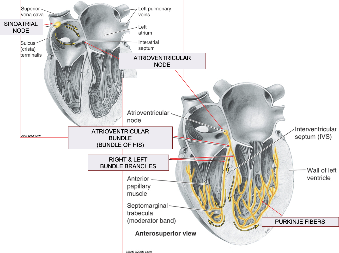
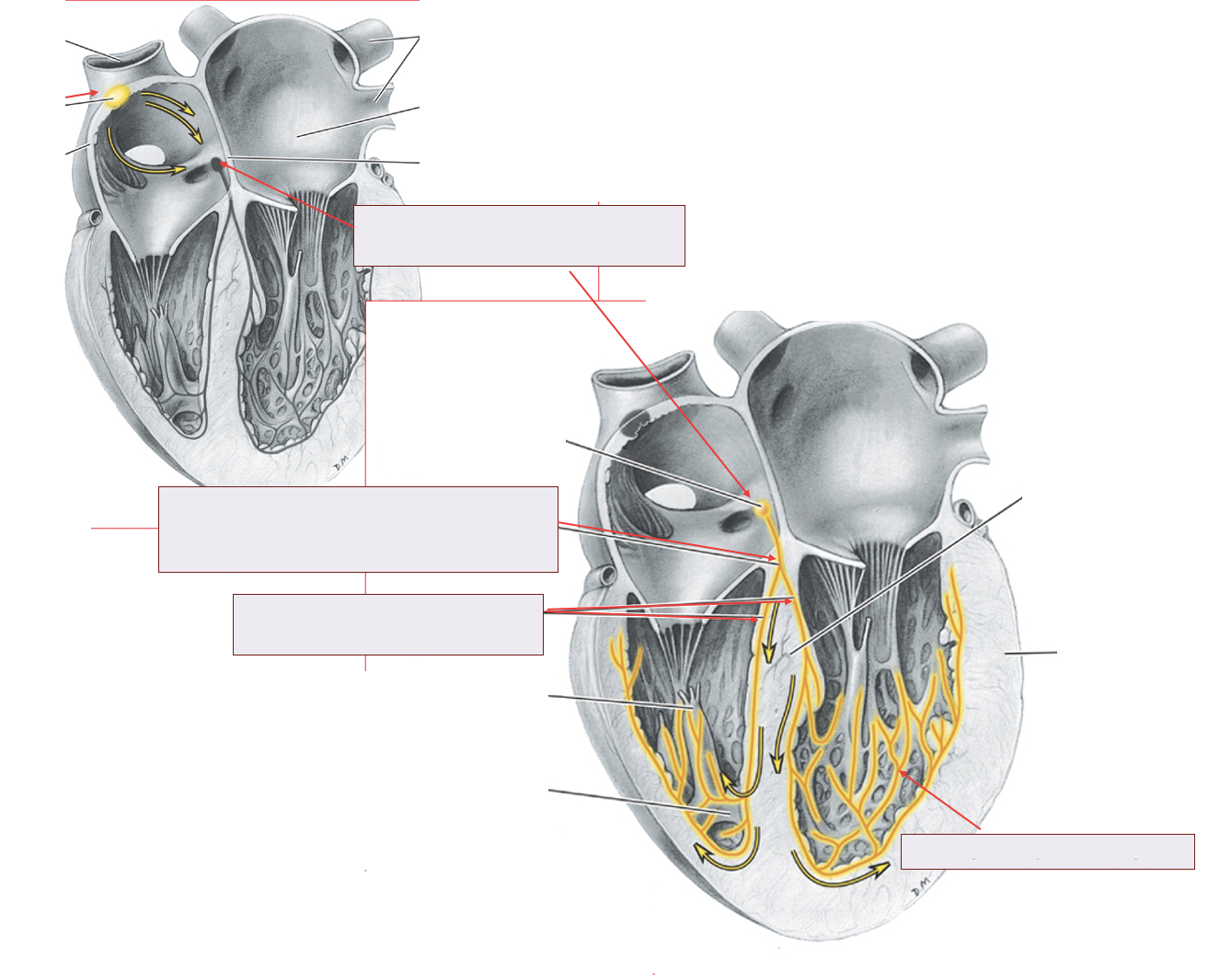
<p><span>•<strong>Sinoatrial node </strong>– Specialized cardiac muscle cells, <strong>initiates conducting impulse</strong></span></p><p><span>•Impulse transmits to <strong>Atrioventricular node</strong></span></p><p><span>•Impulse is distributed through<strong> Atrioventricular bundle (Bundle of His)</strong></span></p><p><span>•<strong>Purkinje Fibers</strong> then transmit the impulse to the right and left ventricle for simultaneous contraction</span></p><p><span>–Fibers also run through the <strong>Moderator Band </strong>found in the right ventricle between the interventricular septum and the anterior papillary muscle</span></p>