Chapter 10 - Cells divide for growth, repair, replacement and reproduction
1/74
There's no tags or description
Looks like no tags are added yet.
Name | Mastery | Learn | Test | Matching | Spaced | Call with Kai | Chat |
|---|
No analytics yet
Send a link to your students to track their progress
75 Terms
Average life span of human cells

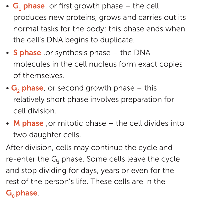
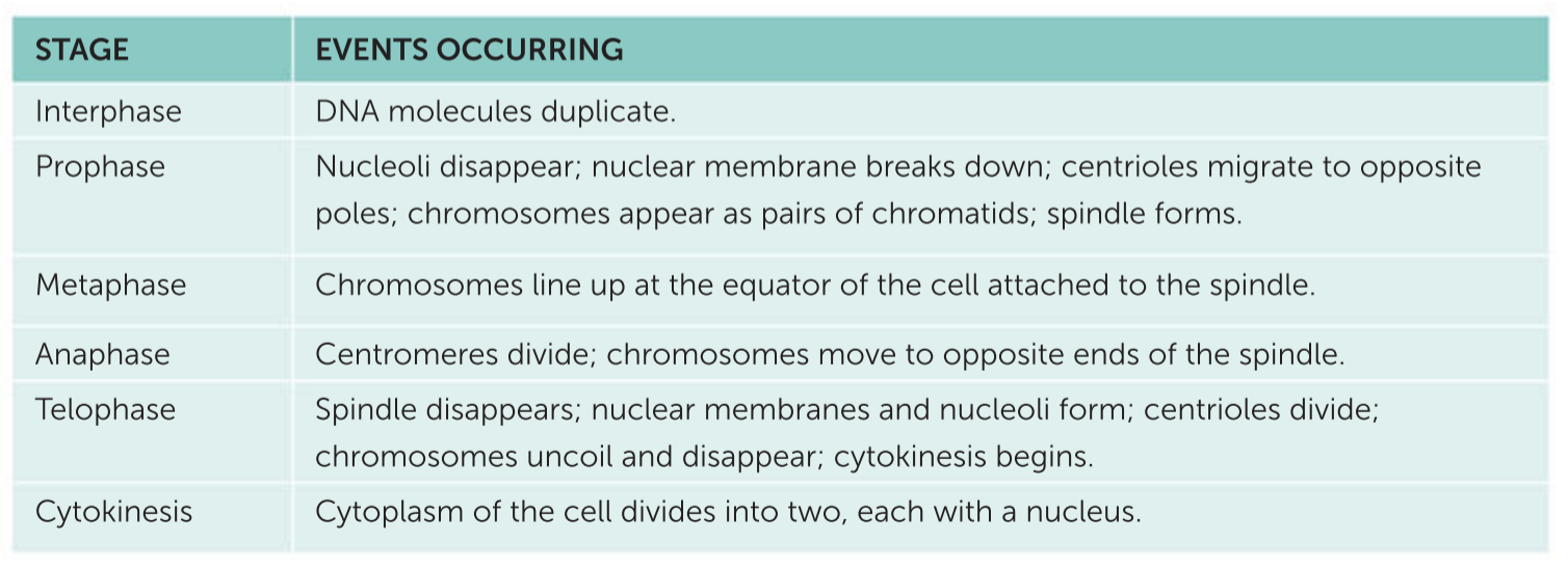
The events that occur in the cell cycle have been divided into a number of phases:
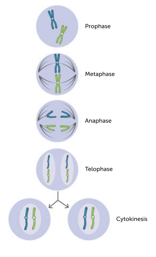
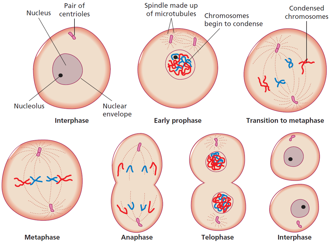
The stages of mitosis in a cell with two chromosomes
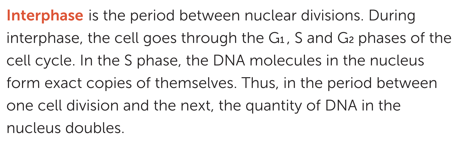
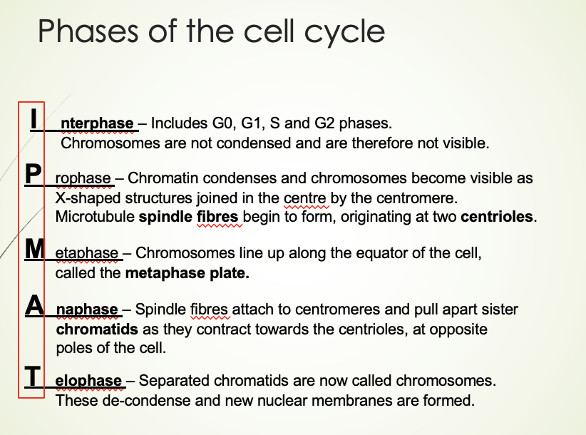
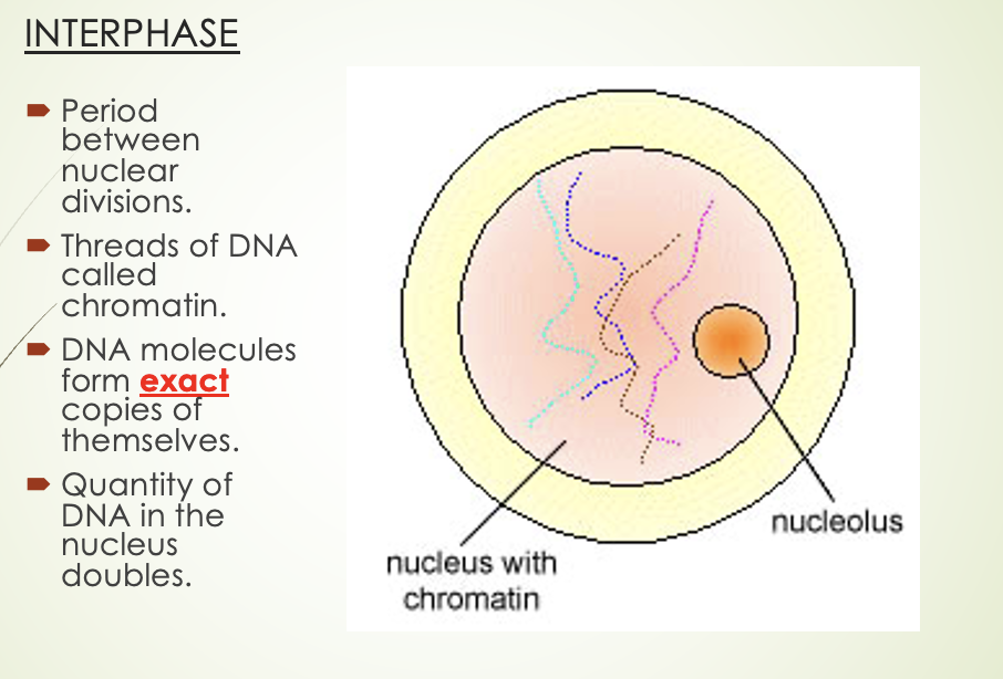
Interphase
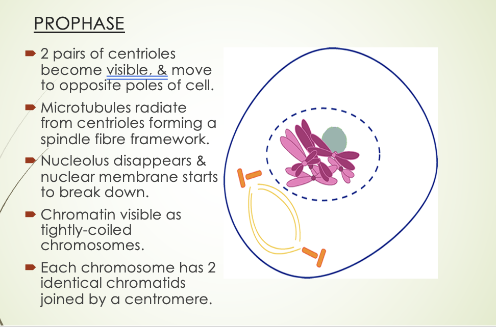
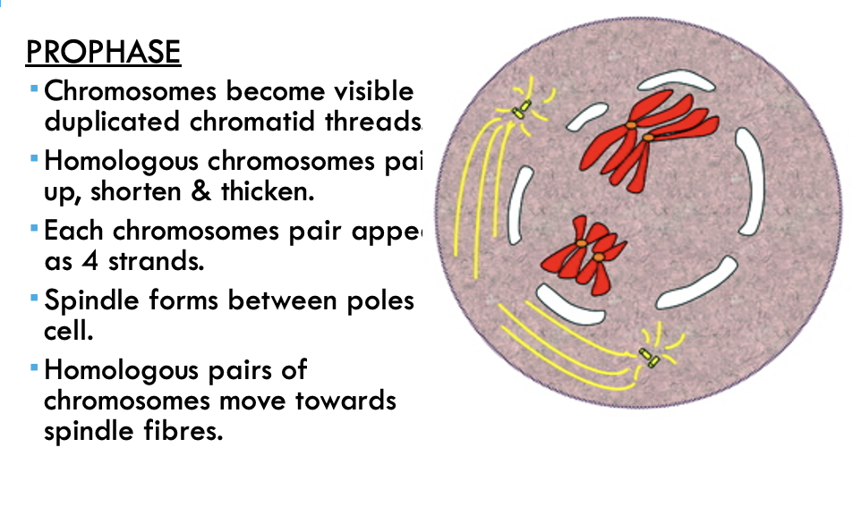
Prophase
Two pairs of centrioles become visible early in prophase. They move to opposite ends of the cell and microtubules begin to radiate from them. At the same time, the nucleolus disappears and the nuclear membrane begins to break down. The chromatin threads of DNA become tightly coiled and can be seen as chromosomes. Coiling the long, delicate DNA molecules makes it easier to distribute the DNA to the daughter cells. Each chromosome consists of two chromatids, which are joined at a point called the centromere. The two chromatids are identical, tightly coiled DNA molecules produced from DNA replication during interphase.
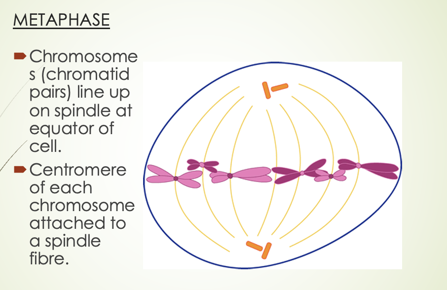
Metaphase
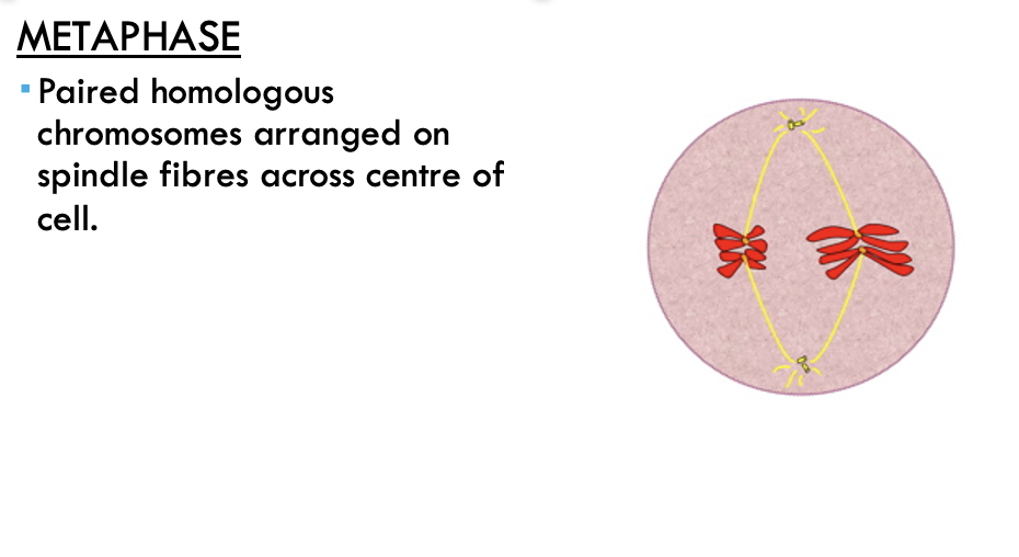
During metaphase, the chromatid pairs line up on the equator of the spindle. The centromere of each pair is attached to a spindle fibre.
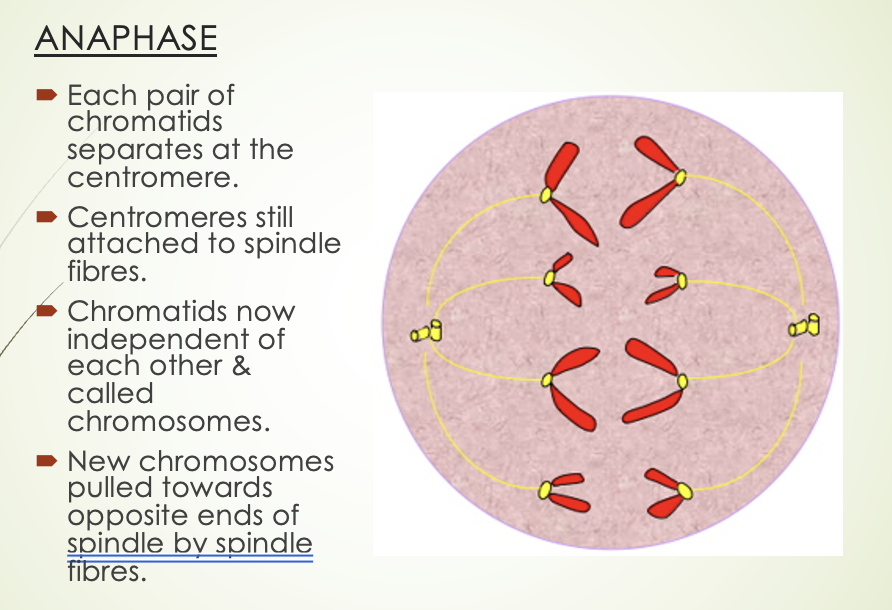
Anaphase
In anaphase, each pair of chromatids separates at the centromere. As the chromatids have become independent of each other, they are now each called chromosomes. The new chromosomes are then pulled away from one another towards opposite poles of the cell. The centromeres are still attached to the spindle fibres, and it seems that the spindle fibres pull the chromosomes apart in some way.
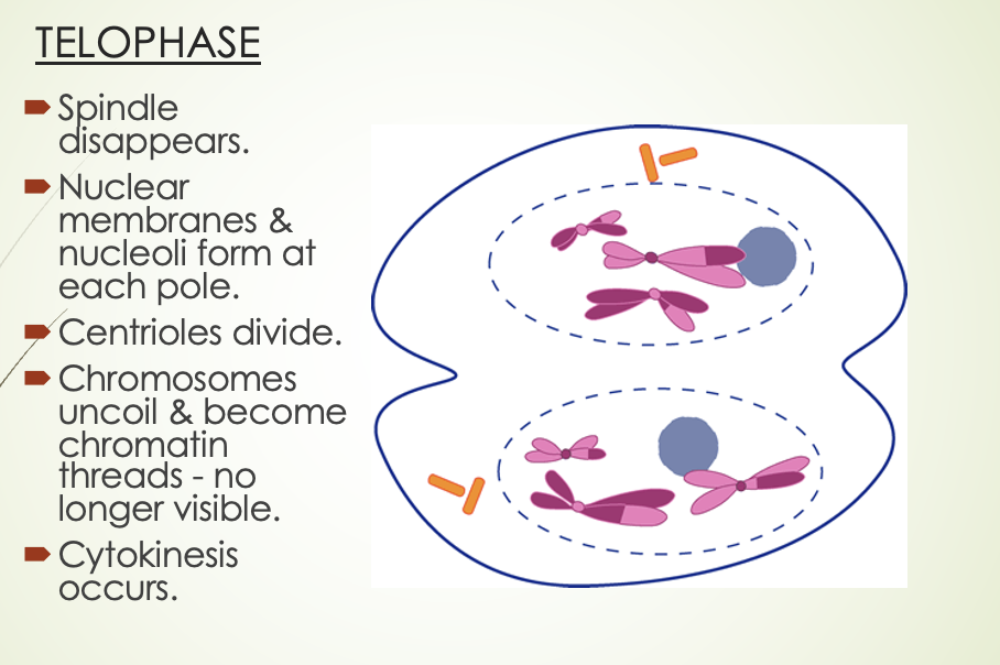
Telophase
During telophase, the two sets of chromosomes form tight groups at each pole of the cell. A nuclear membrane forms around each group, and a nucleolus appears in each new nucleus. The spindle fibres disappear, and the chromosomes gradually uncoil to become chromatin threads once more.
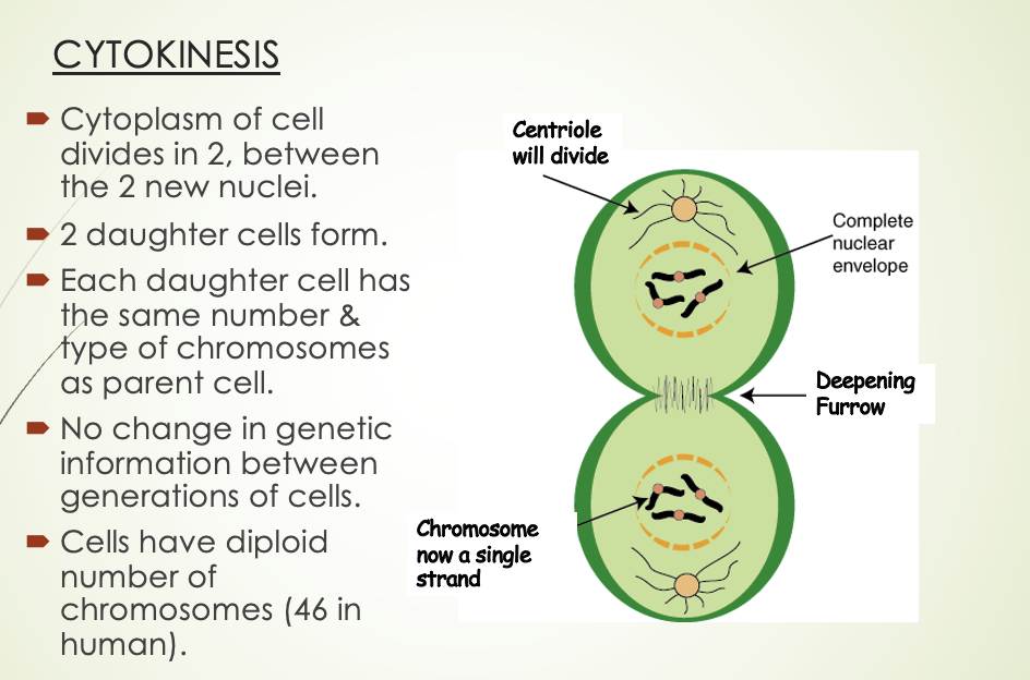
Cytokinesis
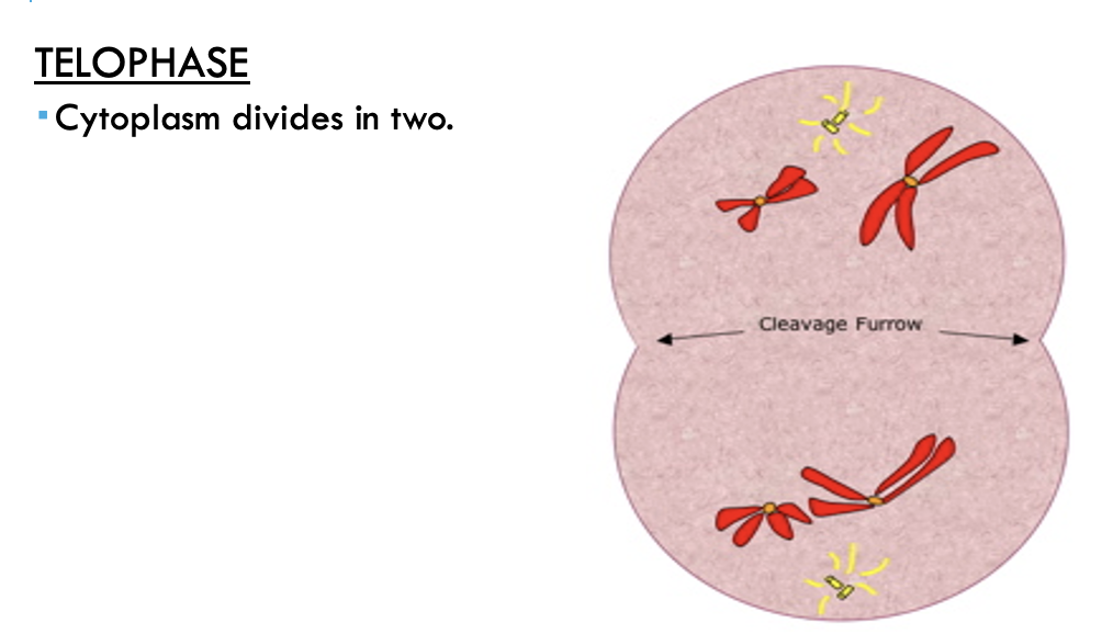
Division of the cytoplasm is called cytokinesis. A furrow develops in the cytoplasm between the two nuclei. The furrow gradually deepens until it cuts the cytoplasm into two parts, each with its own nucleus. (Note: Although the term ‘mitosis’ is commonly used to refer to cell division, it technically refers just to the division of the nucleus.)
Summary of cell division
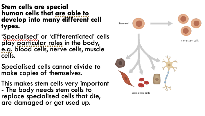
differentiation.
cells undergo division by mitosis, different genes become activated. This makes the cells differentiate into specialised cells that can perform particular functions
Stem cells
The cells that can undergo differentiation are called stem cells.
Totipotent stem cells
have the ability to form the embryo and the membranes that will surround, support and nourish it. An example is the early embryo before the formation of the inner cell mass.
Pluripotent stem cells
are capable of giving rise to most, but not all, tissues of an organism. An example is the inner cell mass.
Multipotent stem cells
are able to give rise to cells that have a specific function. An example is blood stem cells.
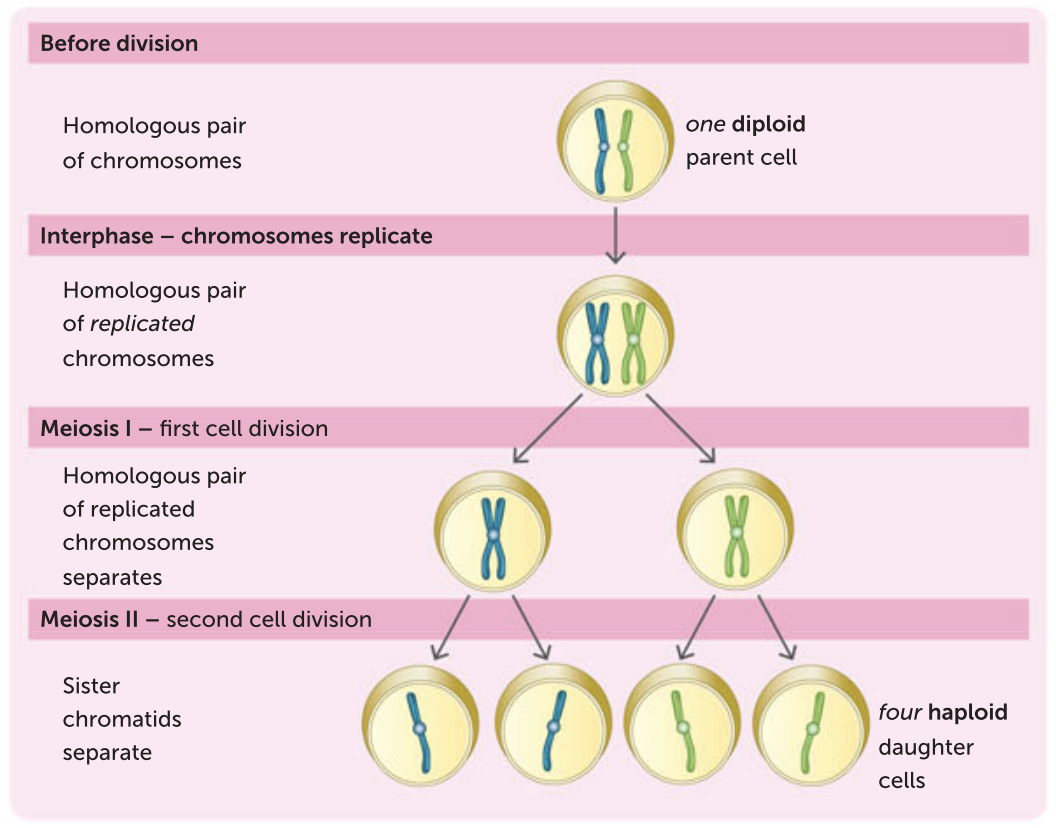
First division:
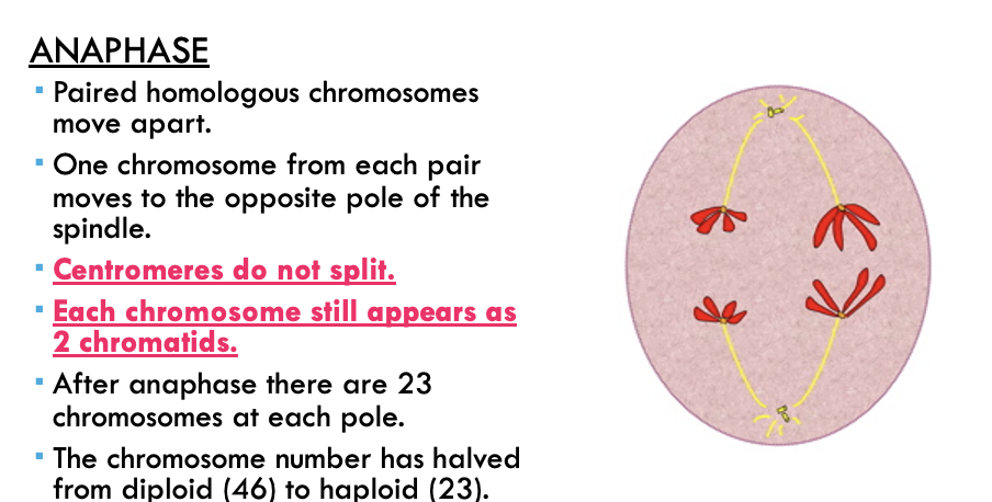
The homologous pairs separate and two daughter cells form with 23 chromosomes, each with two chromatids.
Second division:
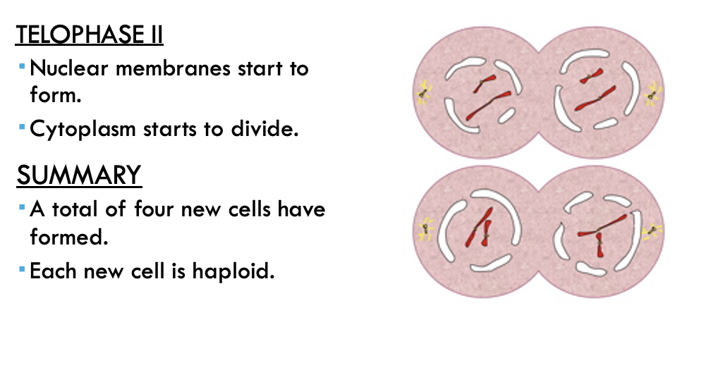
The chromatids separate, resulting in four daughter cells with 23 chromosomes, each with one chromatid.
Summary of meiosis: one diploid parent cell produces four haploid daughter cells
Second meiotic division

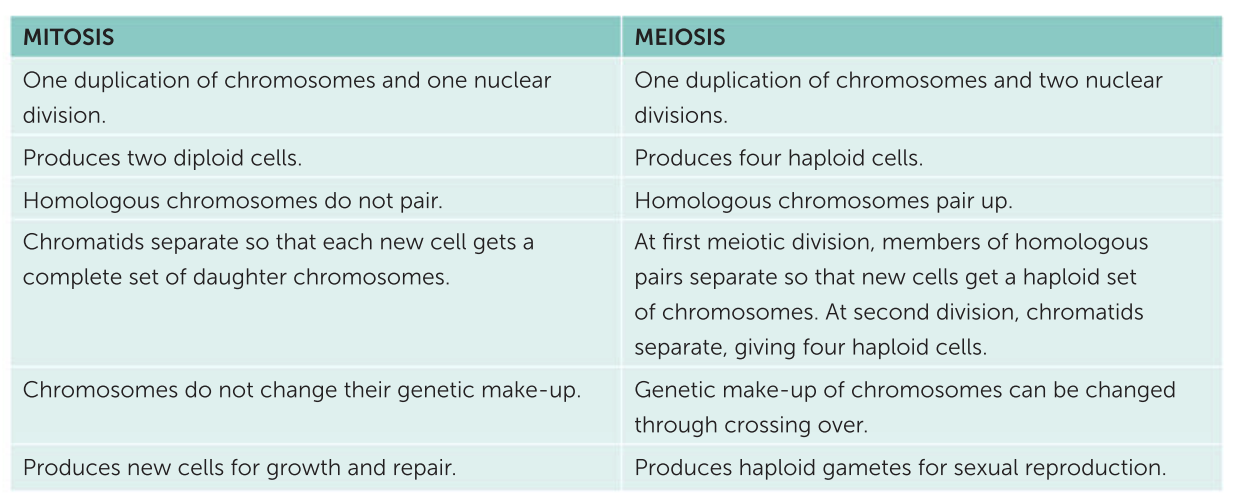
Comparison of mitosis and meiosis
Crossing over
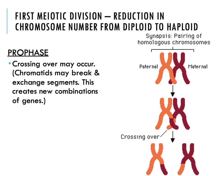
An important feature of meiosis occurs during the prophase of the first meiotic division. When the homologous chromosomes are paired, the chromatids may cross, break and exchange segments. This is called crossing over and the point where two chromatids cross is called a chiasma. Crossing over can result in a new combination of alleles along the chromosome. This is called recombination. Therefore, crossing over creates new combinations of genes so that the chromosomes passed on to the offspring are not exactly the same as those inherited from the parents.
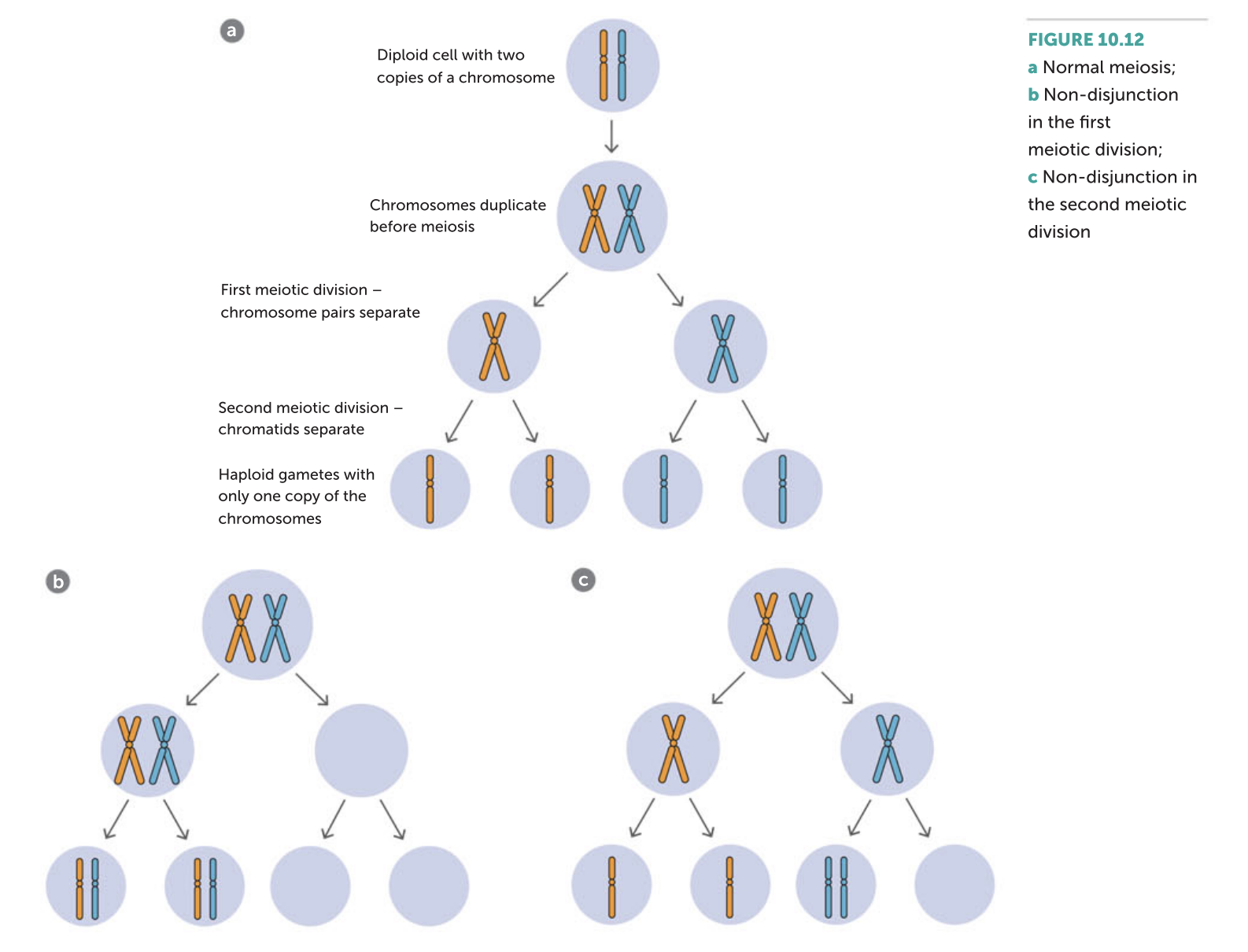
Non-disjunction
During the first division of meiosis, the homologous chromosomes pair and then separate. Sometimes one or more of the chromosome pairs may fail to separate when the cell divides. In the second meiotic division, one or more of the chromatids may fail to separate. These situations are called non-disjunction, and they result in one of the daughter cells receiving an extra chromosome and the other daughter cell lacking that chromosome
Monosomy
Monosomy is where an individual is missing a chromosome – they have only one copy instead of the normal two. Like trisomy, monosomy usually results in severe malformations and often miscarriage.
Haploid number
The number of chromosomes in a cell with only one chromosome from each homologous pair; half the diploid number
Homologous chromosomes
The pairs of chromosomes containing genes that control the same characteristics
Independent assortment
The random combination of alleles due to allele pairs separating independently of one another
THE ROLE OF DNA IN CELL PROTEIN SYNTHESIS
The genetic code in DNA provides the instructions for making cell proteins.
As well as instructions, the cell needs reactants (amino acids) and energy (from respiration).
Proteins are formed on the ribosomes in the cytoplasm.
DNA does not leave the nucleus of the cell.
The cell has a special way of transferring the genetic code to the ribosome.
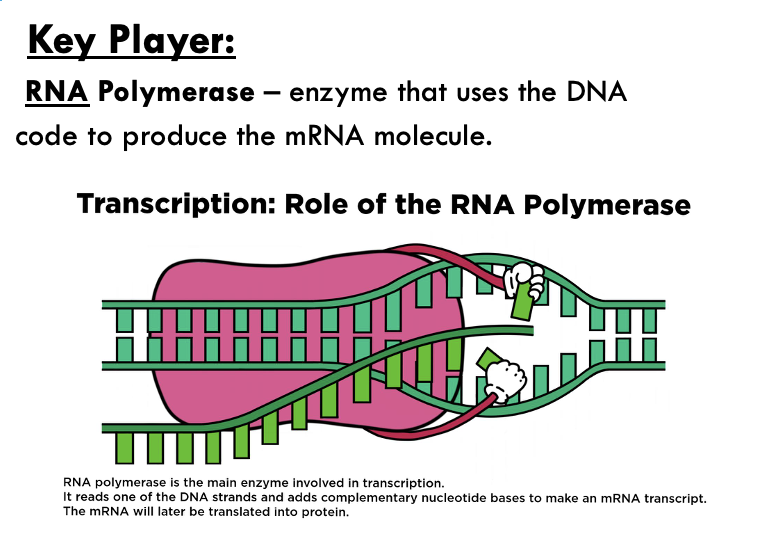
Creating a useful copy of dna - mrna
Translation
building the protein
LOCATION: Ribosomes
KEY PLAYER: tRNA – brings the corresponding amino acid to the ribosome.
Ploidy
´Ploidy is the number of pairs of chromosomes in a cell.
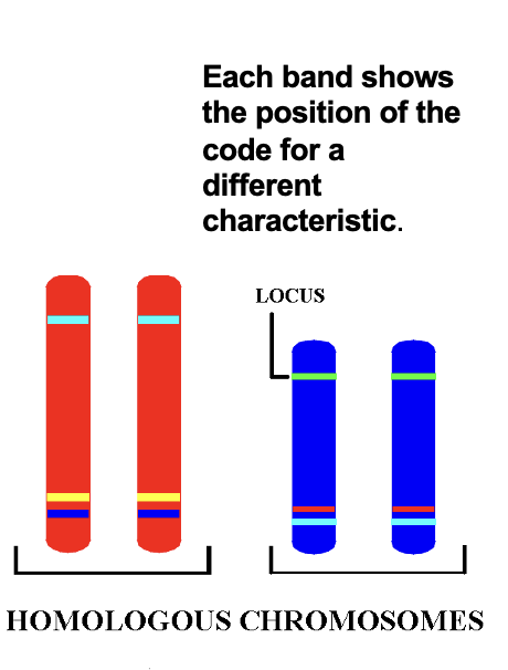
Genetic loci
A genetic location is called a locus.
A locus is the position of a gene, a cluster of genes or even a single nucleotide on a chromosome.
Since there are two of each chromosome in a somatic cell, there are two copies of each gene.
CELL DIVISION BY MITOSIS
´In mitosis cells are produced for growth, maintenance and repair of the body.
´The more wear & tear on a cell, the shorter its life span.
´Intestinal cells last 1.3 days
´Brain nerve cells last a lifetime.
The cell cycle
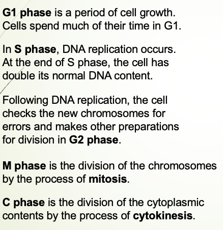
G0 phase occurs
G0 phase occurs when the cell has
exited the cell cycle and is not preparing
to undergo division.
Cells in G0 phase can sometimes
re-enter the cell cycle if they receive the
correct stimuli.
Cells that have terminally
differentiated (i.e. reached their
most specialised, final form such
as a nerve cell) are usually in
G0 phase.
Interphase
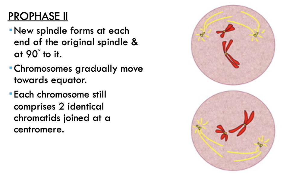
PROPHASE
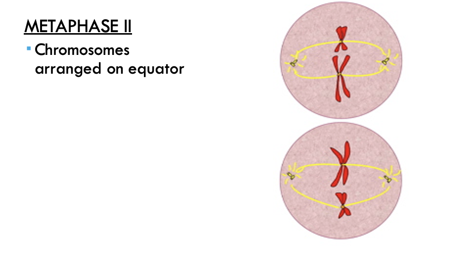
METAPHASE
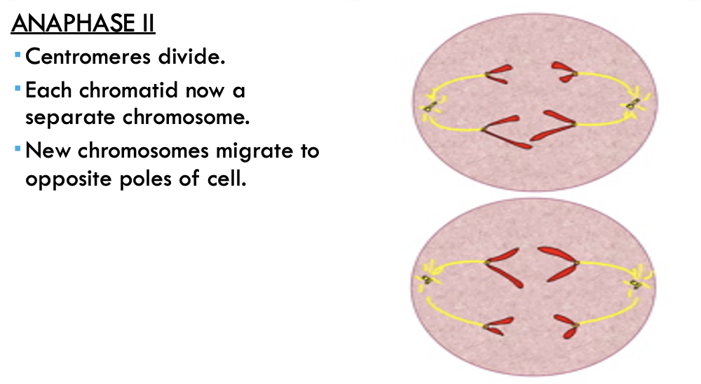
ANAPHASE
TELOPHASE
CYTOKINESIS
Gene expression
Protein synthesis is also known as gene expression.
Only certain genes are expressed in a cell at any given time.
Gene expression can be switched ‘on’ or ‘off’, depending on the cell’s requirements.
Epigenetics and gene expression
Epigenetics refers to differences in traits caused by environmental factors that can be inherited.
The differences are not due to changes in the DNA sequence, but due to modifications, such as methylation of the DNA or histone modification.
This changes the gene expression without changing the DNA i.e. genes are more likely to be turned on or off.
Dna methylation
Methyl groups are clusters of hydrocarbons which attach to DNA strands
When a methyl group is attached to a particular gene, the gene cannot be expressed. i.e. the gene is “switched off”.
Histone modification
Refers to how tightly the chromatin is wound around the histone protein.
Tightly wound chromatin stops access for RNA polymerase, stopping gene expression.
What are stem cells?
three main types of stem cells
Multipotent
Pluripotent
Totipotent
Mutipotent
§Multipotent – differentiate in to more than one type of body cell
Pluripotent
able to differentiate, or mature, into the three primary groups of cells that form a human being: (ectoderm — giving rise to the skin and nervous system, endoderm — forming the gastrointestinal and respiratory tracts, endocrine glands, liver, and pancreas and mesoderm — forming bone, cartilage, most of the circulatory system, muscles, connective tissue, and more)
Totipotent
§Totipotent - give rise to any of the 220 cell types found in an embryo as well as extra-embryonic cells (placenta).
Source of stem cells:
embryonic stem cells
Embryonic stem cells are pluripotent. They can differentiate into all types of body cell.
Taken from the inner cell mass of a 5-7 day old embryo (formed by in-vitro fertilisation).
Embryo is destroyed in the process.
One embryo can give rise to a line of pluripotent stem cells.
Strict controls for their use.
SourceS of adult stem cells
1. Umbilical blood & Placental cells
Adult stem cells refers to a human of any age after cell differentiation.
Multipotent adult stem cells can be obtained from umbilical cord blood and placental stem cells.
These tissues can be donated to the national cord blood blank or stored in case the child needs it in the future.
Sources of adult stem cells
2. body organs
These multipotent cells are found in most organs of the body.
They usually only form the tissues of the organ from which they are found.
Harvesting and culturing a patient’s own stem cells prevents tissue rejection.
TERMS ASSOCIATED WITH GAMETE PRODUCTION
Gametogensis
Spermatogenisis
Oogenesis
Gametogenesis
Gametogenesis – process of gamete development.
Spermatogenesis
Spermatogenesis – process of spermatozoa development in male testis.
Oogenesis -
process of ova development in female ovary.
HOMOLOGOUS CHROMOSOMES
At fertilisation, 23 chromosomes from a (haploid) ovum unite with 23 chromosomes from a (haploid) spermatozoon to form a (diploid) human cell with 46 chromosomes.
Thus diploid cells have 23 chromosome pairs.
Each member of a pair carries genetic information for the same features.
They are called homologous chromosomes.
Humans have 23 pairs of homologous chromosomes.
MEIOSIS
Specialised form of cell division used only in gamete production (ova & sperm).
Results in halving of cell chromosome number in humans from 46 to 23.
Diploid cell chromosome number = 46.
Haploid cell chromosome number = 23.
At fertilisation, ova & sperm nuclei fuse to restore diploid chromosome number.
New human develops with 46 chromosomes.
FIRST MEIOTIC DIVISION – REDUCTION IN CHROMOSOME NUMBER FROM DIPLOID TO HAPLOID
Secound MEIOTIC DIVISION – REDUCTION IN CHROMOSOME NUMBER FROM DIPLOID TO HAPLOID
Third MEIOTIC DIVISION – REDUCTION IN CHROMOSOME NUMBER FROM DIPLOID TO HAPLOID
Fourth MEIOTIC DIVISION – REDUCTION IN CHROMOSOME NUMBER FROM DIPLOID TO HAPLOID
Fifth MEIOTIC DIVISION – REDUCTION IN CHROMOSOME NUMBER FROM DIPLOID TO HAPLOID
SECOND MEIOTIC DIVISION – SEPARATION OF CHROMATIDS. 1
SECOND MEIOTIC DIVISION – SEPARATION OF CHROMATIDS. 2
SECOND MEIOTIC DIVISION – SEPARATION OF CHROMATIDS. 3
SECOND MEIOTIC DIVISION – SEPARATION OF CHROMATIDS. 4
ALZHEIMER’S DISEASE
Disease
Alzheimer’s disease results from the gradual destruction of brain tissue, caused by a build-up of an unusual protein both inside (“tangles) and outside (‘plaques”) the nerve cells of the brain.
This affects the ability of the brain to process information and transmit nerve impulses.
OVERALL RESULT
•degeneration of nerve cells
•shrinkage of the brain
•decreases acetylcholine
SYMPTOMS:
•Memory loss
•confusion
•mood swings
•aggression
Parkinson’s disease
Parkinson disease is:
•a degenerative disorder of the brain.
•caused by the destruction of dopamine-producing cells in the basal nuclei of the cerebrum.
In Parkinson's disease nerve cells in the part of the brain that produces dopamine, (a region in the mid-brain - the substantia nigra), begin to decrease in number.
This causes a decrease in the amount of the available dopamine.
Also, the chemical in the synapse that breaks down the dopamine continues to deplete what little dopamine is left.
symptoms
Parkinson’s disease:
typically affects people over the age of 50.
Is characterised by:
slowed physical and mental responses
muscular tremors
stiffness of the limbs and trunk
impaired balance and coordination
plus a variety of other symptoms .
Treatment
At present there is no cure for Parkinson’s disease.
Symptoms of the disease can be treated using:
•a variety of medications
•Surgery in some severe cases
•deep brain stimulation (using a small electrical pulse generator) has had encouraging results in some cases.
EMBRYONIC STEM CELL THERAPY
•Fertilised egg matures until it reaches blastocyst stage
•blastocyst cells from the inner cell mass are harvested
•these are unspecialised stem cells
•cells stimulated with growth factors
•the cells then mature into adult nerve cells which can be cloned (multiplied)
•nerve cells are then injected back into patient
•new cells replace damaged nervous tissue (Parkinson’s – produce dopamine; in Alzheimer’s new cells for neural networks
•symptoms of disease reduced