HISTEM W14/15 Prenatal Development: Development of Orofacial Structures
1/56
There's no tags or description
Looks like no tags are added yet.
Name | Mastery | Learn | Test | Matching | Spaced |
|---|
No study sessions yet.
57 Terms
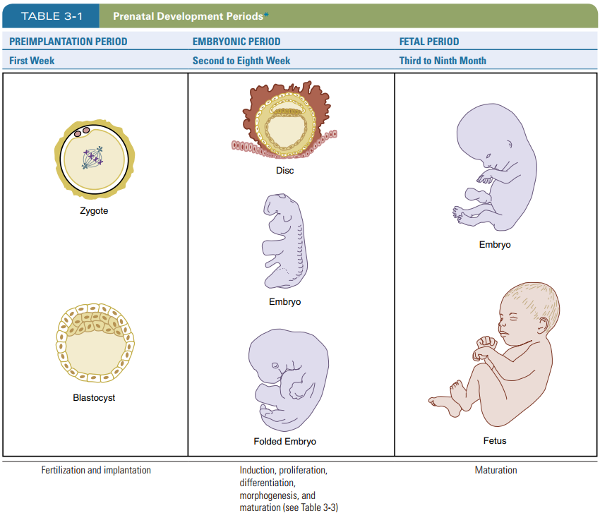
What are the 3 periods of prenatal development?
Pre-implantation period → 1st trimester; zygote to blastocyst
blastocyst implants and then embryonic period starts
Embryonic period → 1st trimester
Fetal period → second and third trimester
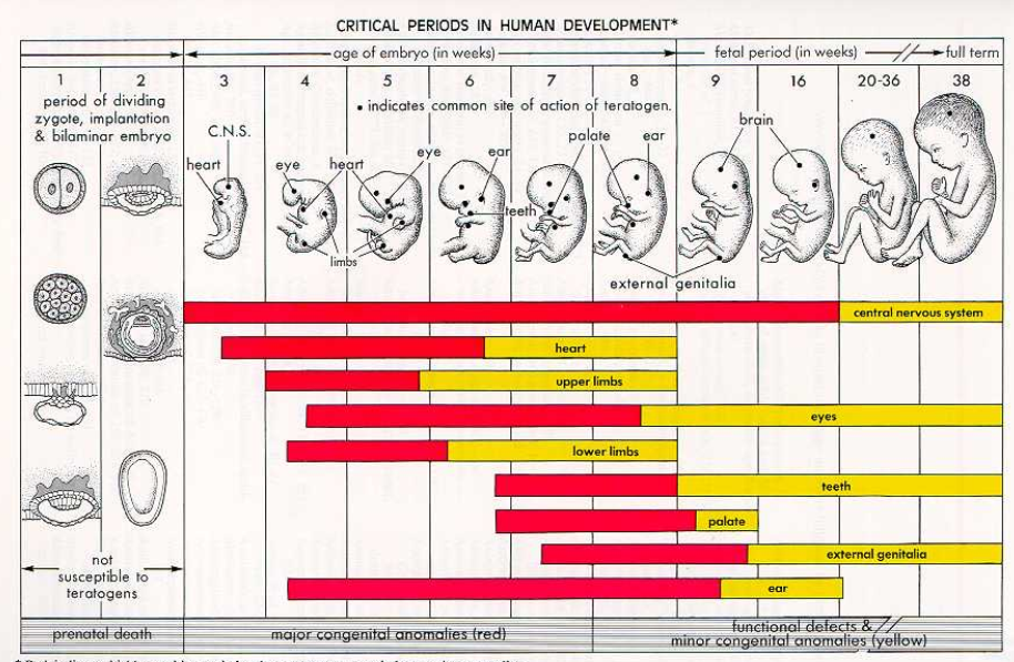
Which weeks of development is susceptible to prenatal death?
Week 1-2
Sperm (23) + ovum (23) = Zygote (46 chromosomes)
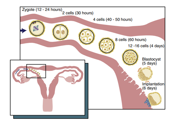
What happens in the first week after fertilization/conception?
Fertilized ovum, now called zygote undergoes mitosis and forms initial ball of cells called a Morula
Morula undergoes more mitosis and becomes blastocyst/blastula(vesicle)
Blastocyst travels from fallopian tube to the uterus
Implantation occurs at the END of week 1.
Trisomy 21 aka Downsyndrome is a major congenital malformation that occurs due to …?
disturbances occuring during the basic process of meiosis during fertilization.
Once implantation begins, what happens to the blastocyst?
Blastocyst is embedded in the endometrium and continues dividing (cleavage) forming a peripheral layer of cells called the Trophoblast layer and a small inner mass of embryonic cells/embryoblast layer
What are the 5 developmental processes? Describe each briefly
Induction → action of one group of cells on another group leading to establishment of developmental path as a response
Proliferation → controlled cellular growth and accumulation of byproducts (interstitial growth/appositional)
Differentiation → embryonic cells become distinct in structure and function
Morphogenesis → development of specific tissue structure/shape; increase in complexity of structure and function of cells
Maturation → attaining adult function/size of tissues/organs by proliferation, differentiation and morphogenesis.
The blastocyst grows by proliferation of the embryonic cells. What are the 3 layers formed within the blastocyst?
Ectoderm, mesoderm, endoderm.
What is the Bilaminar embryonic disc?
Bilaminar embryonic disc → developed from the blastocyst; 3D flattened, circulate plate of bi-layered cells.
will later develop into the embryo
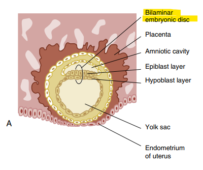
What are the 2 layers that make up the bilaminar embryonic disc?
Superior epiblast → high columnar cells
Inferior hypoblast → small cuboidal cells
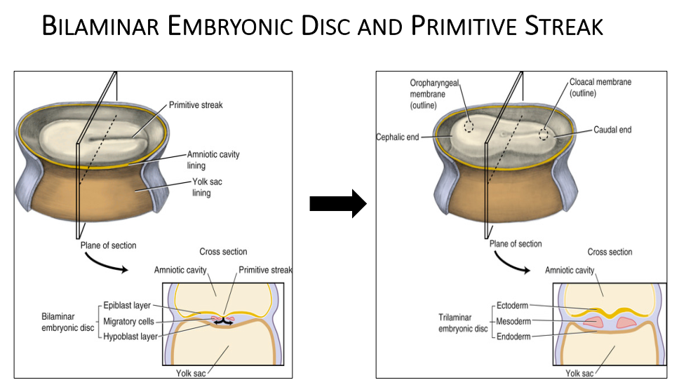
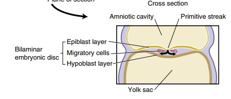
What is the primitive streak? When does it form?
Primitive streak = furrowed, rod-shaped thickening in the middle of the bilaminar disc as a result from increased cell proliferation in the midline area.
FORMS IN THIRD WEEK OF DEVELOPMENT
Primitive streak gives rise to bilateral symmetry

In week 3 of development, what happens to the epiblast layer and hypoblast layer?
Cells from epiblast layer migrates towards the hypoblast layer in the area of the primitive streak
Migrated epiblast layer cells form the mesoderm and endoderm → 3 layers present
Embryonic disc is now called trilaminar embryonic disc
Which embryonic cell layer is considered the ectoderm?
Epiblast layer is now the ectoderm.
Hypoblast layer becomes displaced by migrating cells and is now called extra-embryonic endoderm.
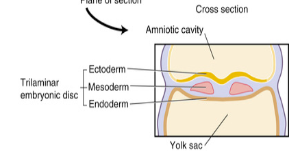
The 3 primary germ layers aka primary embryonic layers
Ectoderm - outer
Mesoderm - middle
Endoderm - inner
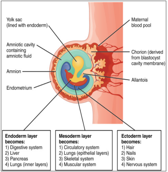
What organs arise from the endoderm? (4)
Hypoblast layer → cuboidal
Epithelial lining of the GI tract → pharynx, stomach, intestines
Respiratory and digestive lining
Liver
Pancreatic cells
What organs arise from the Mesoderm? (4)
Kidneys
Skeletal system
Muscle system
Blood, lymph cells, bone marrow
Some reproductive and excretory organs
Tooth: dentin, cementum, pulp, PDL, alveolar process
What organs arise from the Ectoderm? (4)
Epithelium of outer body → columnar epithelium
Epithelium lining of the oral and nasal cavity, sinuses
Tooth Enamel
Nervous system
The development of the face happens during which weeks of development?
weeks 4-12
begins during the 4th week. Wha
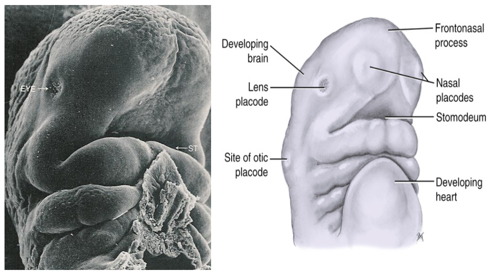
What is a Stomodeum?
The primitive mouth formed during the 4th week of development.
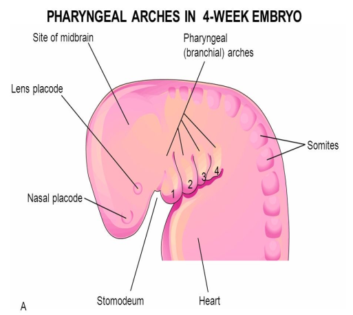
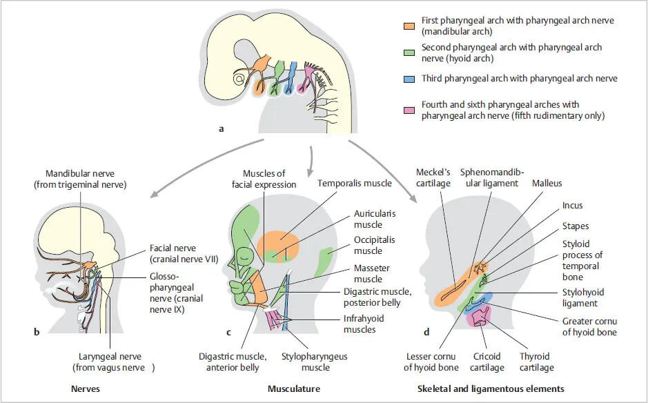
There are 6 branchial arches that form below the stomodeum, which arches are involved in the development of the face and oral cavity? What are their names?
First arch: Mandibular arch
Second arch: Hyoid arch
Third arch
What are the names of the cartilages associated with the first and second branchial arches? What do these cartilage develop into?
1st Branchial arch = Meckle’s cartilage → important for formation of the mandible (template)
2nd Branchial arch = Reichert’s cartilage → forms upper body of the hyoid bone
What part of the 1st branchial arch form the 2 maxillary processes? What does the remainder of the arch form?
Buddings on both ends of the 1st branchial arch become the 2 maxillary processes, the rest of the arch forms the mandibular process.
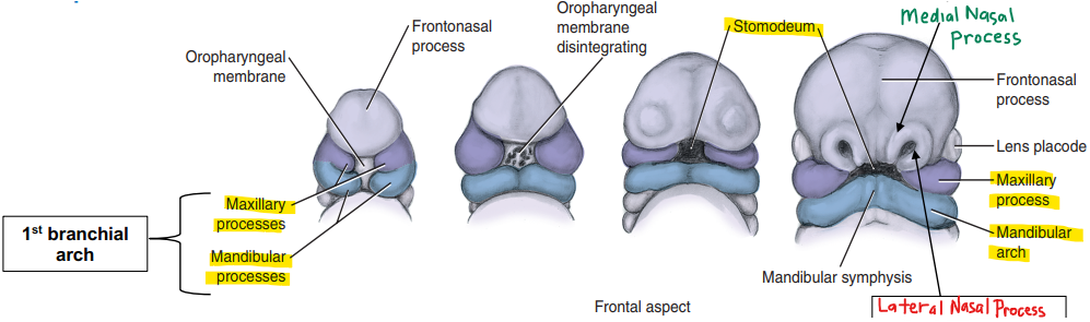
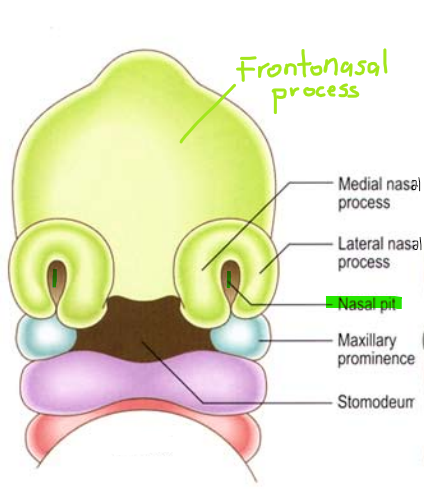
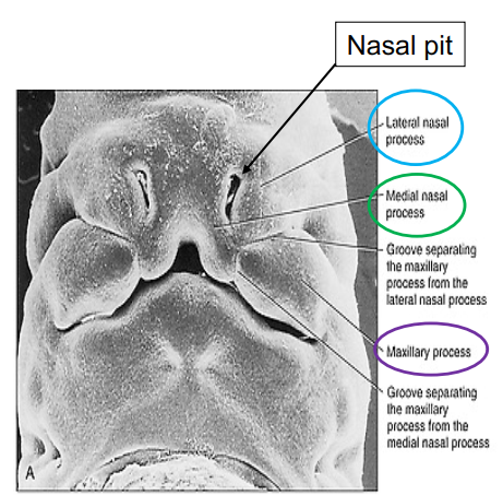
The frontonasal process is divided into the medial nasal process and the left/right lateral nasal processes. What facial structures do they develop into?
Frontonasal process → medial and lateral nasal process →
Medial nasal process → forms globular process (aka intermaxillary segment to form the part of the palate), center of the upper lip (philtrum)
Left and right lateral process → forms the ala of the nose (side of the nose/nostrils)
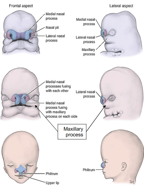
What fuses with the globular process to complete the development of the upper lip?
left and right maxillary process → grows medially from the corners of the primitive mouth and fuse to the globular process forming the upper lip.

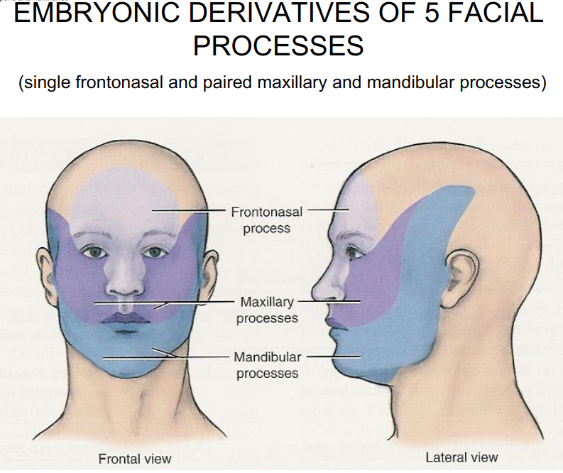
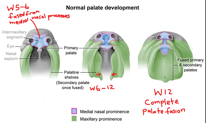
By the 5th - 6th week of fetal development, what structures have formed in regards to the face? (5 origins)
Frontonasal process → forms forehead, bridge of nose, nasal pits.
Maxillary processes (from 1st branchial arch) → form upper cheeks, lateral upper lip, most of upper jaw.
Mandibular processes → fuse to form lower jaw, lower lip, chin.
Medial nasal processes → merge to form philtrum and middle portion of upper lip.
Lateral nasal processes → form sides (ala) of nose.
By week 6, major facial prominences have appeared and start fusing to establish basic facial structure.
Which cranial nerve arises from the first branchial arch?
Trigeminal nerve CN 5
The muscles for mastication is developed from which branchial arch?
1st (mandibular) branchial arch
Which cranial nerve arises from the 2nd branchial arch?
Facial nerve CN 7
The muscles for facial expression are developed from which branchial arch?
2nd (hyoid) arch.
What causes this developmental disturbance during the embryonic period?
Ectodermal Dysplasia → abnormal development of one or more structures from the ectoderm leading to partial/complete anodontia (absence of some teeth or all teeth)
Caused by: mutation in genes leading to abnormal ectodermal structure development.
How does Rubella affect fetal development?
Rubella aka measles (viral infection) affects on unborn infant:
cataracts
deafness
damage to heart or brain
How does fetal alcohol syndrome affect facial development of the fetus?
Fetal alcohol syndrome causes:
crowding of teeth
mouth breathing
anterior open bite
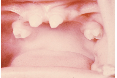
How does Syphilis affect prenatal teeth development?
Syphilis can cause Hutchinson’s incisors (notched incisors) and Mulberry molars (globular shaped enamel)
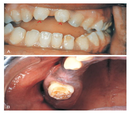
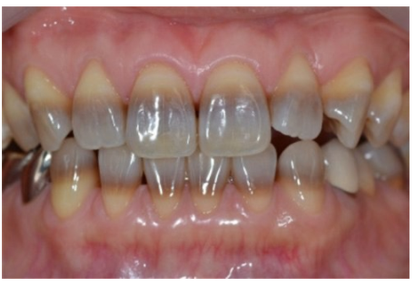
What is this?
Tetracycline staining
Intrinsic yellow-brown stain
chemically bound to dentin
most common in children under 8yo or if drug was taken during pregnancy
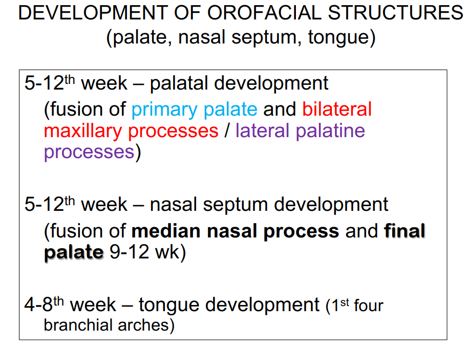
Palate development starts during which week of prenatal development? What are the 3 structures that give rise to the palate?
5th week.
Palate arises from:
2 maxillary processes
globular process AKA intermaxillary segment
T or F - The development of the intermaxillary segment is from the fusion of the medial nasal processes on the inside of the stomodeum.
TRUE
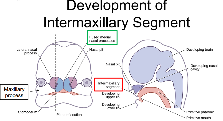
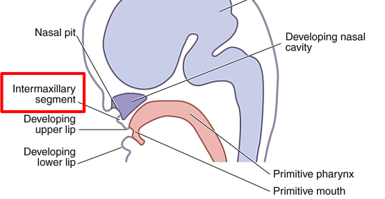
The intermaxillary segment forms during which weeks of development? What does the intermaxillary segment give rise to?
Intermaxillary segment forms during week 5-6 → by fusion of medial nasal processes
Develops into:
Floor of nasal cavity and nasal septum
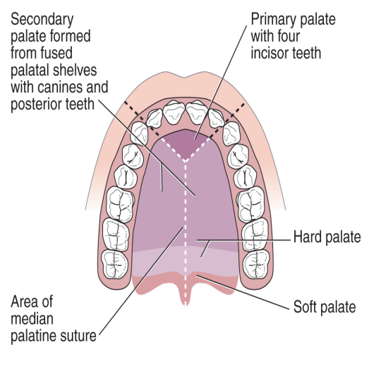
primary palate (anterior 1/3rds of the hard palate) which then forms the premaxilla
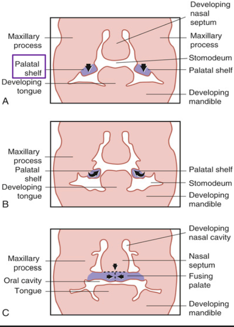
Describe the formation of the secondary palate.
Timing: Begins around week 6–7 of embryonic development.
Origin: From the maxillary processes → form lateral palatine shelves (outgrowths on either side of the tongue).
Process:
2 shelves form vertically alongside the tongue from the 2 lateral maxillary processes
As the tongue begins to move, shelves elevate (flip-up) to a horizontal position above it.
Shelves grow medially and fuse at the midline → forming the secondary palate.
Fusion also occurs anteriorly with the primary palate and superiorly with the nasal septum.
Result:
Separates oral and nasal cavities posteriorly.
Forms hard and soft palate (posterior 2/3rds of the final palate), uvula
T or F - During the 12th week of development, the Primary palate and the secondary palate fuse in the roof of the mouth in a U-shaped pattern.
FALSE - fuse in a Y-shaped pattern.
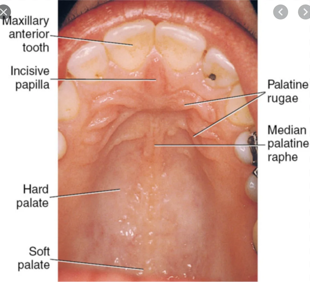
What is medial palatine raphe? how is it formed?
Median palatine raphe: A midline ridge running along the hard palate, from the incisive papilla to the uvula.
Formation:
Formed when the lateral palatine shelves (from the maxillary processes) fuse at the midline during secondary palate development.
The line of fusion becomes the median palatine raphe, covered by mucosa.
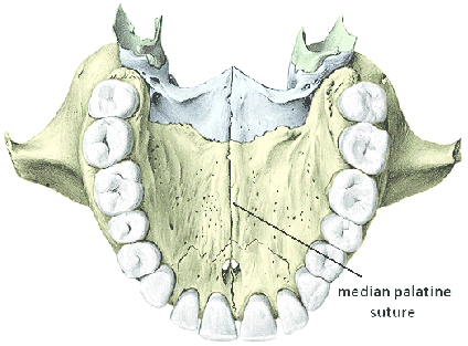
What is the median palatine suture?
Median palatine suture: The bony union where the right and left palatine processes of the maxilla fuse in the midline.
Location: Runs along the center of the hard palate, beneath the median palatine raphe.
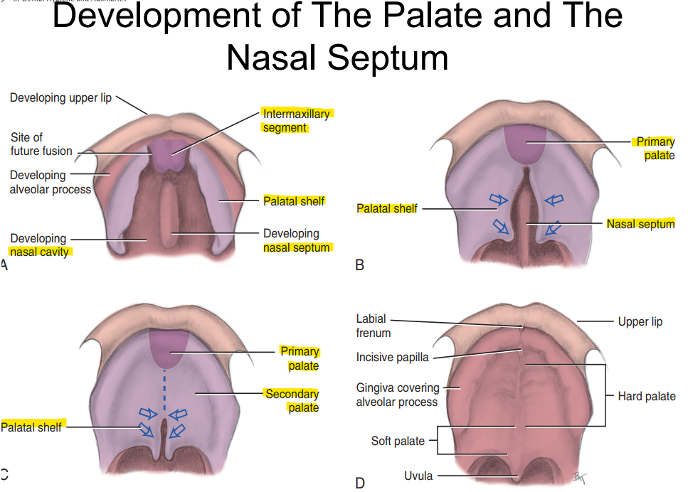
Development of the Palate and nasal septum
Write out the Palatal development timeline.
5-6th week = primary palate development → intermaxillary segment fuses with medial nasal process
6th-12th week = Secondary palate development → fused palatal shelves from maxillary processes
12th week = finalizing palate → fusion of all 3 processes
During 4th week of development, the frontonasal process undergoes growth and creates _____ which will later develop into the nasal cavity.
creates nasal pits which form future nasal cavity
*tissue around nasal placodes on the frontonasal process grow to form the pits.
The nostrils of the nose are formed from…?
fusion of the lateral nasal, maxillary and medial nasal processes
Which developmental process fuse together and form:
middle part of the nose
tubercle of the upper lip
philtrum
Medial Nasal Processes
What is cleft lip? How is it formed?
Cleft lip is the partial or complete failure of fusion between the medial nasal processes and 1 or both maxillary processes.
Incidence is 1:1000, twice as common in males
mandibular cleft lip is very rare
Most common cleft lip is on UNILATERAL on the left side.
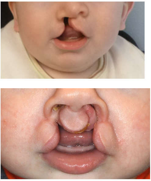
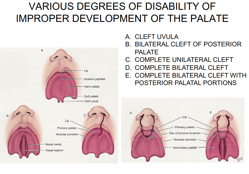
What is cleft palate? How is it formed?
Cleft palate is the failure of lateral palatine processes fusion with each other or with the premaxilla (primary palate)
causes nasal cavity to be exposed to oral environment
more common in females
hereditary influence may be a factor
other etiological factors may include: mechanical interference by the tongue, stress, infectious disease, malnutrition, increasing maternal age.
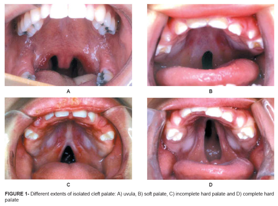
Various degrees of disability due to improper development of the palate.
Which branchial arch is the body and root of the tongue developed from?
Body of tongue = first branchial arch (week 4) → develops first
Base/root of tongue = 2nd, 3rd, 4th branchial arches → developed later
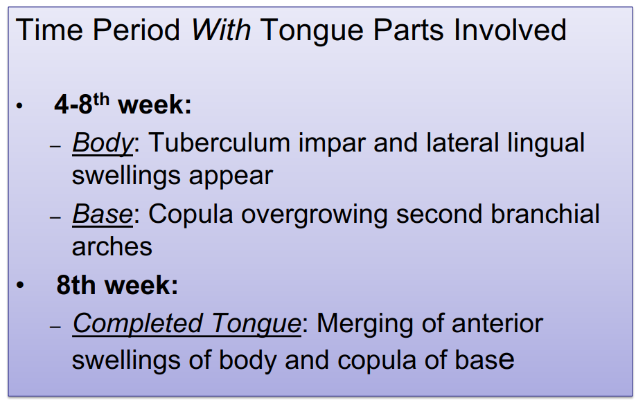
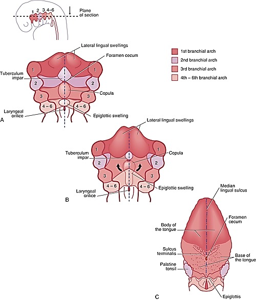
Describe the development of the tongue.
Development of the Tongue — Summary:
Begins with a small midline swelling called the tuberculum impar.
Lateral lingual swellings form on each side of it (from the 1st branchial arch).
These lateral swellings grow, merge, and overtake the tuberculum impar → forming the anterior two-thirds (body) of the tongue.
Their line of fusion forms the median lingual sulcus.
Behind this area, the copula (from 3rd and part of 4th arches) develops → forming the posterior one-third (base) of the tongue.
Posterior to the copula, the epiglottic swelling appears → becomes the epiglottis.
Development of orofacial structures timeline

What is this anomaly?
Cleft tongue
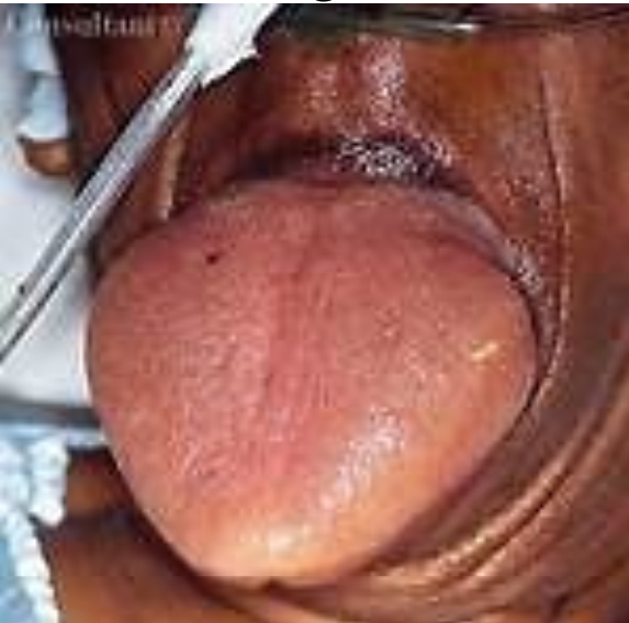
Macroglossia - enlarged tongue
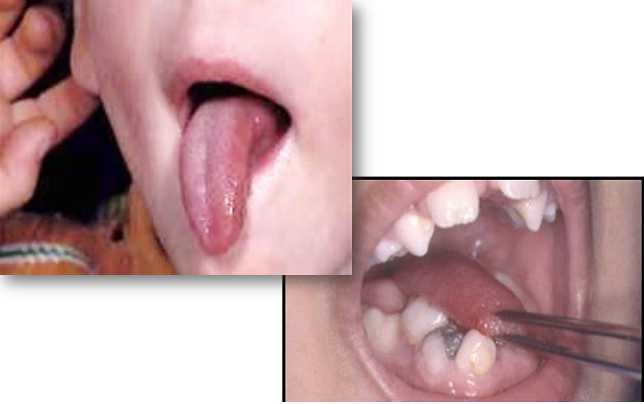
Microglossia - abnormally small tongue
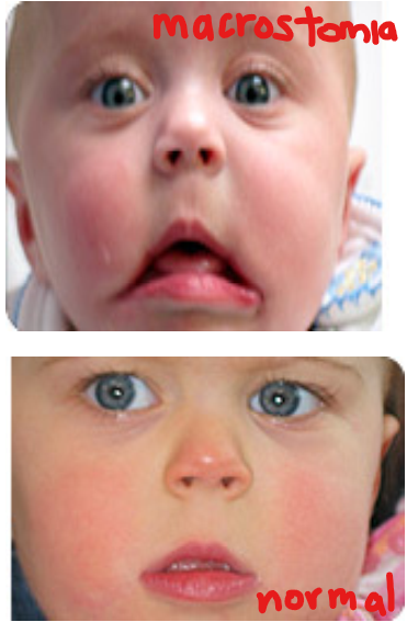
What is Macrostomia? How does it compare to microstomia?
Macrostomia → Extremely large mouth opening that is a result from a failure of fusion between maxillary process and mandibular arch at the corners of the mouth.
Microstomia → extremely small mouth opening caused by merging of the maxillary process and mandibular arch at the corners of the mouth.
37% of people with orofacial clefts also have additional anomalies or hereditary diseases such as…?
Congenital heart disease
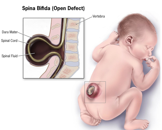
Spina bifida → A neural tube defect where the vertebral arches fail to fuse over the spinal cord during embryonic development.