Case 4: Alessandra W. - CHF
 Call Kai
 Learn
Learn Practice Test
 Spaced Repetition
 Match
Match Flashcards
 Knowt Play
1/50

There's no tags or description
Looks like no tags are added yet.
Name | Mastery | Learn | Test | Matching | Spaced | Call with Kai | Chat |
|---|
No analytics yet
Send a link to your students to track their progress
51 Terms
Atrial Contraction
Signal propagation through atria cause depolarization and contraction
SA node → AV node
During P wave
Ventricular Contraction
Signal propagation through ventricles cause depolarization and contraction
AV node → His-Purkinje system
End of QRS to start of T wave
Ejection Fraction (EF)
Percentage of blood pumped out of left ventricle per heartbeat
Normal: 55-70%
Low: <40% → Heart failure
Left Heart Pressure
Higher
Pump to systemic circulation
Wiggers diagram
Right Heart Pressure
Lower
Pump to pulmonary circulation
Swan-Ganz (pulmonary artery) catheter
Pressure Variables
Depend on compliance and volume
High Pressure: Low compliance, high volume
Low Pressure: High compliance, low volume
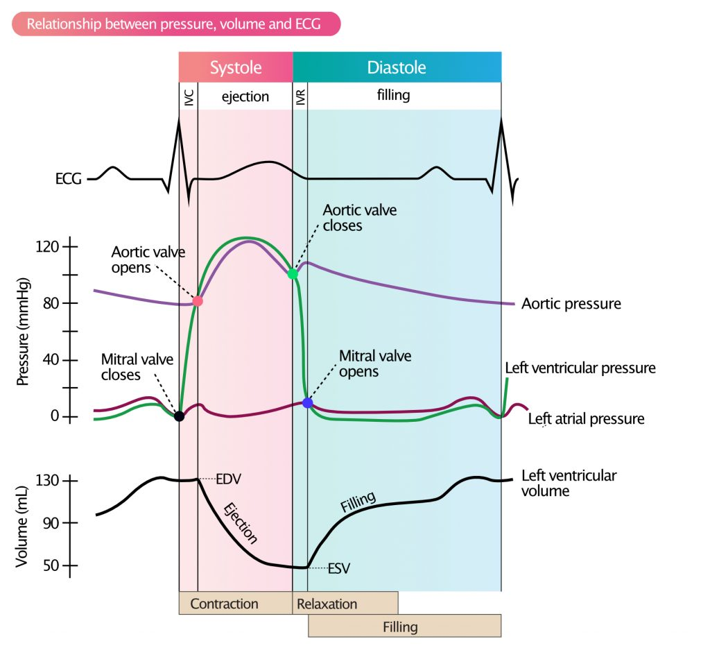
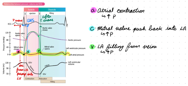
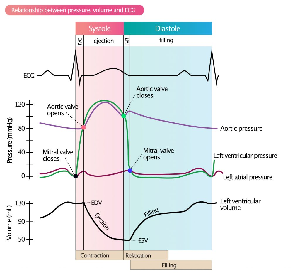
Left Atrium Pressure
a Wave: Atrial contraction
Increase pressure
During P wave
c Wave: Mitral valve moving into atrium
Increase pressure
During QRS
v Wave: Venous atrial filling
Increase pressure
After T wave
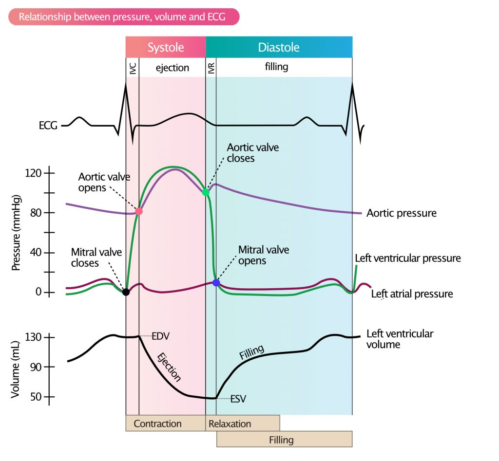
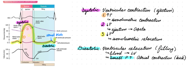
Left Ventricle Pressure
Systolic Pressure: Ventricular contraction
Increase pressure
All valves closed (isovolumic contraction)
Ejection
Aortic valve opens
Decrease pressure
All valves closed (isovolumic relaxation)
Diastolic Pressure: Ventricular relaxation
Mitral valve opens
Passive blood flow into ventricle
Small pressure increase: Atrial contraction (atrial kick)
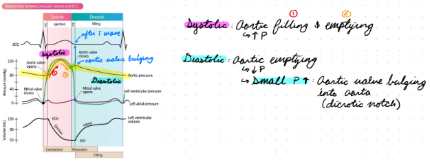
Aorta Pressure
Systolic Pressure: Aortic valve opens for ventricular emptying
Increase pressure
Diastolic Pressure: Aortic valve closes
Decrease pressure
Slow from elastic artery stretch
Small pressure increase: Aortic valve closes and bulges into aorta
Dicrotic notch
After T wave and during diastole
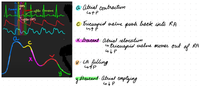
Right Atrium Pressure
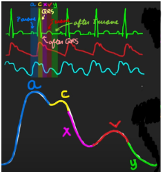
a Wave: Atrial contraction
Increase pressure
During P wave and late diastole
c Wave: Tricuspid valve moving into atrium
Increase pressure
During QRS and systole
x Descent: Atrial relaxation + tricuspid valve moves out of atrium
Decrease pressure
After QRS and during systole
v Wave: Venous atrial filling
Increase pressure
During T wave and late systole
y Descent: Tricuspid valve opens for atrial emptying
Decrease pressure
After T wave and early diastole
Right Ventricle Pressure
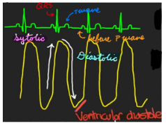
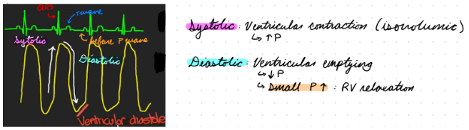
Systolic Pressure: Ventricular contraction
Increase pressure
Higher than right atrium
During QRS and systole
Diastolic Pressure: Pulmonic valve opens for ventricular emptying
Decrease pressure
Same as right atrium
During T wave and late systole
Small pressure increase: Ventricular relaxation
Before P wave
Pulmonary Artery Pressure
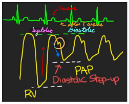
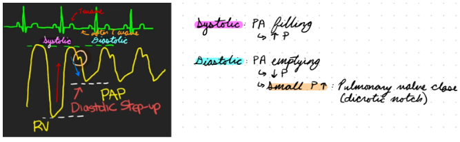
Systolic Pressure: Pulmonary artery filling
Increase pressure until same as right ventricle
During T wave
Diastolic Pressure: Pulmonary artery emptying
Decrease pressure
Higher than right ventricle (diastolic step-up)
Small pressure increase: Pulmonary valve closes
Dicrotic notch
After T wave and during diastole
Ventricular Performance
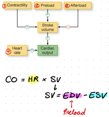
Determined by stroke volume (SV) and cardiac output (CO)
CO = HR x SV
SV = EDV - ESV
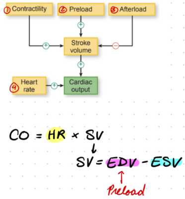
Determinants of Ventricular Performance
Preload
Afterload
Contractility (inotropic state)
HR
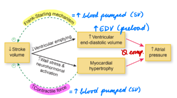
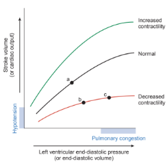
Frank-Starling Mechanism
LV adjust SV to match EDV
1: Preload
Volume of blood in ventricle at end of diastole (EDV)
Myocardial stretch at end of diastole
Greater distension during diastole = Greater volume ejected during systole
Frank-Starling relationship
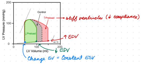
Preload: PV Loop Changes
Increase Preload:
Increase EDV
Increase SV = Constant ESV
Decrease Preload:
From stiff ventricles (low compliance)
Decrease EDV
Decrease SV = Constant ESV
2: Afterload
Force ventricles need to overcome to empty (aortic pressure)
Myocardial tension during systole
Determined by wall stress (Laplace relationship)
Increased wall thickness = Decreased wall stress
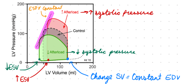
Afterload: PV Loop Changes
Increase Afterload:
Increase ejection/systolic pressure = Increase ESV
Decrease SV = Constant EDV
Decrease Afterload:
Decrease ejection/systolic pressure = Decrease ESV
Increase SV = Constant EDV
3: Contractility
Inotropic state
Change in myocardial force
Independent from prelaod and afterload
Frank-Starling Relationship:
Increased contractility = Curve shift up = Increase SV
Decreased contractility = Curve shift down = Decrease SV
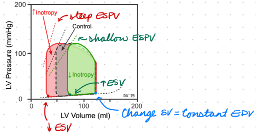
Contractility: PV Loop Changes
Increase Contractility:
Steep end-systolic pressure-volume relationship (ESPVR)
Up and left
Increase SV = Decrease ESV
Decrease Contractility:
Shallow ESPVR
Down and right
Decrease SV = Increase ESV
4: HR
Heartbeats/contractions per minute
High HR:
Decrease diastolic filling time
Increase diastolic filling pressure
Decrease SV
Low HR:
Increase diastolic filling time
Decrease diastolic filling pressure
Increase SV
Pulmonary Edema
Fluid accumulation in lung interstitium and alveolar spaces
Medical emergency = Require immediate treatment
Clinical Presentation: Bibasilar lung crackles (auscultation) and dyspnea
Pulmonary Edema From CHF
Frank-Starling mechanism
Increased EDV (preload) in LV = Ventricle cannot increase SV
Increase pulmonary vein pressure = Increase capillary hydrostatic pressure = Fluid leak out of capillaries
Peripheral Edema
Fluid accumulation in interstitium of extremities
Peripheral Edema From CHF
Frank-Starling Mechanism
Same as left heart (pulmonary edema)
Neurohormal Activation
Baroreceptors sense effective arterial blood volume (to perfuse tissues)
Increase vascular resistance and Na+/water retention
Adrenergic NS
RAAS
Increase ADH production
Maintain BP (perfusion) → BP = CO x TPR
CO decreased in HF = Increase TPR
Impaired Lymphatic Drainage
Increased interstitial fluid > Lymphatic system drainage capacity = Fluid accumulates
Congestive Heart Failure (CHF): Description
Structural/Functional impairment of ventricular filling or ejection fraction
Reduced heart function
CHF Types
HF with reduced EF (HFrEF): “Systolic”
Low SV
Low LV EF
HF with preserved EF (HFpEF): “Diastolic”
Low SV
Normal/low EDV
Preserved LV EF
Increased LV filling pressure
Not often used since both present with systolic and diastolic dysfunction
CHF: Epidemiology
Risk factors:
Older age
Hypertension
Obesity and metabolic syndrome
Impaired renal function
CHF: Etiology
Usually ischemia
Coronary artery disease (low O2 to myocardial tissue)
Valvular diseases
Aortic stenosis
Aortic and mitral regurg
Stages:
A: At risk, asymptomatic
B: Pre-HF, asymptomatic
C: Symptomatic HF
D: Advanced HF
CHF: Presentation
Profile A: Warm and dry
No reduced CO and vasoconstriction
No increased LV filling pressure (congestion)
Profile B: Warm and wet
No reduced CO and vasoconstriction
Increased LV filling pressure
Profile L: Cold and dry
Reduced CO and vasoconstriction
No increased LV filling pressure
Profile C: Cold and wet
Reduced CO and vasoconstriction
Increased LV filling pressure
CHF Pathogenesis: HFrEF
Decreased contractility = Ventricular dysfunction in systole = Decreased blood pumped (LVEF)
Decrease CO

CHF Pathogenesis: HFpEF
Decreased ventricular compliance = Ventricular dysfunction in diastole = Decrease ventricular filling (preload) + Increased diastolic pressure
Decrease CO

CHF Pathogenesis: Left-Sided HF
Increased Afterload:
Increased aortic pressure (arterial hypertension)
Outflow obstruction (aortic stenosis)
Increased Preload:
LV volume overload (aortic regurg)
CHF Pathogenesis: Right-Sided HF
Increased Afterload:
Increased pulmonary artery pressure (pulmonary hypertension)
Increased Preload:
RV volume overload (tricuspid regurg, L to R shunt)
CHF: Investigations
Clinical features
Imaging
Blood test
CHF: Imaging
ECG
CXR
Echo (TTE)
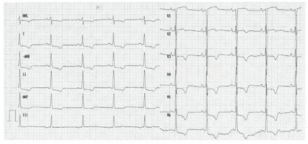
CHF Imaging: ECG
HFpEF: Normal
ST-segment and T-wave changes
Elevation
Depression
P wave changes
Biphasic
Long QTc interval
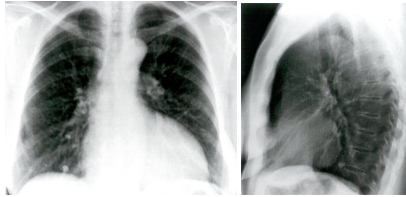
CHF Imaging: CXR
Changes in cardiac silhouette
Cardiomegaly
Pericardial effusion
Pulmonary congestion
Valve calcification
CHF Imaging: Echo
LV systolic/diastolic dysfunction
Reduced filling and contraction
Thick or dilated atria and ventricles
CHF Investigation: Blood Test
Brain natriuretic peptide (BNP)
Vasodilator and diuretic hormone produced by cardiomyocytes during stretch and ventricular wall stress
Elevated
>35 (chronic)
>100 (acute)
CHF: Clinical Presentation
Nocturia (urinating at night)
Fatigue
Tachycardia and arrythmia
S3/S4 sounds
CHF: Treatment/Management
Lifestyle changes
Increase exercise
Weight loss
Healthy eating (low Na+)
Smoking and alcohol cessation
Treat underlying conditions
Pharmacological: Decrease BP
Devices:
Automated implantable cardioverter defibrillator
Cardio resynchronization therapy
CHF Management: Pharmacological
Beta-1 blocker
Diuretics
Sodium-glucose cotransporter 2 (SGLT2) inhibitor
Renin-angiotenin-aldosterone system (RAAS) inhibitor
Mineralocorticoid receptor antagonist
CHF Management: Beta-1 Blocker
First-line
Ex: Metoprolol, Propranolol
MOA:
Inhibit beta-1 receptors in heart = Decreaes HR and contractility = Decrease work for heart during pumping
Decrease BP and energy needs = Reverse pathological remodelling
CHF Management: Diuretics
Ex: Spironolactone, furosemide
MOA: Increase Na+, ion, water excretion = Decrease edema
CHF Management: Sodium-Glucose Cotransporter 2 (SGLT2) Inhibitor
First-line
Ex: Empagliflozin
MOA: Decrease glucose (+ water) reabsorption = Decrease edema
HF Management: Renin-Angiotensin-Aldosterone System (RAAS) Inhibitor
Ex: Ramipril, Losartan
MOA:
Prevent angiotensin-2 binding to receptors to decrease Na+ and water retention
Decrease vasoconstriction and preload
CHF Management: Mineralocorticoid Receptor Antagonist (MRA)
Ex: Spironolactone
MOA:
Inhibit aldosterone effects to decrease Na+ reabsorption and H+/K+ excretion
Decrease water retention
Decrease pathological remodelling
HCF Treatment: Automated Implantable Cardioverter Defibrillators (AICDs)
Detect tachyarrhythmias = Deliver electrical impulse to reset sinus rhythm
CHF Treatment: Cadiac Resynchronization Therapy (CRTs)
Generate pulse to synchronize right and left ventricle contraction