CMS Heme exam
 Call Kai
 Learn
Learn Practice Test
 Spaced Repetition
 Match
Match Flashcards
 Knowt Play
1/379

There's no tags or description
Looks like no tags are added yet.
Name | Mastery | Learn | Test | Matching | Spaced | Call with Kai | Chat |
|---|
No analytics yet
Send a link to your students to track their progress
380 Terms
erythrocytes (RBC)
what is the most abundant cell type that transports O2 and contains hemoglobin?
4 globin chains and 4 heme groups
hemoglobin is composed of______
it is responsible for oxygen and carbon dioxide transport
heme
O2 binds to______ and carries O2 molecules to the tissues
globin
CO2 binds to ____ -->excreted out by the lung
2 a-chains
2 b-chains
Hemoglobin A is composed of 4 globin or protein chains
leukocytes (WBC)
___ in the blood have the primary function of immune response
Two categories: granulocytes and agranulocytes
neutrophils, eosinophils, basophils
what are the granulocytes WBC
neutrophils
what granulocyte is involved in acute bacterial infections
◦Phagocytic
eosinophils
what granulocyte is involved with Parasitic worm infections
basophils
what granulocyte is involved in Inflammatory response and vasodilation
◦Histamine containing granules
lymphocytes and monocytes
what are the agranulocytes WBC
lymphocytes
what agranulocyte has two primary populations
◦T cells: acts directly against target
◦B cells: utilizes antibody production against target
monocyte
what agranuloyte has Circulating macrophages
◦Phagocytic
Hematopoiesis
formation of red blood cells that occurs in red bone marrow of long bones and axial skeleton
hematopoietic stem cell
all blood cells arise form the _____
kidneys
hematopoiesis is under the influence of EPO in the ______.
EPO is produced by cells that sense tissue oxygenation
reticulocytes
immature RBCs
takes 4 days to mature---> 120 circulation then removed by macrophages
common myeloid progenitor and common lymphoid progenitor
the hematopoietic stem cells can divide into ______ and _____

megakaryocyte, erythrocyte, mast cell and myeloblast
common myeloid progenitor turns into
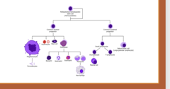
basophils, neutrophils, eosinophils, and monocytes
what does a myeloblast turn into
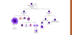
macrophages
monocytes turn into

B and T lymphocyte and natural killer cells
the common lymphoid progenitor turn into

MCV
the size of RBC
reticulocyte count
what lab eval measures for immature RBS, normally seen in vary small amounts
elevated
if bone marrow is working well (normal) there will be _________ levels of reticulocyte count
haptoglobin
molecule that binds free circulating hemoglobin (released form RBCs when it breaks apart)
this prevents toxicity
low
______ haptoglobin is a useful lab marker of hemolysis , because it means RBCs are being destroyed and the haptoglobin is being used up
Ferritin
____ is the main storage form of iron in the body
reflects your total body iron stores.
low
if iron is ______
There's no iron to store, so ferritin levels drop — this is a hallmark of iron deficiency anemia.
iron deficiency
IF FERRITIN IS LOW =
increases
If inflammation is present: Ferritin also behaves like an acute-phase reactant, meaning it ______
transferrin
iron transporter molecule
Total Iron Binding Capacity (TIBC)
measures the blood's capacity to bind iron with transferrin, the main iron transport protein.
increases
If iron is low, the body makes more transferrin to try to pick up more iron → TIBC_____
So TIBC is inversely related to body iron stores.
iron saturation
measures the percentage of transferrin that are carrying iron.
normal is 20-50%
serum iron
measures the amount of free circulating iron in serum
*least helpful*
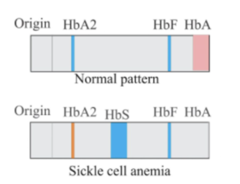
Hemoglobin Electrophoresis
a laboratory test that separates and measures different types of hemoglobin found in the blood.
Coombs (DAT)
test is used to detect antibodies that are bound directly to the surface of red blood cells
These antibodies can cause the RBCs to be destroyed — leading to hemolysis.
agglutination (clumping)
coomb's test:
1. A patient's RBCs are taken from a blood sample.
2. These cells are mixed with Coombs reagent
3. If the patient's RBCs are already coated with antibodies, the Coombs reagent will bind to those antibodies and cause _____
Positive Coombs test,
Agglutination = ________meaning antibody-coated RBCs are present.
immune-mediated hemolysis
what does a postive direct coombs tets mean?
*RBCs are being destroyed because antibodies are attached to them
Bone Marrow Aspiration and Trephine (Core) Biopsy
A bone marrow exam helps evaluate how blood cells are being produced.
It typically includes two components:
anemia
condition characterized by:
-A decreased number of circulating red blood cells (RBCs) or
-A decrease in total hemoglobin (Hb) → resulting in reduced oxygen-carrying capacity of the blood.
anemia
Because oxygen delivery is reduced with _____
-Fatigue
-Weakness
-Malaise
-Pallor (pale skin/mucous membranes)
-decreased production of RBC (bone marrow not making enough)
-increased destruction of RBC (hemolysis)
-blood loss
what can cause anemia
Microcytic
size of RBC
MCV ≤ 80 fL =
Normocytic
size of RBC
MCV 81-99 fL =
Macrocytic
size of RBC
MCV ≥ 100 fL =
problem in the bone marrow
reticulocyte counts in anemia
if reticulocytes (immature RBC) are LOW =
problem is not in the bone marrow
reticulocyte counts in anemia
if reticulocytes (immature RBC) are HIGH =
microcytic anemia
anemia with MCV
iron deficiency
what is a major cause of microcytic anemia
duodenum
where is iron usually absorbed?
*our body likes to hold onto iron (only small amount is lost throughout the day*
-occult or GI loss
-failure to meet increased requirements (menstruation or pregnancy)
-inadequate dietary intake
-malabsorption of iron
-chronic hemolysis
Iron deficiency develops when iron loss or demand exceeds intake and absorption.
The main causes can be grouped into 5 categories:
heme
patho behind iron deficiency:
Ferritin drops as stored iron in the liver, spleen, and bone marrow is used up
iron is required to make ______
microcytic
less heme = less hemoglobin per RBC ---> RBC appears more pale (hypochromic)
the bone marrow tries to make RBCs but with insufficient hemoglobin the marrow compensates and produces ________ RBC
iron deficiency anemia
Hypochromic, (pale) microcytic anemia due to underproduction of RBCs caused by insufficient iron.
Results in low hemoglobin and small, pale red blood cells.
blood loss like cancer (GI) or menstruation
what are the most causes iron deficiency anemia
asymptomatic
iron deficiency is mostly ________
but other symptoms include
-Fatigue
-weakness
-pallor
-shortness of breath
-heart palpitations
-headaches
-tinnitus
-glossitis
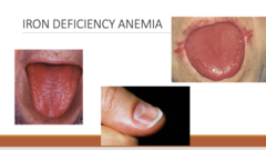
-angular cheilitis
-pica (craving for non nutritious food, ice or dirt)
-koilonychias (fingernails are thin)
what are som unique manifestations of iron deficiency anemia?
low
low
low
High variation
CBC of Iron deficiency anemia:
Hemoglobin:______
MCV:______
MCH:___(hypochromic)
RDW ( variation in RBC)
low
low
High
iron studies in iron deficiency anemia:
Ferritin:___
serum iron:____
TIBC (total iron binding capacity):____
iron deficiency anemia
a low ferritin level =
ALWAYS****
the body compensates by making more transferrin (iron transport protein in blood)
why would TIBC be elevated in iron deficiency anemia:
low
what would the reticulocyte count be in iron deficiency anemia?
bone marrow cannot respond appropriately due to iron deficiency
Iron stores are absent
if we did a bone marrow biopsy what would we see?
confirms depleted iron in the marrow
This Rarely needed unless diagnosis is unclear*
normal
*A ______ferritin does not exclude iron deficiency anemia because it is an acute phase reactant and may be elevated in inflammation, malignancy, or infection*
oral ferrous sulfate
treatment for iron deficiency anemia
(IV if more severe)
blood transfusion
treatment severe iron deficiency anemia (profound) HGB
thalassemia
inherited disorders causing reduced or absent globin chains, leading to globin imbalance, hemolysis, and ineffective RBC production
Microcytosis out of proportion (small RBC)
what is a classic finding of thalassemia?
________ to the degree of anemia
MCV is often much lower than expected given the hemoglobin level
no beta globin made
some beta globin made
beta thalassemia:
You have two β-globin genes (one from each parent). on chromosome 11
mutations:
β⁰=
β⁺=
silent carrier with mild anemia
β/β⁰ or β/β⁺
is you inherit only one abnormal beta mutation=
βeta Thalassemia Major
severe profound anemia with hemolysis
β⁰/β⁰
if you inherit both abnormal beta mutation=
transfusion
if you have profound anemia what is the treatment
silent carrier, no symptoms
alpha thalassemia:
there are 4 alpha globin genes on chromosome 16 (2 from each parent)
severity depend how many genes are deleted
deletion of 1 alpha gene=
thalassemia trait or minor thalassemia
Mild anemia
deletion of 2 alpha genes
Hgb H disease
moderate severe microcytic anemia
deletion of 3 alpha chains
san see splenomegaly
Thalassemia major, hydrops fetalis
total hemolysis of RBC
deletion of 4 alpha chains
hemoglobin electrophoresis
what is the gold standard diagnostic test for thalassemia
beta thalassemia:
Hemoglobin electrophoresis results:
look for low HbA and increased HbA2 and HbF
alpha thalassemia
Hemoglobin electrophoresis results:
look for HbH or Hb Bart's,
-chronic transfusion
-iron chelation, folic acid
-splenectomy
treatment for thalassemia is only required if severe symptoms (e.g., β-thalassemia major, Hgb H disease)
what are the treatments
Gene therapy (e.g., Zynteglo / betibeglogene autotemcel) and Allogeneic stem cell transplant
what is a definitive cure for thalassemia
this is new
microcytosis hypochromia, target cells, elliptical cells.
what type of cells will we see with thalassemia
stippled cell
what specific cell will we see with beta thalassemia
increased
will reticulocyte count be increased of decreased with thalassemia
lead poisoning anemia
Microcytic hypochromic anemia with basophilic stippling
lead poisoning anemia
Chronic or repeated exposure
Routes: GI tract, respiratory tract, skin, transplacental
Common sources: batteries, solders, paints, pottery, plumbing, gasoline, some homeopathic remedies
Zinc protoporphyrin (ZPP)
Lead displaces iron in heme synthesis and
_______ forms instead of heme → defective hemoglobin
lead poisoning anemia
patient presents with ◦Loss of appetite
◦Vomiting
◦Weight Loss
◦Learning Disabilities
◦Behavior Problems
Acute or Late Signs/Symptoms
◦Cranial nerve paralysis
◦Encephalopathy
◦Seizures
◦Coma
what is the diagnosis
basophilic stripping with tear drop cell
with lead poisoning anemia what would we see on a peripheral smear
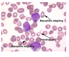
chelation like succimer and dimercarpol
treatment for lead poisoning anemia
sideroblastic anemia
Defect in incorporating iron into heme → iron accumulates in mitochondria → microcytic, hypochromic anemia.
*****we have the iron but we cant take the iron and put in into the heme molecule**
ringed sideroblasts
what would we see on a bone marrow biopsy for sideroblastic anemia
(iron-loaded mitochondria around the nucleus)

elevated
what would we see on on a serum iron test (measures free circulating iron) for sideroblastic anemia?
we have the iron we just cant get it into the heme
normocytic anemia
Anemia with MCV between 80-100
reticulocyte count
If MCV is normal, then first step is to evaluate how the marrow is responding using the ______
indicates if marrow is compensating properly.
exogenous
if reticulocyte count is HIGH ->>>>the cause is ______ and the Marrow is responding appropriately and compensating
(ex: blood loss, autoimmune hemolytic anemia and G6PD)
intrinsic bone marrow problem
if reticulocyte count is LOW/ NORMAL ---> the cause is _________
Marrow not responding adequately