Dental Radiology keyterms I
1/126
Earn XP
Description and Tags
These flashcards cover key vocabulary terms and concepts related to digital imaging, dental film, and the processing of radiographs as discussed in the lecture.
Name | Mastery | Learn | Test | Matching | Spaced |
|---|
No study sessions yet.
127 Terms
Digital Radiography
A modern imaging technique used in dentistry since 1987, where digital images replace traditional radiographs and x-rays.
Latent Image
The invisible image on film produced after exposure to x-rays, which becomes visible after processing.
Film Speed
Refers to the amount of radiation required to produce a radiograph of standard density, determined by silver halide crystal size.
Intraoral Film
Film specifically designed for taking dental x-rays inside the mouth.
Extraoral Film
Film placed outside the mouth used to examine larger areas of the head or jaws.
Digitizing Images
The process of converting film-based radiographs for viewing on a computer through scanning.
Processing Solutions
Chemical solutions used in film processing that come in forms like powder, ready-to-use liquid, or liquid concentrate.
Automatic Processor
A machine that handles all film processing steps automatically, requiring only 4 to 6 minutes for developing.
Beam Alignment Devices
Tools that assist in correctly positioning the x-ray beam relative to the tooth and film or sensor.
Film Composition
Intraoral film consists of a cellulose acetate film base coated with radiation-sensitive emulsions.
Digital Sensor Components
The active imaging components in direct digital radiography, typically either a Charge-Coupled Device (CCD) or a Complementary Metal-Oxide-Semiconductor (CMOS) sensor, comprising a grid of x-ray sensitive pixels.
Phosphor Storage Plates (PSPs) Operation
A type of indirect digital imaging where reusable plates coated with photostimulable phosphors capture x-ray energy, which is then read by a laser scanning device to convert the latent image into a digital image.
Advantages of Digital Radiography
Key benefits include reduced patient radiation exposure, elimination of chemical processing, immediate image display, improved image enhancement tools, and simplified archiving and sharing.
Role of Film Base in Intraoral Film
The flexible, transparent polyester or cellulose acetate layer that serves as the foundation for the emulsion, offering support and dimensional stability to the dental x-ray film.
Disadvantages of Digital Radiography
Higher initial equipment expense, larger and more rigid digital sensors that can be less comfortable for patients, and the need for careful handling to prevent sensor damage.
X-ray Tube Head Purpose
The component of the dental x-ray unit responsible for housing the x-ray tube and generating x-ray photons.
Cathode Function in X-ray Tube
Consists of a tungsten filament that, when heated, produces a cloud of electrons (thermionic emission) which are then accelerated towards the anode.
Anode Function in X-ray Tube
Made of a tungsten target embedded in a copper block, it attracts the electrons from the cathode and converts their kinetic energy into x-ray photons and heat upon impact.
Collimator Purpose
A lead diaphragm positioned at the exit of the tube head to restrict the size and shape of the x-ray beam, thereby reducing patient exposure to unnecessary radiation.
Filtration in X-ray Tube Head
Aluminum discs placed in the path of the x-ray beam to remove low-energy (soft) x-ray photons, which are harmful to the patient and do not contribute to image formation.
Steps of X-ray Production
Heating the Cathode: Filament heats up, releasing electrons.
Electron Acceleration: High voltage pulls electrons toward the anode.
Impact on Anode: Electrons strike the tungsten target.
X-ray Generation: Kinetic energy converts to x-rays (1%) and heat (99%) upon impact.
Beam Filtration & Collimation: X-rays are filtered and collimated as they exit the tube head.
Film Size in Dentistry
Different sizes of dental film are used for specific purposes: #0 (pediatric), #1 (anterior teeth), #2 (standard adult, posterior bitewings and periapicals), #3 (long bitewing), and #4 (occlusal for larger areas).
Density of a Radiograph
The overall blackness or darkness of a processed radiograph. It is influenced by the milliamperage (mA), exposure time, and kilovoltage (kVp).
Quality (Contrast) of a Radiograph
Refers to the range of shades of gray between the lightest and darkest areas of a radiograph. It is primarily controlled by the kilovoltage (kVp) settings of the x-ray unit.
ALARA Principle
An acronym meaning 'As Low As Reasonably Achievable,' emphasizing that all radiation exposure should be kept to a minimum to protect patients and operators.
Developer Solution Purpose
Chemically reduces the exposed silver halide crystals on the film into black metallic silver, thus creating the visible radiographic image.
Fixer Solution Purpose
Removes unexposed and undeveloped silver halide crystals from the film emulsion and hardens the emulsion, making the image permanent and light-resistant.
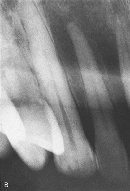
Radiographic Error: Elongation
Distortion where tooth images appear longer than their actual size due to insufficient vertical angulation of the x-ray beam.
Digital Radiography
A modern imaging technique used in dentistry since 1987, where digital images replace traditional radiographs and x-rays.
Latent Image
The invisible image on film produced after exposure to x-rays, which becomes visible after processing.
Film Speed
Refers to the amount of radiation required to produce a radiograph of standard density, determined by silver halide crystal size.
Intraoral Film
Film specifically designed for taking dental x-rays inside the mouth.
Extraoral Film
Film placed outside the mouth used to examine larger areas of the head or jaws.