Seasons LG LO's Wk3 pt.1
 Call Kai
 Learn
Learn Practice Test
 Spaced Repetition
 Match
Match Flashcards
 Knowt Play
1/30

Earn XP
Description and Tags
Ions and Heart Rhythms: Ion Channels, Transport & Electrochemical Gradient
Name | Mastery | Learn | Test | Matching | Spaced | Call with Kai | Chat |
|---|
No analytics yet
Send a link to your students to track their progress
31 Terms
LO 1: describe how the heart muscle contracts since it cannot have multiple individual muscle cells contracting at random times
the heart must act as a single, coordinated unit.
LO 1: intercalated discs
what they are
what types of connections do they contain
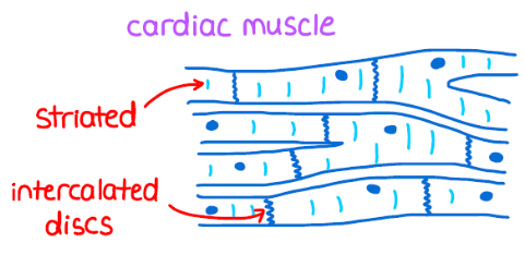
Intercalated Discs:
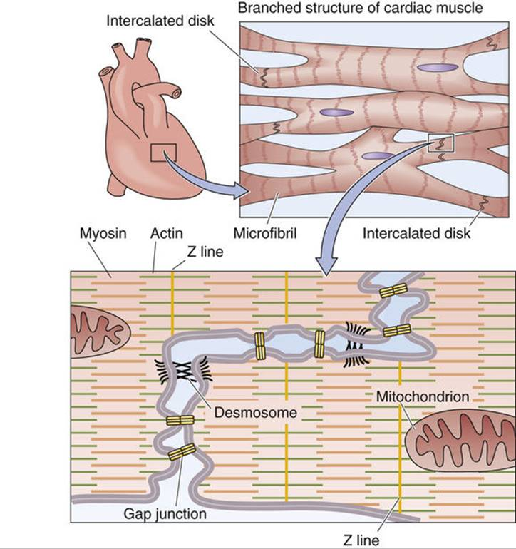
specialized junctional regions that join cardiac muscle cells (cardiomyocytes) end-to-end
These discs contain two vital types of connections: desmosomes and gap junctions.
LO 1: desmosomes
what they are
their purpose in the heart muscles
mneumonic
Physical Stability (Desmosomes):
Think of desmosomes as "mechanical staples".
specialized complexes that hold the cells tightly together so that when the heart contracts with great force, the cells do not pull apart or tear away from each other.
mnuemonic: desmosomes don’t separate
LO 1: gap junctions
what they are/function
what are they composed of
Electrical Continuity (Gap Junctions):
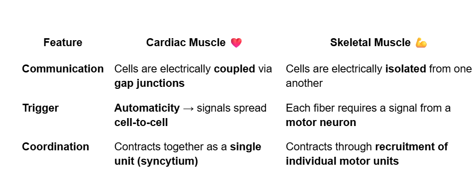
These are the true communicators.
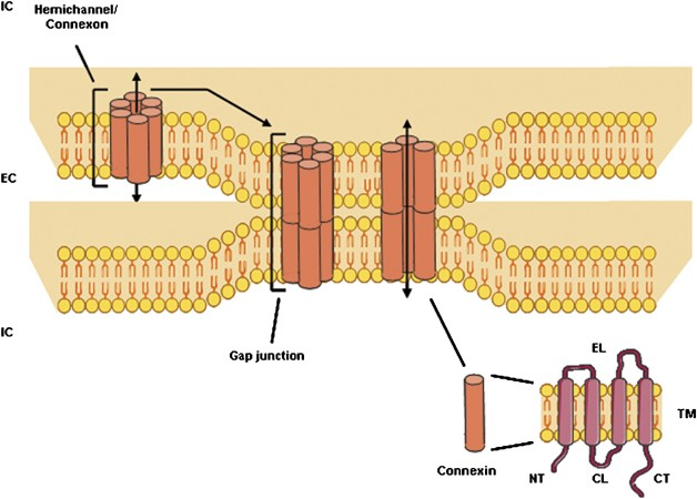
specialized protein channels that directly connect the cytoplasm of adjacent animal cells.
Gap junctions are composed of proteins called connexins that form tubular channels between adjacent cells.
LO 1: ion flow
what do ion channels create
where do the ions flow
Ion Flow:
These gap junction channels (formed by connexin) create a bridge of low electrical resistance, allowing ions (like Na+ and Ca2+) to flow directly from the cytoplasm of one cell into the next.

LO 1: functional syncytium
what causes it
what is it
Ions move through gap junctions → this depolarizes the neighboring cardiac cell → then the signal keeps spreading to other connected cells
Functional Syncytium:
Because ions move freely through these bridges without having to cross a cell membrane, a depolarization event in one cell triggers a chain reaction in all connected cells.
This allows the entire myocardium to behave as a functional syncytium—a community of cells acting as a single unit.

LO 1: cardiac muscle
describe how communication occurs
what is the trigger & the coordination
LO 1: what happens if gap junctions are disrupted ex.) MI
In a healthy patient, this rapid communication ensures that the atria contract first to fill the ventricles, followed by a coordinated ventricular contraction to eject blood to the body.
If these "tunnels" (gap junctions) are disrupted—which can happen during a myocardial infarction (heart attack) or due to electrolyte imbalances—the electrical wave becomes chaotic.
This can lead to arrhythmias, where the heart cells "wiggle" independently (fibrillation) instead of squeezing together, resulting in a failure to pump blood.
LO 1:
__________ (_________) provide the low-resistance pathway for electrical spread.
________ provide the mechanical strength to prevent cell separation during contraction.
_________is the term for the heart acting as a single unit due to these connections.
Gap junctions (connexins) provide the low-resistance pathway for electrical spread.
Desmosomes provide the mechanical strength to prevent cell separation during contraction.
Functional Syncytium is the term for the heart acting as a single unit due to these connections.
LO 2: what are the 2 types of electrical signals that the heart uses
contractile myocytes (the muscle that does the heavy lifting)
conductible pacemaker cells (the specialized cells that set the beat).
LO 2: what are myocytes vs pacemakers designed for
myocytes: long, powerful contractions, generate force
pacemakers: automaticity- the ability to fire on their own without external help. initiate and distribute the impulse
LO 2: action potential phases diagram

LO 2: action potential phases 0-4 cardiomyocyte
what they are called & what occurs
phase 4 myocytes vs SA node/pacemaker
Phase 4 of the Sinoatrial (SA) node action potential is the spontaneous diastolic depolarization (pacemaker potential) that initiates each heartbeat. It is driven by the opening of HCN (hyperpolarization-activated cyclic nucleotide-gated) channels, which generate the "funny" current (\(I_{f}\)). This allows a slow influx of sodium and potassium ions that steadily raises the membrane potential
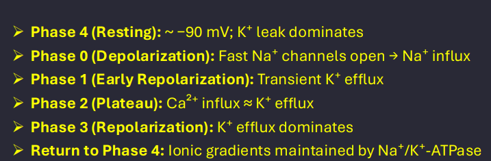
LO 2: what phases do myocytes vs pacemaker go though
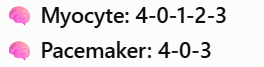
LO 2: phase 4 myocytes vs pacemakers
Phase 4 (Resting vs. Diastolic Depolarization):
Myocytes: Have a stable resting membrane potential (RMP) at -90 mV. They stay quiet until a signal arrives from a neighbor.
Pacemakers: Have no stable resting potential. Instead, they use a "funny current" (If)—an inward sodium current through HCN (hyperpolarization-activated cyclic nucleotide-gated) channels, that causes the voltage to slowly "drift" upward toward the threshold automatically.
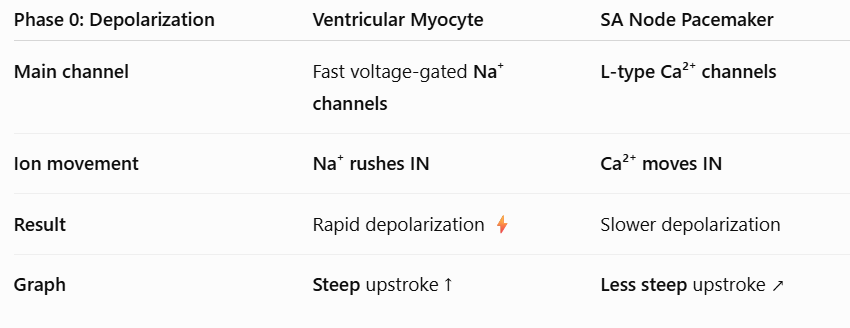
LO 2: phase 0 myocytes vs pacemakers
Phase 0 (Upstroke/Depolarization):
Myocytes: Use fast voltage-gated sodium channels. This causes a near-vertical, lightning-fast spike in voltage.
Pacemakers: Use slower L-type calcium channels for their upstroke. Because calcium moves slower than sodium, the upstroke is more "lazy" and slanted on a graph. Fast sodium channels are inactivated in these cells.
LO 2: phase 1&2 myocytes vs pacemakers
Phase 1 & 2 (The Plateau):
Myocytes: Have a distinct plateau phase (Phase 2) where calcium influx perfectly balances potassium efflux. This keeps the cell depolarized (contracted) for a long time.
Pacemakers: Completely lack Phase 1 and Phase 2. They go straight from depolarization to repolarization.
LO 2: phase 3 myocytes vs pacemakers
Phase 3 (Repolarization):
Both cells use potassium (K+) exit to return the cell to its negative starting voltage.
LO 2:
__________ is due to the unstable Phase 4 funny current in pacemaker cells.
Myocytes use ______ _______channels for depolarization; pacemakers use ______ _____
The _____ (Phase ___) only exists in myocytes to ensure a long contraction.
Automaticity is due to the unstable Phase 4 funny current in pacemaker cells.
Myocytes use fast sodium channels for depolarization; pacemakers use slow calcium channels.
The plateau (Phase 2) only exists in myocytes to ensure a long contraction.

LO 3:
why can’t heart cells stay continuously contracted like neurons
what phase helps this
the heart must contract → relax → refill → contract again
A neuron just needs to send a quick "zap," but a heart cell needs to squeeze and stay squeezed to push blood out.
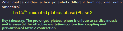
phase 2: heart cells add a massive calcium-driven plateau to slow everything down by keeping depolarization for longer and delaying repolarization
LO 3: duration of action potential neurons vs cardiomyocytes and why
Duration:
Neurons: The signal is incredibly brief, lasting only 1–2 milliseconds.
Cardiomyocytes: The action potential is "marathon-length," lasting 200–300 milliseconds.

LO 3: plateau neurons vs cardiomyocytes
The "Why" Behind the Plateau:
Neurons: No plateau. They fire and reset instantly.
Cardiomyocytes: The Phase 2 plateau is caused by calcium entering while potassium leaves. This long duration creates a long refractory period, meaning the heart cell cannot be shocked again until it has finished its current contraction.
LO 3: tetanus neurons vs cardiomyocytes
tetanus: sustained contraction
Tetanus (The Safety Switch):
Neurons/Skeletal Muscle: Can be stimulated repeatedly and very fast, leading to tetanus (a sustained, frozen contraction).
Cardiomyocytes: Because the action potential lasts as long as the contraction itself, tetanus is impossible. This is vital—if your heart had a tetanic contraction, it would stay frozen in a squeeze and never refill with blood.
The heart’s contraction is triggered by an action potential, but because the plateau creates a long refractory period, another action potential cannot occur before the muscle relaxes, preventing tetanus.
LO 3:
Plateau (Phase 2) : ion & function
The long cardiac action potential ensures the heart has time to ______ and ______.
Tetanus can happen in ________/______ muscle but is impossible in the______.
Plateau (Phase 2) : Ca²⁺ influx keeps ventricular myocytes depolarized longer.
The long cardiac action potential ensures the heart has time to relax and refill.
Tetanus can happen in neurons/skeletal muscle but is impossible in the heart.
LO 4:
what is excitation-contraction coupling in simple terms
what is the essential messenger
Excitation-contraction (E-C) coupling = how a cardiac action potential (electrical signal) causes the heart muscle to contract (physical squeeze).
calcium is the essential messenger
LO 4: the signal
what does the action potential travel down in cardiac muscle cells
The Signal:
The action potential travels down T-tubules, which are tunnels that bring the electrical signal deep into the center of the cell.

LO 4: trigger calcium
what phase, what channels, & what occurs
Trigger Calcium:
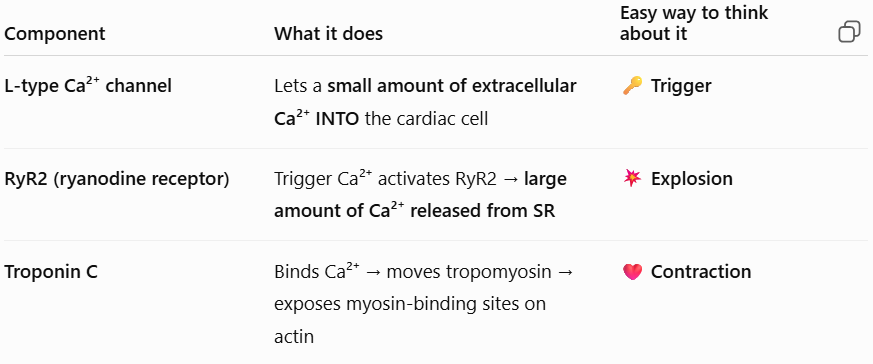
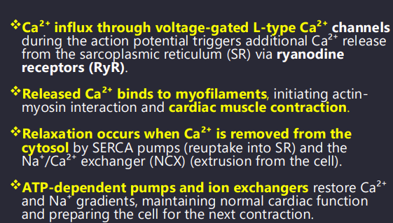
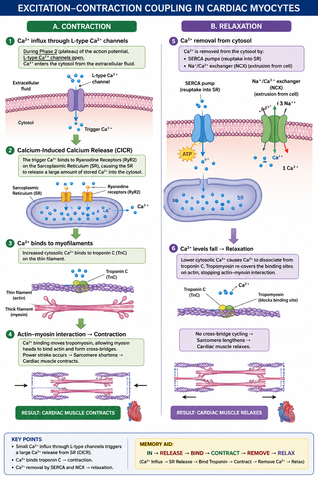
During the Phase 2 plateau, L-type calcium channels open in the T-tubule membrane, letting a small amount of extracellular calcium into the cell.
LO 4: calcium-induced calcium release
what does the calcium from the trigger bind [receptors] and where
what does the sarcoplasmic reticulum do & what is this process called
(CICR):
This small "trigger" calcium binds to Ryanodine receptors (RyR2) on the Sarcoplasmic Reticulum (SR).
This causes the SR to dump a massive amount of stored calcium into the cytoplasm—a process called Calcium-Induced Calcium Release (CICR).
LO 4: contraction
what does calcium bind
what does this lead to
what then leads to muscle contraction
Contraction:
Calcium binds to Troponin C. This causes tropomyosin to move out of the way, exposing binding sites on actin. Myosin heads then grab actin and pull, shortening the muscle (contraction).
Ca²⁺ binds troponin C → tropomyosin moves → myosin binding sites on actin exposed → myosin heads bind to actin & pull

LO 4: relaxation
what must occur for the muscles to relax
what 2 options can accomplish this
Relaxation:
To relax, calcium must be sucked back into the SR by the SERCA pump or pushed out of the cell by the NCX (Sodium-Calcium Exchanger).
SERCA: Sarcoplasmic/Endoplasmic Reticulum Ca²⁺-ATPase
NCX: Na⁺/Ca²⁺ Exchanger

LO 4:
_________________: A small amount of extracellular calcium triggers a large release from the SR.
_______ channels provide the trigger; ________ receptors provide the large release of calcium.
___________is the specific protein calcium binds to initiate contraction.
CICR (Calcium-Induced Calcium Release): A small amount of extracellular calcium triggers a large release from the SR.
L-type channels provide the trigger; Ryanodine receptors provide the large release of calcium.
Troponin C is the specific protein calcium binds to initiate contraction.