Biology Exam 2
1/103
Earn XP
Description and Tags
Name | Mastery | Learn | Test | Matching | Spaced | Call with Kai |
|---|
No analytics yet
Send a link to your students to track their progress
104 Terms
Epithelial Tissue
Found throughout the body both inside and out. Main glandular tissue. Attached to underlying connective tissue at the basement membrane. Usually has no vascular tissue-blood supply. Cells reproduce rapidly results in rapid healing. Functions are protection, secretion, absorption, excretion, and sensory receptors.
Connective Tissue
Most abundant tissue in your body. Bonds structures together. Provides support, protection, framework, fill, space, stores fat, produces blood cells, and fights infection. Composed of scattered cells with an inter cellular matrix. Made up of ground substance in fiber. Cells can reproduce.
Muscle Tissue
Skeletal: skeletal muscles-voluntary (striated)
Smooth: In hollow organs, stomach-involuntary
Cardiac: Wall of heart
Nerve Tissue
Found in brain, spinal cord and nerves.
Fibrous
Supports, protects, and holds bones, muscles, and other tissues and organs in place.
Supportive
Provides structure and strength to the body to protect soft tissues.
Fluid
Mains the continuity of the body by connecting different parts of the body.
Compare the structure and function of bone and cartilage
Bone is hard, inelastic, and a tough organ that forms part of the vertebral skeleton. Cartilage is a soft, elastic, and flexible connective tissue that protects the bone from rubbing against each other
Differentiate between blood and lymph
Blood carries substances such as nutrients, oxygen, and hormones, to the tissues and cells. Lymph comes from plasma but is made up of more water, sugar, and electrolytes
State the role of epithelial cells in the body.
Epithelial cell functions include serving as a protection barrier, secreting substances, and absorbing substances
Define homeostasis and provide an example.
Homeostasis: ability to maintain a relatively constant internal environment in the body.
Example: regulation of body temperature
Distinguish between positive and negative feedback mechanisms
Positive Feedback: brings about the an increasing change in the same direction as the original stimulus.
Negative Feedback: output of the system, resolves or corrects the original stimulus.
Compare the function of the epidermis and dermis.
The epidermis is the outermost layer of the skin and provides a waterproof barrier and contributes to skin tone. The dermis is below the epidermis and contains connective tissue, hair follicles, sweat glands, blood vessels, and lymphatic vessels.
Describe the structure and function of human skin.
The skin is made up of multiple layers including the epidermis, subcutaneous, and dermis. The skin provides a waterproof barrier and protects the inside organs.
Distinguish between the different forms of epithelial tissue with regard to location and function.
Simple columnar epithelium lines the stomach and intestines. Pseudostratified columnar epithelium lines portions of the respiratory tract and some of the tubes of the male reproductive tract. Transitional epithelium can be stretched. Glandular epithelium is specialized to produce and secrete substances.
Identify the two components of the cardiovascular system.
The heart and blood vessels.
Summarize the functions of the cardiovascular system.
Generate blood pressure, transport blood, regulate blood flow as needed, and exchanges nutrients and wastes at the capillaries.
Explain the purpose of the lymphatic system in circulation.
Draining excess fluids and proteins from tissues back in to the bloodstream which prevents the tissue from swelling.
Systolic Pressure
-Normal level is 120mmhg
- Highest arterial pressure
- Reached during ejection of blood from heart
- Decreases with distance from left ventricle
Diastolic Pressure
- Lowest arterial pressure
- Normal level is 80mmhg
- Decreases with distance from left ventricle
- Heart ventricles are relaxed
Understand how the pulse relates to heart rate.
Every time the heart beats, it squeezes and propels blood through the network of arteries in your body. Your pulse is the pressure in your arteries going up briefly as your heart pushes out more blood to keep circulation going.
Describe the structure and function of the three types of blood vessels.
Arteries carry blood away from your heart. Veins carry blood back toward your heart. Capillaries, the smallest blood vessels, connect arteries and veins.
Identify the structures and chambers of the human heart.
The heart is a cone-shaped, muscular organ located between the lungs, directly behind the sternum. Septum separates the heart into a right side and a left side. Atriums are the two upper and thin walled atria on the right and left side. They are wrinkled, ear like flap on the outer surface of an auricle. The valves line between the atria and the ventricle which are called the atrioventricular AV valves. These barrels are supported by strong fibrosis strings. The remaining two valves are the semilunar valve, with flaps shaped like half moons. These valves lie between the ventricles and their attached vessels.
Describe the flow of blood through the human heart
Deoxygenated blood comes into the right atrium from the body, moves into the right ventricle and is pushed into the pulmonary arteries in the lungs. After picking up oxygen, the blood travels back to the heart through the pulmonary veins into the left atrium, to the left ventricle and out to the body's tissues through the aorta.
Explain the internal and external controls of the heartbeat
Internal control:
-Impulses trace between gap junctions at intercalated disks
-SA node in R atrium initiates heartbeat, causes atria to contract
-Impulse reaches AV node, also in R atrium, sends signal down AV bundle and Purkinje fibers causes ventricular contraction
External control:
-Heartbeat is controlled by cardiac center in brain and hormones such as epinephrine and norepinephrine
Explain how blood pressure differs in veins, arteries, and capillaries.
Blood pressure decreases from arteries to veins, and this is because of the pressure overcoming the resistance of the vessels. The greater the change in resistance at any point, the greater the loss of pressure at that point.
Compare blood flow in the pulmonary and systemic circuits.
Systemic circulation transports oxygenated blood from the heart throughout the body. Pulmonary circulation brings deoxygenated blood back to the heart.
Identify the major arteries and veins of both the pulmonary and systemic circuits.
The major artery in the pulmonary circulatory system is the pulmonary artery which carries deoxygenated blood back to the heart from the lungs. In contrast, the major artery in the systemic circulatory system is the aorta which transports blood to tissues from the heart.
Compare the oxygen content of the blood in the arteries and veins of the pulmonary and systemic circuits.
The blood returned to the heart through systemic veins has less oxygen, since much of the oxygen carried by the arteries has been delivered to the cells. In contrast, in the pulmonary circuit, arteries carry blood low in oxygen exclusively to the lungs for gas exchange.
Describe the processes that move materials across the walls of a capillary.
Substances pass through the capillary wall by diffusion, filtration, and osmosis. Oxygen and carbon dioxide move across the capillary wall by diffusion. Fluid movement across a capillary wall is determined by a combination of hydrostatic and osmotic pressure.
Explain what happens to the excess fluid that leaves the capillaries.
The fluid turns into interstitial fluid.
List the functions of blood in the human body.
Transportation, defense, and regulatory functions.
Compare the composition of formed elements and plasma in the blood.
Blood Plasma:
Straw colored, sticky fluid
90% water
Contains:
100+ molecules: ions, nutrients, wastes, oxygen, hormones, and vitamins
3 types of proteins
Albumin: keeps water from diffusing out of the bloodstream into the extracellular matrix of tissues
Globulins: include antibodies and blood proteins that transport lipids, iron, and copper
Fibrinogen: involved in a series of reactions that achieves blood clotting
Formed Elements (blood cells):
Cannot divide
Most formed elements do not survive (only remain in blood for short time)
Replaced by new cells produced in the bone marrow
Buffy coat: leukocytes and platelets
Erythrocytes
Explain the role of hemoglobin in gas transport.
Hemoglobin contains iron, which allows it to pick up oxygen from the air we breathe and deliver it everywhere in the body
Compare the transport of oxygen and carbon dioxide by red blood cells.
Oxygen is carried both physically dissolved in the blood and chemically combined to hemoglobin. Carbon dioxide is carried physically dissolved in the blood, chemically combined to blood proteins as carbamino compounds, and as bicarbonate.
Summarize the role of erythropoietin in red blood cell production.
Erythropoietin is a hormone, produced mainly in the kidneys, which stimulates the production and maintenance of red blood cells.
Explain the function of white blood cells in the body.
White blood cells are part of the body's immune system. They help the body fight infection and other diseases. Types of white blood cells are granulocytes (neutrophils, eosinophils, and basophils), monocytes, and lymphocytes (T cells and B cells).
Distinguish between granular and agranular leukocytes
Granular Leukocytes:
Contain granules
Function: phagocytic- engulf and digest foreign cells or molecules
3 types:
Neutrophils:
- most abundant
- nucleus: 6 lobes interconnected by chromatin
- function: destroy bacteria by phagocytosis
Eosinophils
- relatively rare
- nucleus: 2 lobes interconnected by a broad band
- large granules
- function: turn off allergic responses and kill parasites
Basophils
- most rare
- nucleus: 2 lobed, bent into a U or S shape
- contains large granules
- function: release histamine and other mediators of inflammation
Agranular Leukocytes:
Lack granules
2 types
Lymphocytes
- most important cell of the immune system
- nucleus: spherical or indented
- function: mount immune response by direct cell attack or via antibodies
Monocytes
- largest leukocyte
- nucleus: U or kidney shaped
- function: phagocytosis; develop into macrophages in tissue
Explain how blood clotting relates to homeostasis.
Hemostasis refers to normal blood clotting in response to an injury.
List the steps in the formation of a blood clot.
1) Constriction of the blood vessel. 2) Formation of a temporary “platelet plug." 3) Activation of the coagulation cascade. 4) Formation of “fibrin plug” or the final clot.
Explain what determines blood types in humans.
Blood types are based on surface antigens on red blood cells, which may be proteins, carbohydrates, glycoproteins, or glycolipids.
Summarize the role of Rh factor in hemolytic disease of the newborn.
Rh is short for the "rhesus" antigen or blood type. People are either positive or negative for this antigen. If the mother is Rh-negative and the baby in the womb has Rh-positive cells, her antibodies to the Rh antigen can cross the placenta and cause very severe anemia in the baby.
Summarize how the cardiovascular system interacts with other body systems to maintain homeostasis.
The cardiovascular system maintains homeostasis by delivering oxygen and nutrients to cells, removing waste products, distributing hormones and other signals, and helping to regulate body temperature. It does this through a complex network of blood vessels and the heart, which pumps blood.
Describe the structure of the lymphatic system.
The lymphatic system includes numerous structural components, including lymphatic capillaries, afferent lymphatic vessels, lymph nodes, efferent lymphatic vessels, and various lymphoid organs. Lymphatic capillaries are tiny, thin-walled vessels that originate blindly within the extracellular space of various tissues.
Summarize how the lymphatic system contributes to homeostasis.
The primary function of lymphatic system is to maintain interstitial fluid homeostasis. Lymphatic vessels absorb fluids that extravasate from blood vessels and return them to blood circulation. In addition, lymphatic vessels absorb digested lipids from the intestine and regulate inflammation.
Explain how the lymphatic system interacts with the circulatory system.
The lymphatic system supports the circulatory system by draining excess fluids and proteins from tissues back into the bloodstream.
List examples of the body’s innate defenses.
lysozymes, phospholipase (tears and saliva), histatins (saliva), acidic pH, and microbial peptides (alpha and beta defensins secreted by cells of skin, respiratory, and GI tract), surfactant proteins of the lungs.
Summarize the events in the inflammatory response.
The response consists of changes in blood flow, an increase in permeability of blood vessels, and the migration of fluid, proteins, and white blood cells (leukocytes) from the circulation to the site of tissue damage.
Explain the role of the complement system.
It helps with the direction against foreign antigens, because it "complements" the action of antibody in destroying microorganisms.
Explain the role of an antigen in the acquired defenses.
The antigens on the infectious virus signal an immune response, causing the body to create antibodies for the specific strain of viral infection. These antibodies then utilize what is known as immunological memory to help you fight the infection if you are exposed again.
Summarize the process of antibody-mediated immunity and list the cells involved in the process.
When an antigen enters the body, helper T (thymus) cells assist B cells to activate and recognize the antigen. B cells then differentiate into plasma B cells. These plasma B cells produce antibodies against the antigen to kill them and protect the body from harmful pathogen.
Summarize the process of cell-mediated immunity and list the cells involved in the process.
Cell-mediated immunity is primarily driven by mature T cells, macrophages, and the release of cytokines in response to an antigen. T cells involved in cell-mediated immunity rely on antigen-presenting cells that contain membrane-bound MHC class I proteins in order to recognize intracellular target antigens.
Distinguish between active and passive immunity.
Active immunity occurs when our own immune system is responsible for protecting us from a pathogen. Passive immunity occurs when we are protected from a pathogen by immunity gained from someone else.
List and describe two examples each of positive AND negative feedback. Can you come up with an explanation and justification of why negative feedback is easier to control than
positive feedback?
NEGATIVE:
Blood Temp. Regulation - When body temp rises above normal, our body uses negative feedback mechanisms to bring it back to homeostasis. Sweat is produced, gets evaporated, and cools the body. As body temp decreases, sweat decreases, and our body is at homeostasis.
POSITIVE:
Childbirth - The head of the baby presses on the cervix causing a nerve signal to be sent. Oxycontin is secreted by the pituitary gland as a result of and will continue to do so until labor is over.
Negative feedback is easier to control than positive feedback because it’s easier to self-correct. Negative feedback strives for stability in the body by working to maintain a set condition. Negative feedback also exhibits more predictable behavior when it comes to disturbances.
Compare and contrast the main similarities and differences among supportive, fibrous and fluid connective tissue. What are the three types of fibers, and how do they differ?
The similarities of these tissues are the types of cells involved, the extracellular matrix, and the fact that they all function to support and structure in parts of the body. The main differences between these tissues are their composition, how they support the body, and the type of tissue they are. The three types of fibers are collagen, elastic, and reticular fibers. Collagen fibers provide strength, structure, and support. They are long and resistant to stretching. Elastic fibers contain elastin protein and are more elastic and stretchable than collagen fibers. This elasticity allows tissues to provide flexibility and resilience. Lastly, reticular fibers are thin. They form a network that supports and anchors cells and other tissue fibers.
Research and describe modifications of the apical sides of epithelial cells (such as microvilli and cilia) that allow the cells to function better.
Microvilli increases the cell's surface area to allow for more absorption and secretion as found in some cuboidal cells. Cilia on the other hand move together to sweep dust and mucus out of the body.
Compare and contrast the structure and function of the arteries to that of the veins
Arteries carry oxygenated blood away from the heart to the tissues and organs of the body. Arteries branch into arterioles, which branch into capillaries. The elastic nature of the arterial walls helps maintain blood pressure by expanding and recoiling with the heartbeats. Veins carry deoxygenated blood from the tissues back to the heart. They accomplish this by the contraction of the skeletal muscles and their one-way valves that prevent backflow. Veins also hold a significant portion of the blood’s volume and have thinner walls than arteries.
The number of people in the general population with type AB blood is relatively small. That is fortunate because these people make the least useful donors. Why would that be the case?
People with type AB blood can only donate to others with type AB blood due to the presence of both A and B antigens on their red blood cells. Therefore, their blood is compatible with a minimal amount of the population compared to people with type O blood, who are universal donors. Because they are universal donors, those with type O blood can be transfused safely into people with any blood type without an immune response being triggered.
A lymph node biopsy may be done during or before surgery to remove a tumor. The biopsy results determine the number of nodes that need to be removed. Explain how lymph node
removal influences lymph exchange in blood capillaries.
Without certain lymph nodes present within the body, swelling is likely to occur in the areas missing lymph nodes due to a build-up of interstitial fluid. Lymphatic vessels typically collect interstitial fluid once they accumulate and through the capillaries they are meant to return to the lymph node and return, repeating the process.
Why does vaccination provide long-lasting protection against disease?
Vaccines are substances that contain certain antigens for the immune system to respond to. The vaccine usually contains a non-virulent pathogen which can be issued to prepare the immune system for future exposures. Your antibodies work to attack, weaken, and destroy the pathogen. Afterward, your immune system remembers the pathogen. If the pathogen invades again, your body can recognize it and quickly send out the right antibodies so you don't get sick.
Barrier Defenses (First line of defense): Innate non-specific
Skin, hair, stomach acid, cilia, digestive enzymes in mouth, and mucous membranes.
Internal Defenses (Second line of defense): Innate non-specific
Phagocytic cells, inflammation, natural killer cells, and antimicrobial proteins.
Humoral & Cell-Mediated Defenses (Third line of defense)
Cell-mediated, humoral, B cells, Antibodies, cytotoxic T cells, and helper T cells.
Humoral Immunity Steps
1. Antigen enters the body (first exposure)
2. Antigens bind to the antigen receptors on a small subset of B cells.
3. Activated B cells multiply into clones (“clonal selection”)
4. Some clones become memory cells or some clones become plasma cells, which secrete antibodies.
Cell Mediated Immunity Steps
1. a macrophage (antigen presenting cell) engulfs a pathogen.
2. The macrophage digests the pathogen and presents it on the surface of the cell.
3. A specific helper T cells binds to the macrophage.
4. Cytokines secreted by the macrophage and helper T cell activate the helper T cell.
5. Clones of the activated, specific helper T cell form.
After this there are two possible ways.
First way: An activated helper T cell binds to a B cell to activate it.
Second way:
An activated helper T cell binds to a cytotoxic T cell to activate it.
An activated cytotoxic binds to an infected cell.
The cytotoxic T cell release perforin molecules, which form pores in the infected cell.
The infected cell dies and the cytotoxic T cell can attack other cells.
Cartilage
Has less matrix which makes more weak. Absorbs movement such as jumping
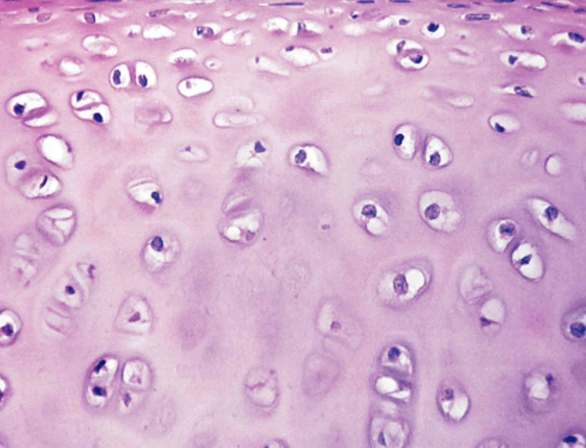
What type of cartilage is this
Hyaline cartilage
What are the two types of fibrous connective tissue?
-Loose: fibers create open, loose framework
-Dense: fibers are densely packed
What are the two types of supportive connective tissue?
-Cartilage: solid yet flexible matrix
-Bone: solid and rigid matrix
What are the two types of fluid connective tissue?
-Blood: contained in blood vessels
-Lymph: contained in lymphatic vessels
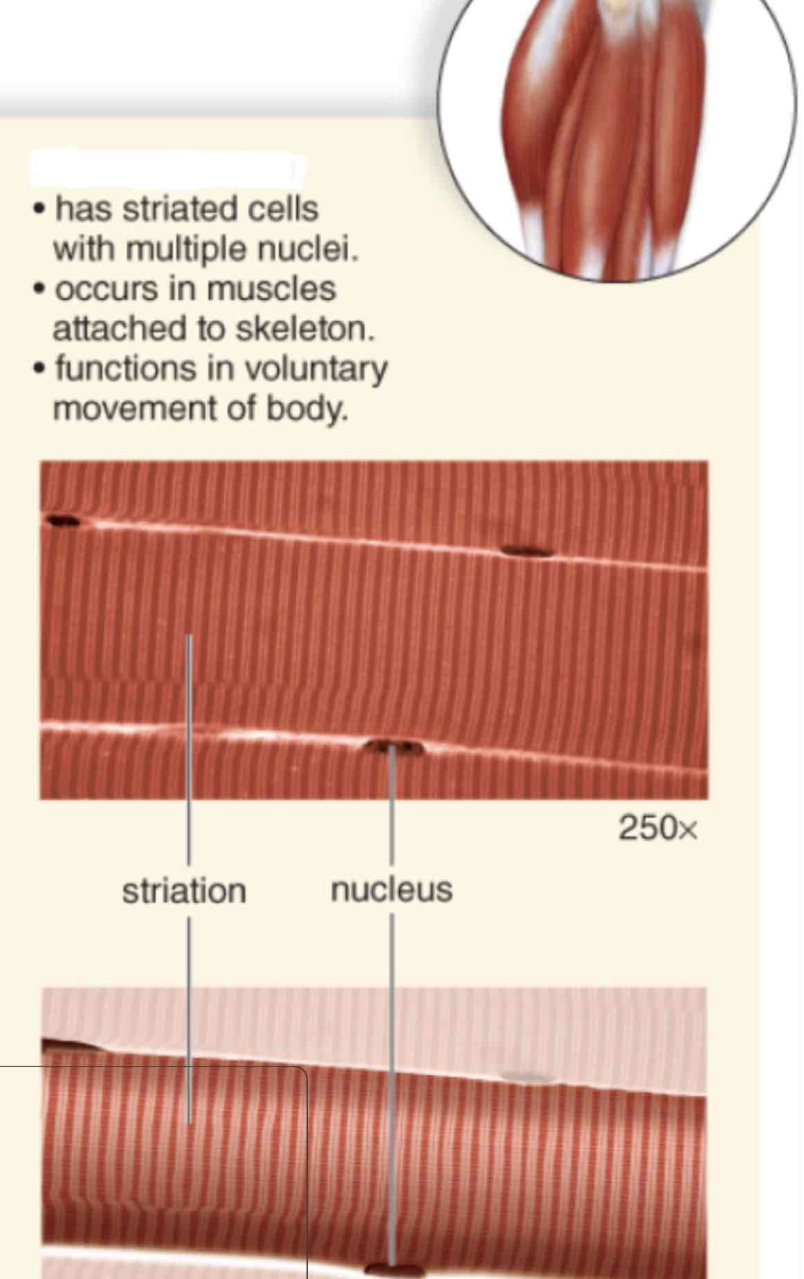
What type of muscle is shown?
Skeletal muscle
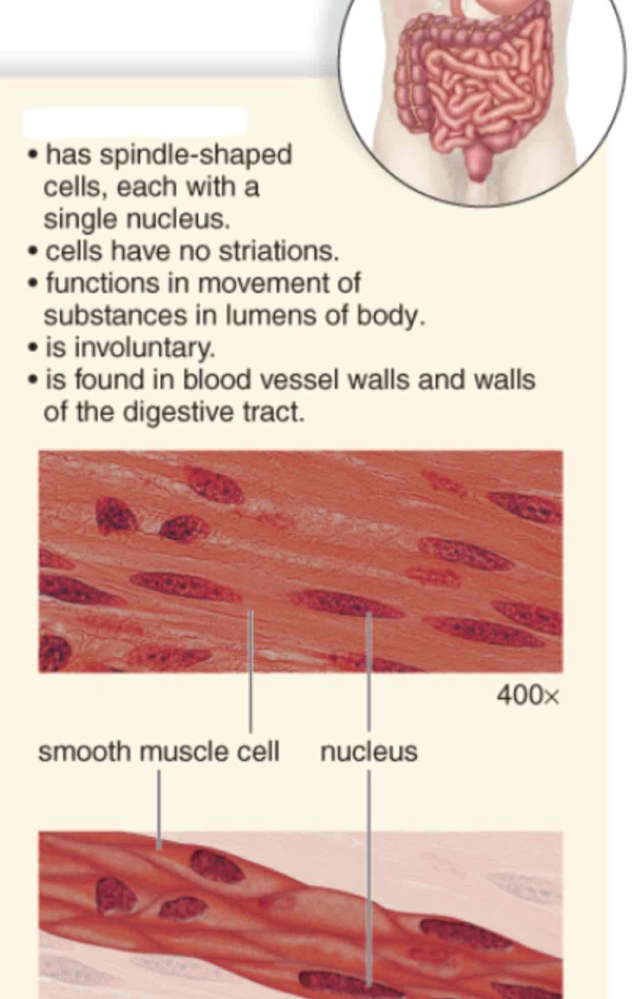
What type of muscle is shown?
Smooth muscle
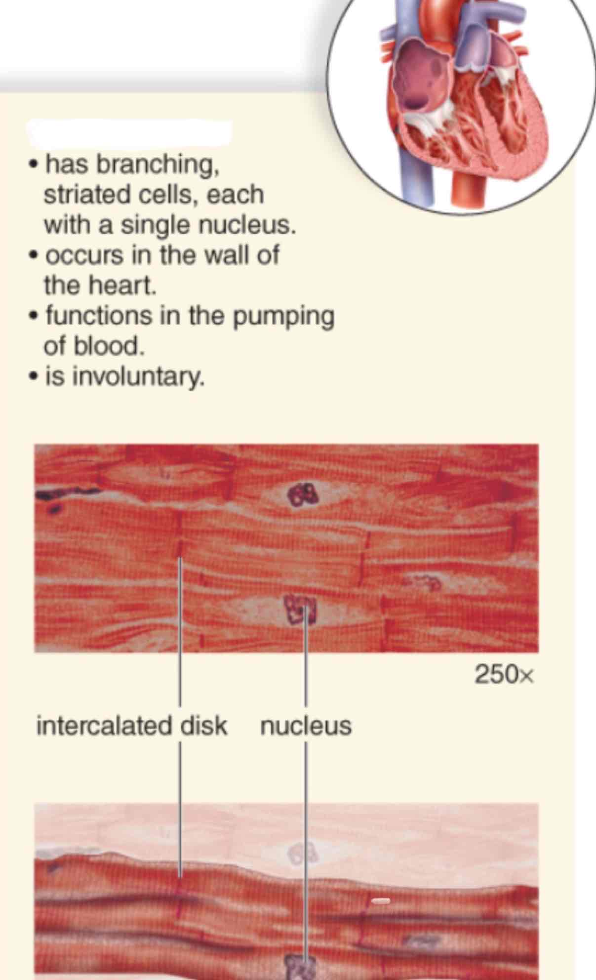
What type of muscle is shown?
Cardiac muscle
Which is more solid loose or dense fiber connective tissue?
Dense has a high number of strong reticular fibers so it is more solid.
Which ventricle is stronger?
The left ventricle is more muscular than the right ventricle because it must pump blood to entire body.
Which is stronger arteries and veins?
Arteries more muscular than veins to withstand higher pressure.
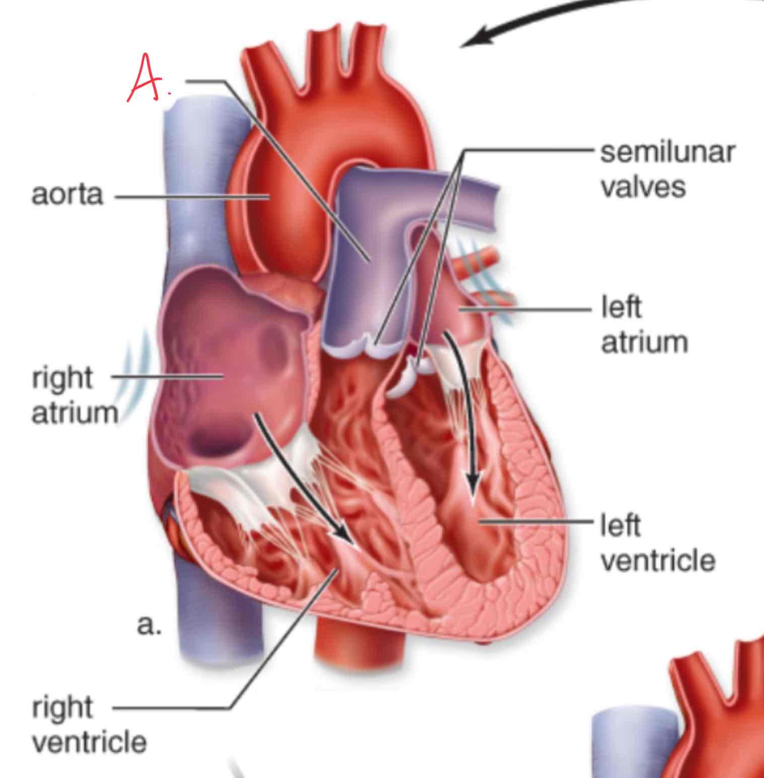
What is letter A?
Pulmonary Trunk
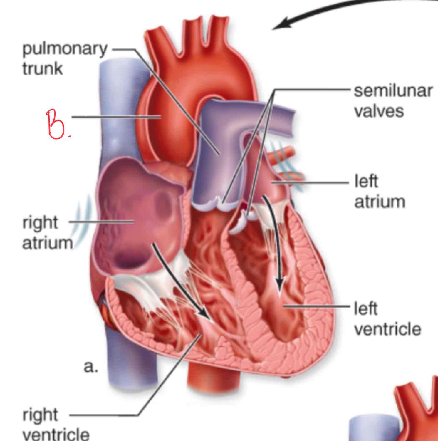
What is letter B?
Aorta
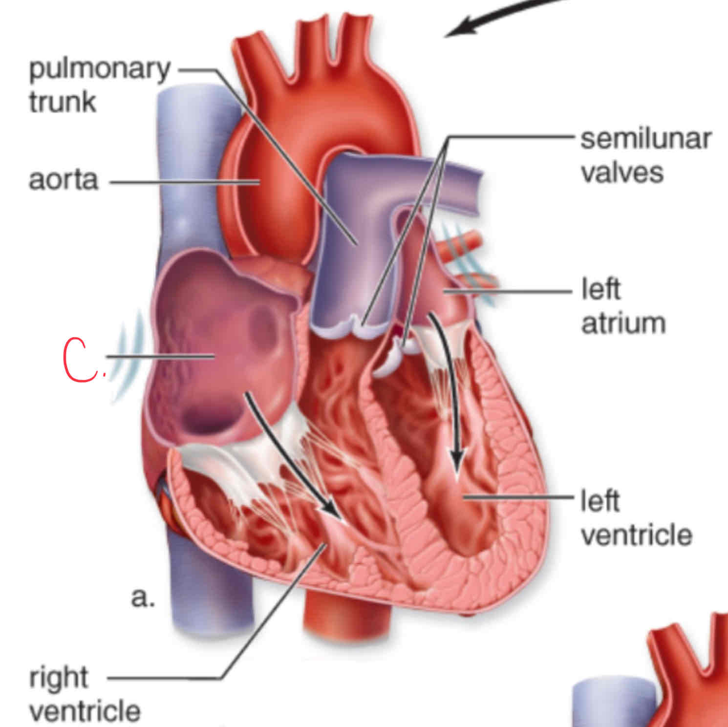
What is letter C?
Right Atrium
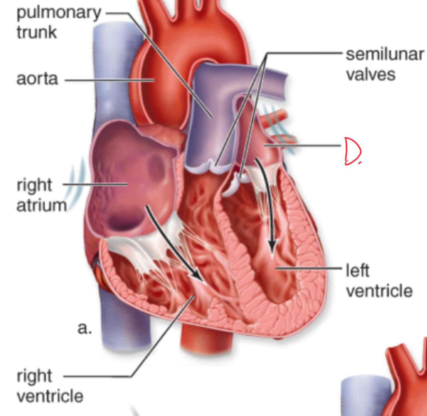
What is letter D?
Left atrium
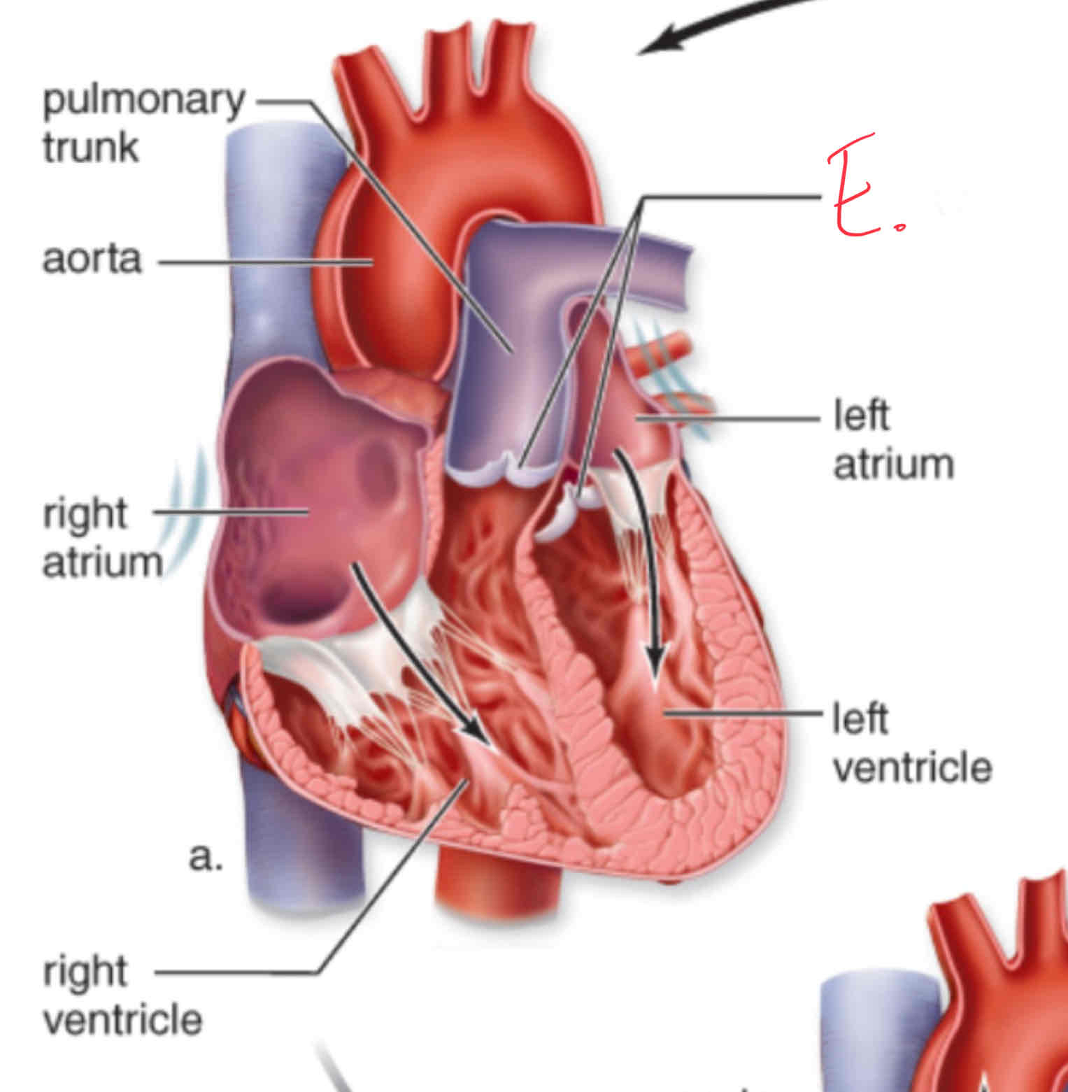
What is letter E?
Semilunar valves
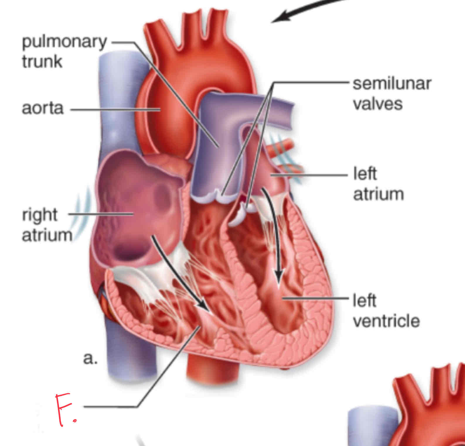
What is letter F?
Right ventricle
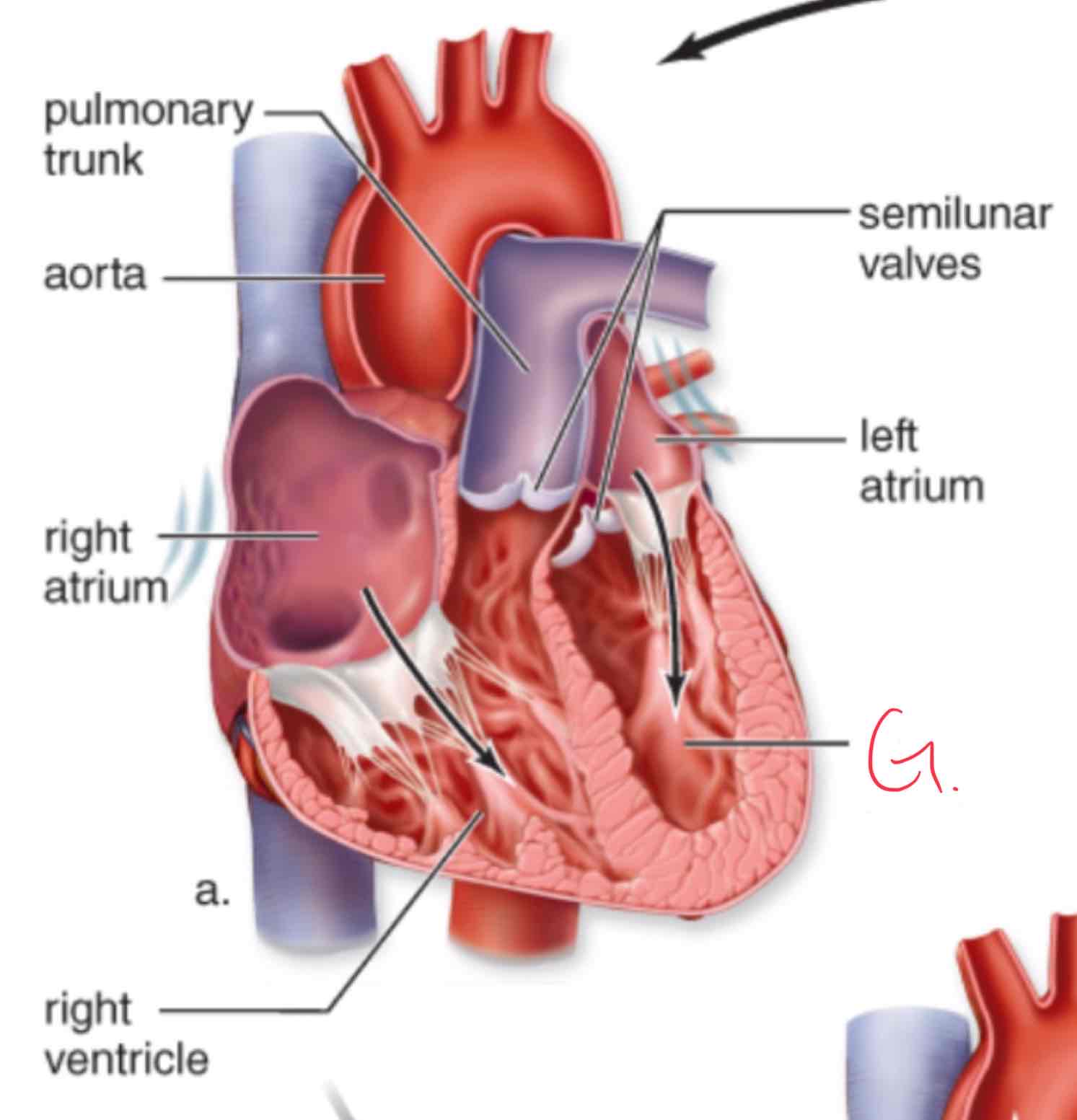
What is letter G?
Left ventricle
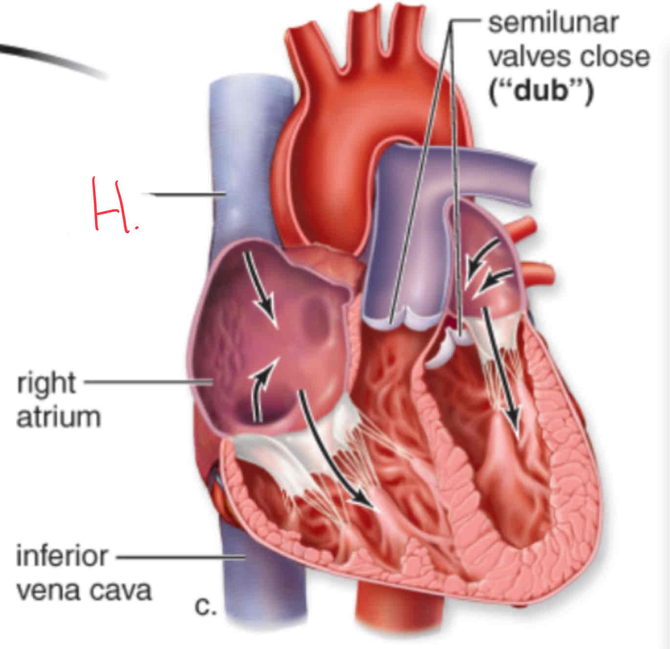
What is letter H?
Superior vena cava
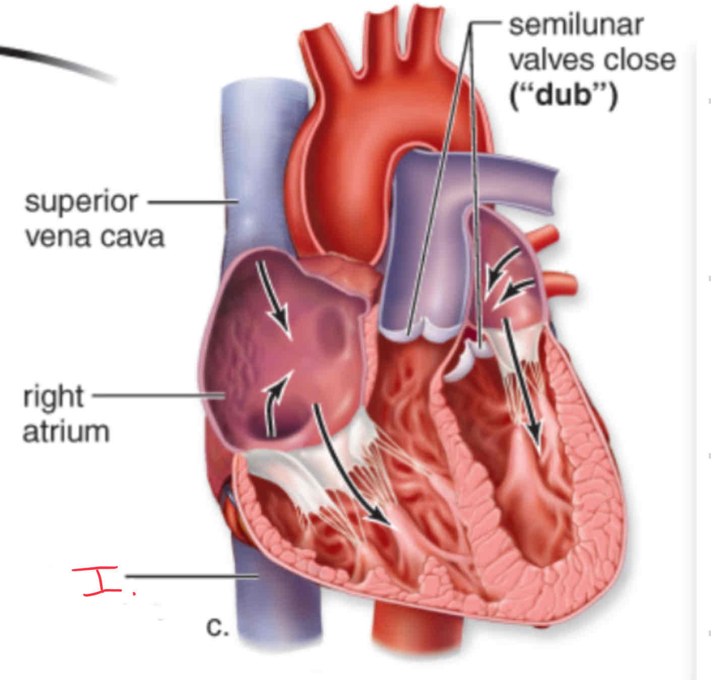
What is letter I?
Inferior vena cava
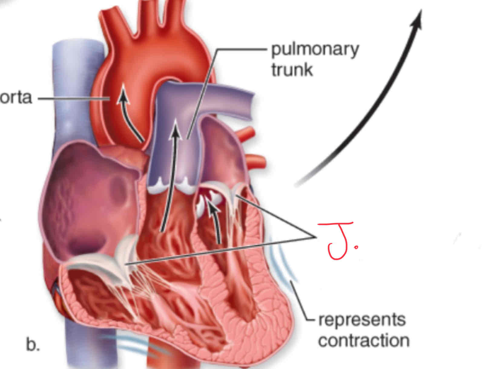
What is letter J?
Atrioventricular value (AV valve)
What is blood pressure controlled by?
Arterioles
What is the difference between pulmonary arteries and pulmonary veins?
Pulmonary arteries are the only arteries that carry deoxygenated blood and pulmonary veins are the only veins that carry oxygenated blood
What is the heart surrounded by?
Pericardium a thick membraneous sac that surrounds the heart in order to protect and support it.
How does the myocardium receive nutrients and oxygen?
Coronary arteries gives the myocardium nutrients and oxygen because it is the first to branch off of the aorta.
Antigen
Foreign substance, often polysaccharide of protein, stimulates immune response
Antibody
Protein made in response to antigen in body which binds specifically to antigen
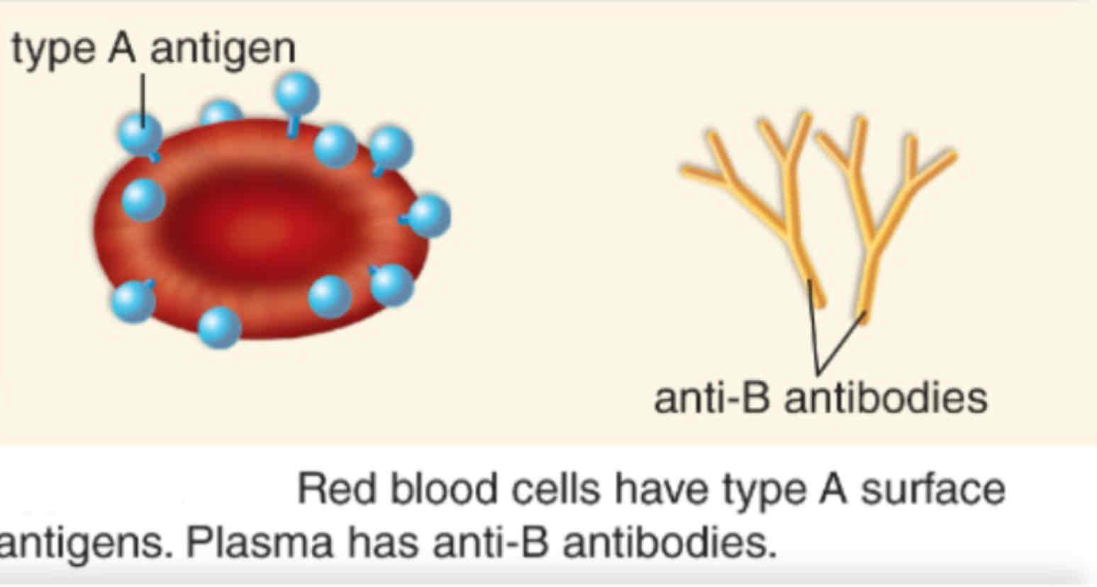
What blood type is shown?
Type A
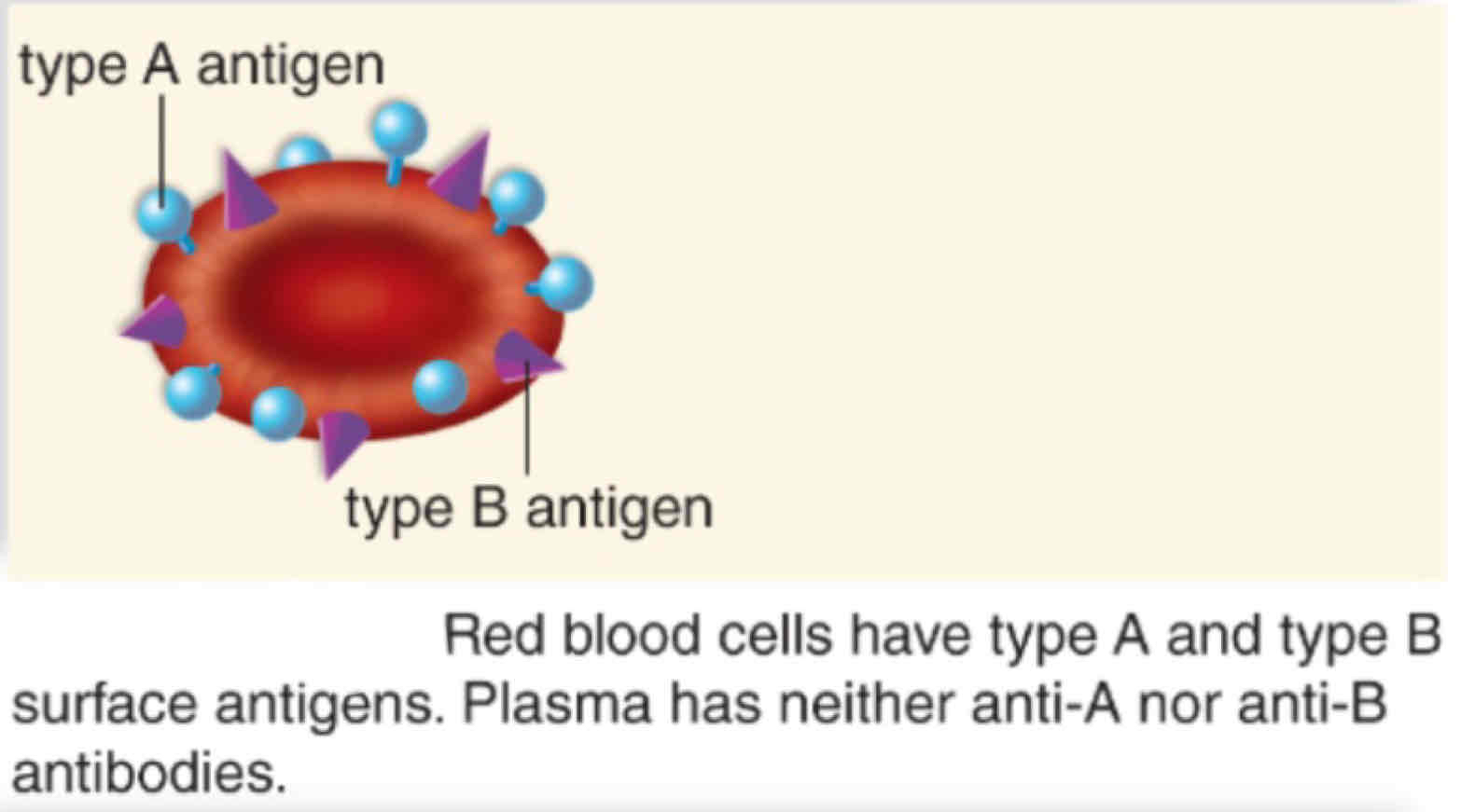
What blood type is shown?
Type AB
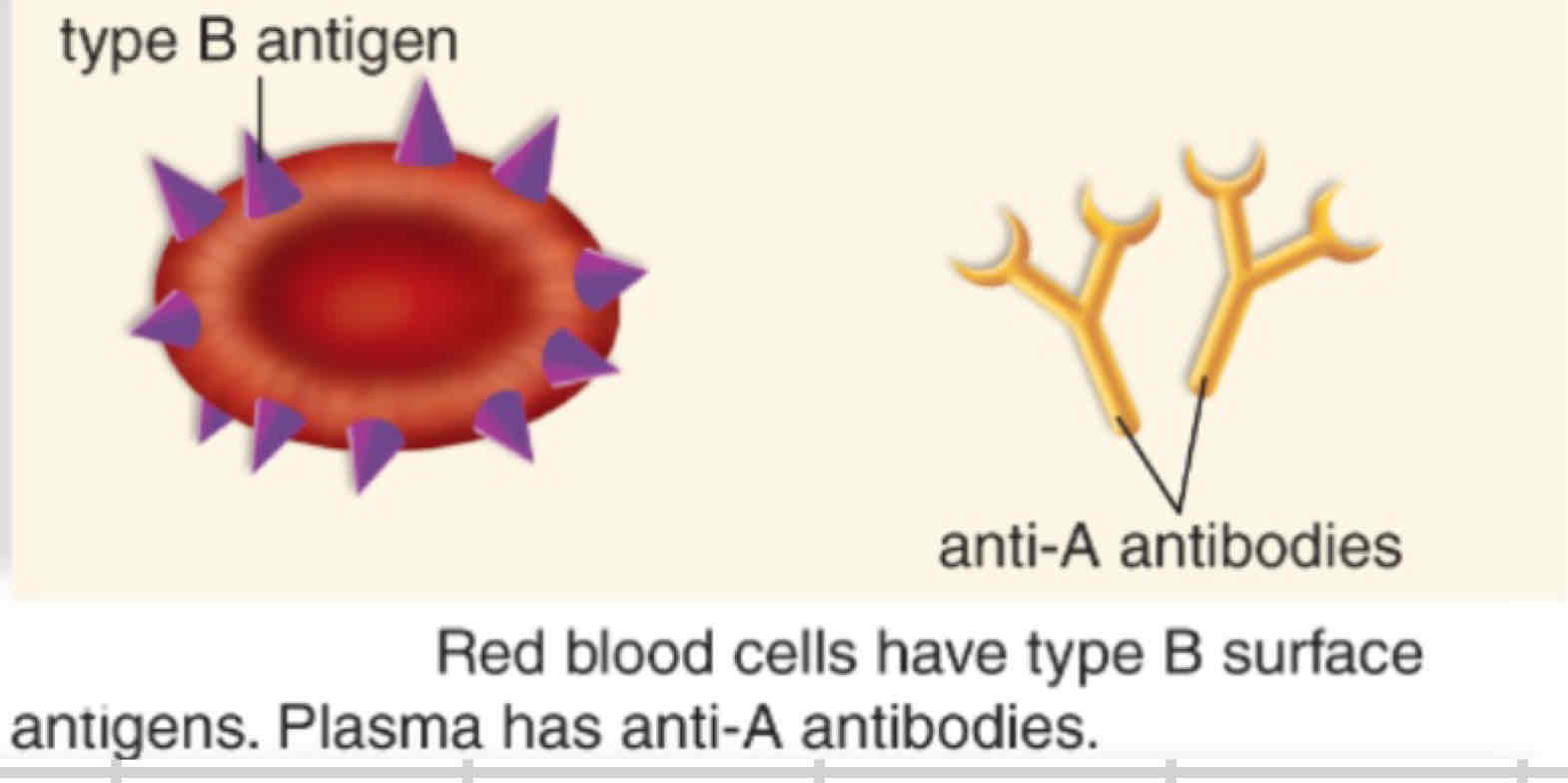
What blood type is shown?
Type B
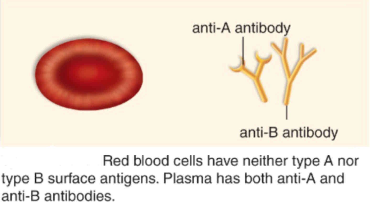
What blood type is shown?
Type O
What are the primary lymphatic organs?
Red bone marrow and thymus
Why are the secondary lymphatic organs?
Spleen and lymph nodes
Where are lymph nodes found and what are the filled with?
Neck, groin regions, and armpit. They are filled with B cells, T cells, and macrophages.
What are the steps of the inflammatory response?
Heat, pain, swelling, and redness