MOINA HEAD & NECK MUSCLES
1/53
There's no tags or description
Looks like no tags are added yet.
Name | Mastery | Learn | Test | Matching | Spaced | Call with Kai |
|---|
No analytics yet
Send a link to your students to track their progress
54 Terms
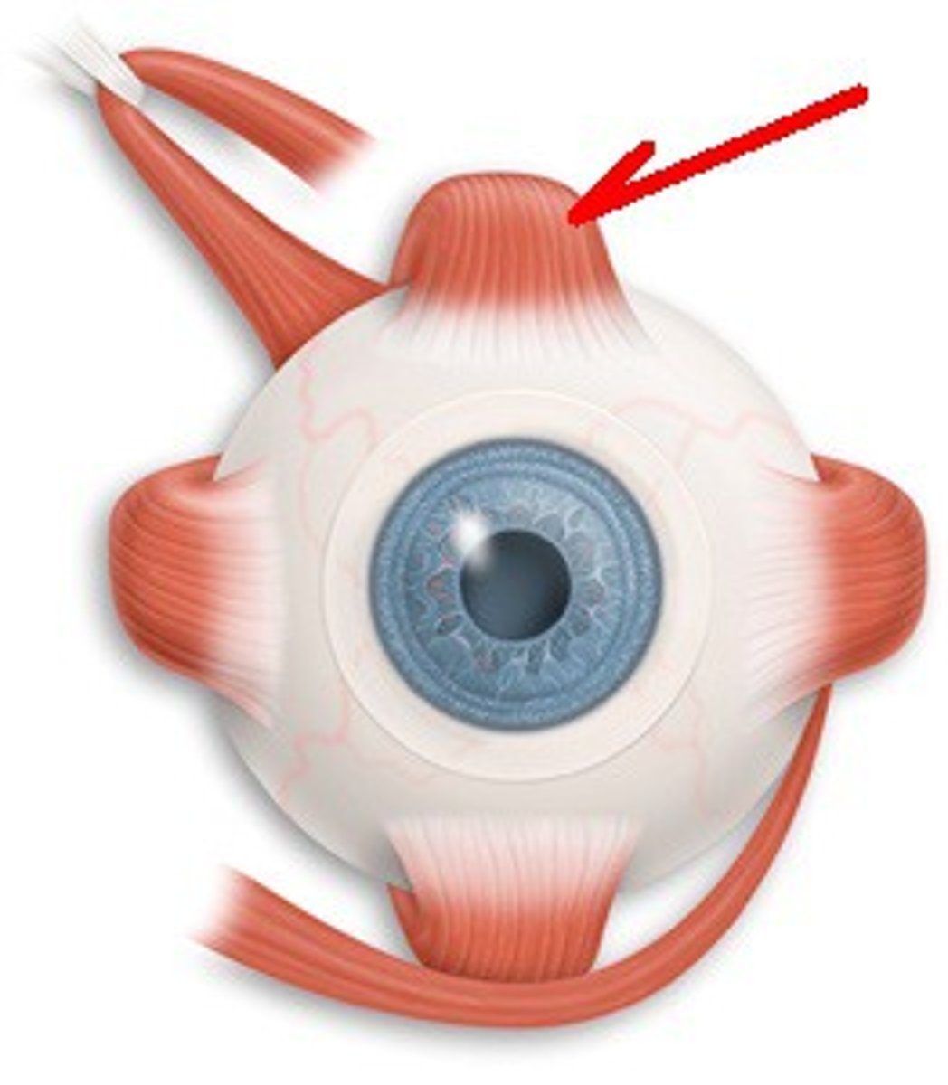
Superior Rectus
O: Tendinous ring on posterior wall of the orbital cavity
I: Superior surface of eyeball just posterior to corneoscleral junction
N: Oculomotor nerve (3rd cranial nerve)
A: Raises cornea upward and medially
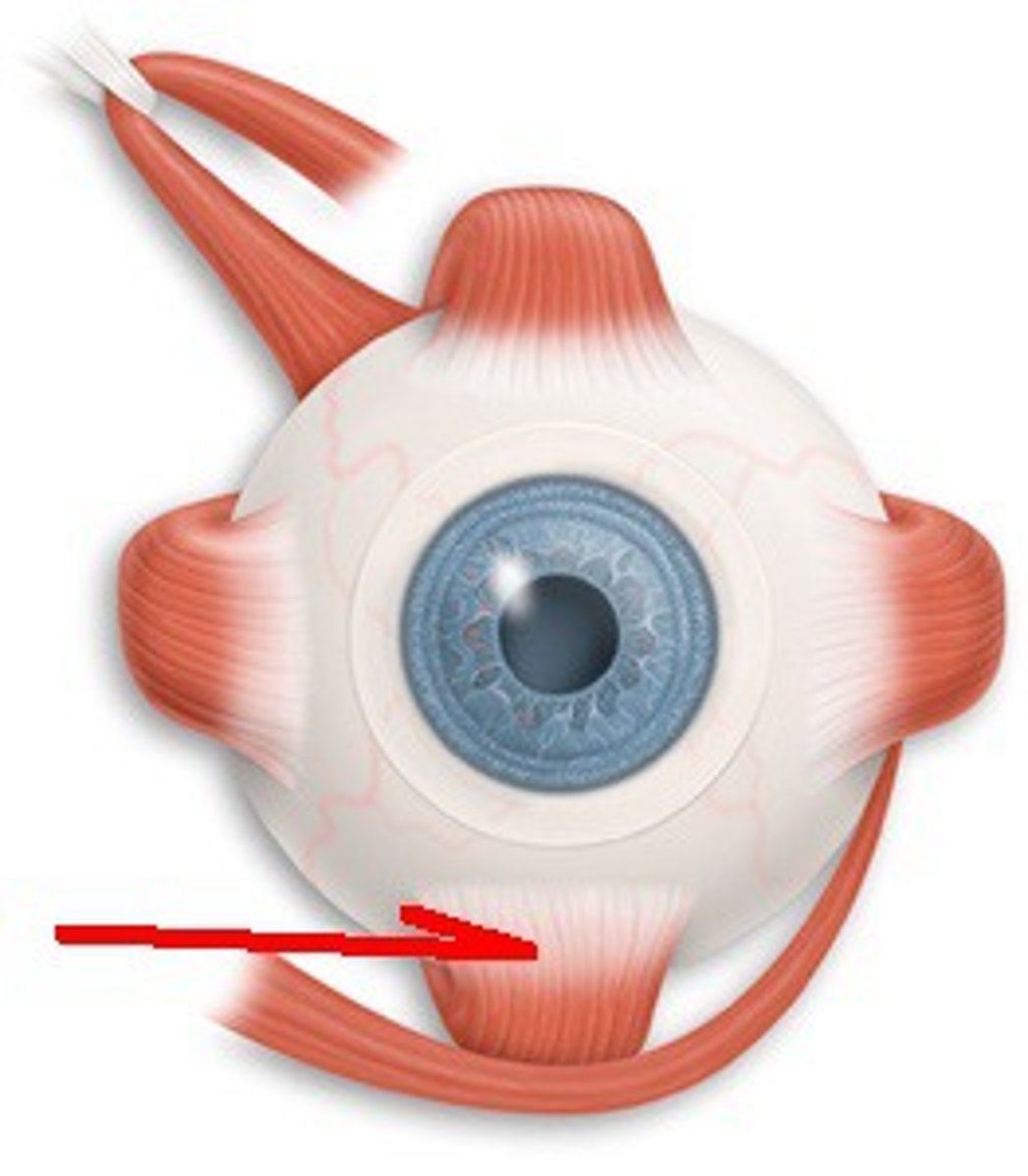
Inferior Rectus
O: Tendinous ring on posterior wall of orbital cavity
I: Inferior surface of eyeball just posterior to corneoscleral junction
N: Oculomotor nerve (3rd cranial nerve)
A: Depresses cornea downward and medially
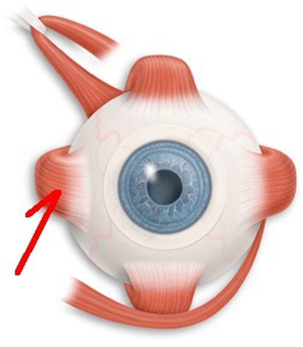
Medial rectus
O: Tendinous ring on posterior wall of the orbital cavity
I: Medial surface of eyeball just posterior to corneoscleral junction
N: Oculomotor nerve (3rd cranial nerve)
A: Rotates eyeball so that cornea looks medially
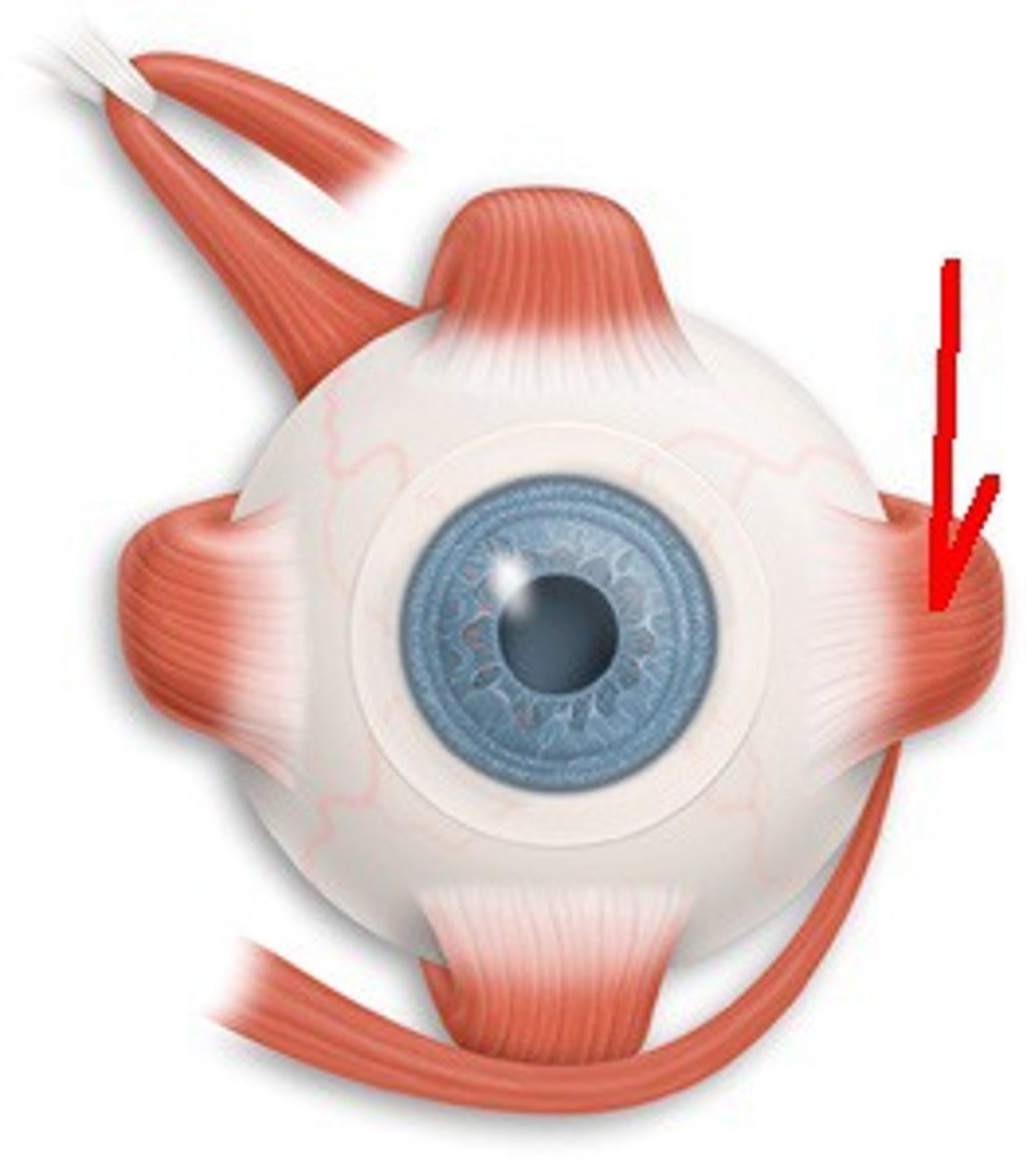
Lateral rectus
O: Tendinous ring on posterior wall of orbital cavity
I: Lateral surface of eyeball just posterior to corneoscleral junction
N: Abducent nerve (6th cranial nerve)
A: Rotates eyeball so that cornea looks laterally
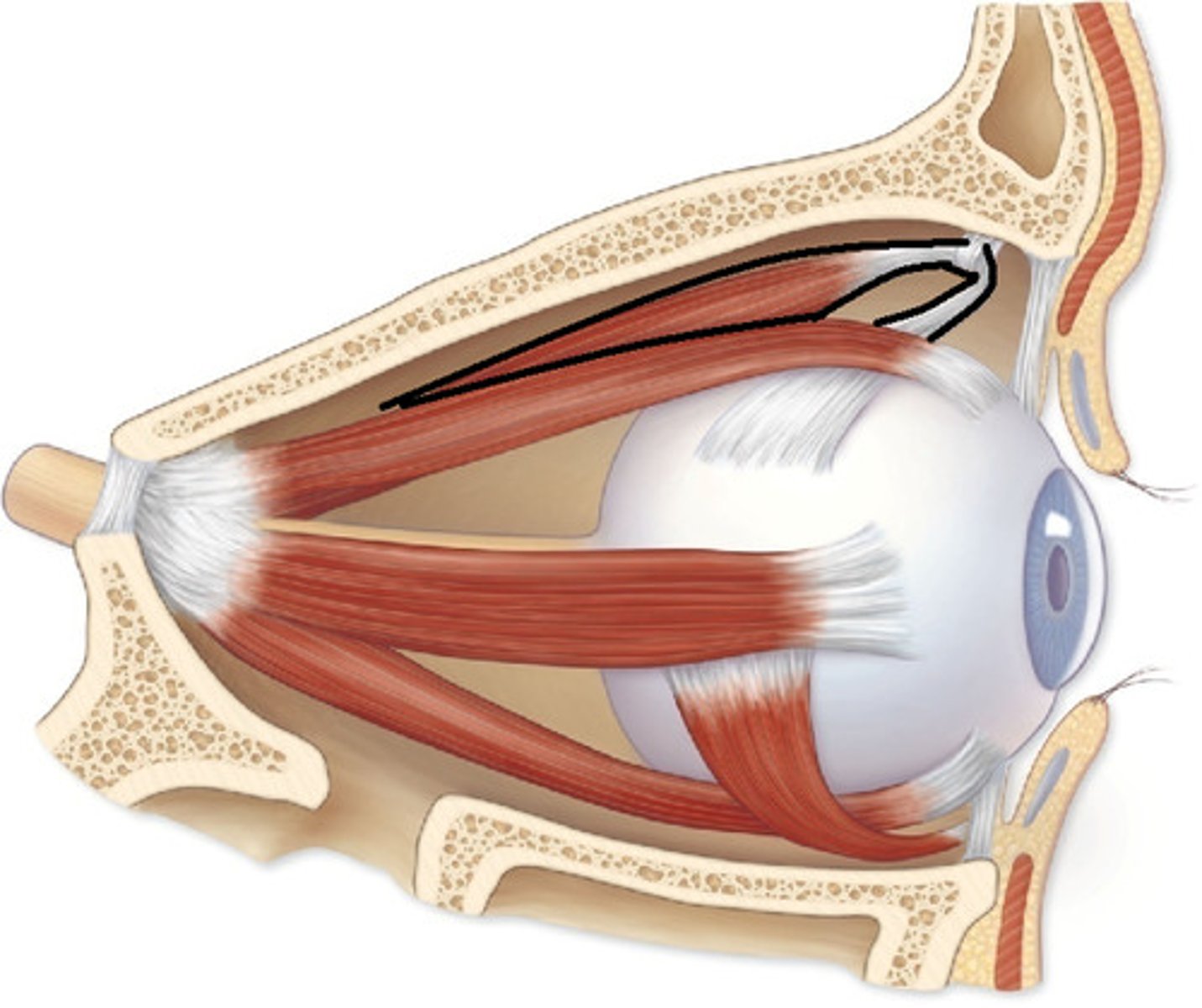
Superior oblique
O: Posterior wall of orbital cavity
I: Passes through pulley and is attached to superior surface of eyeball beneath superior rectus
N: Trochlear nerve (4th cranial nerve)
A: Rotates eyeball so that cornea looks downward and laterally
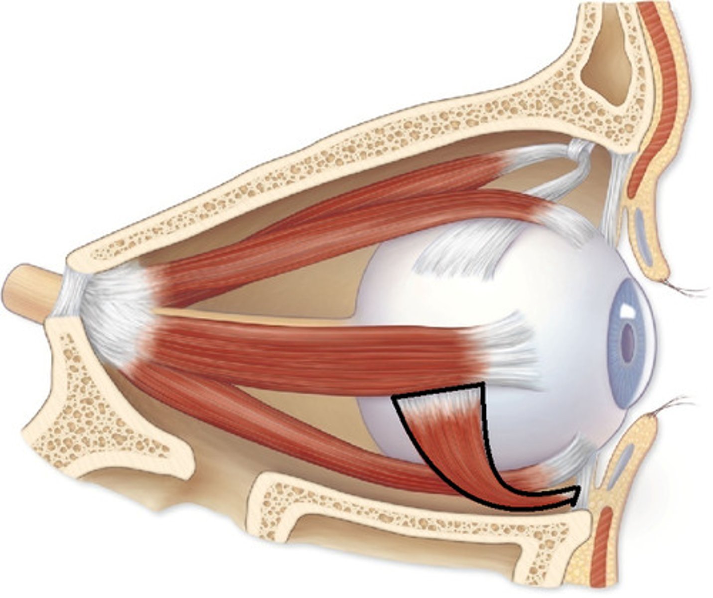
Inferior Oblique
O: Floor of orbital cavity
I: Lateral surface of eyeball deep to lateral rectus
N: Oculomotor nerve (3rd cranial nerve)
A: Rotates eyeball so that cornea looks upward and laterally
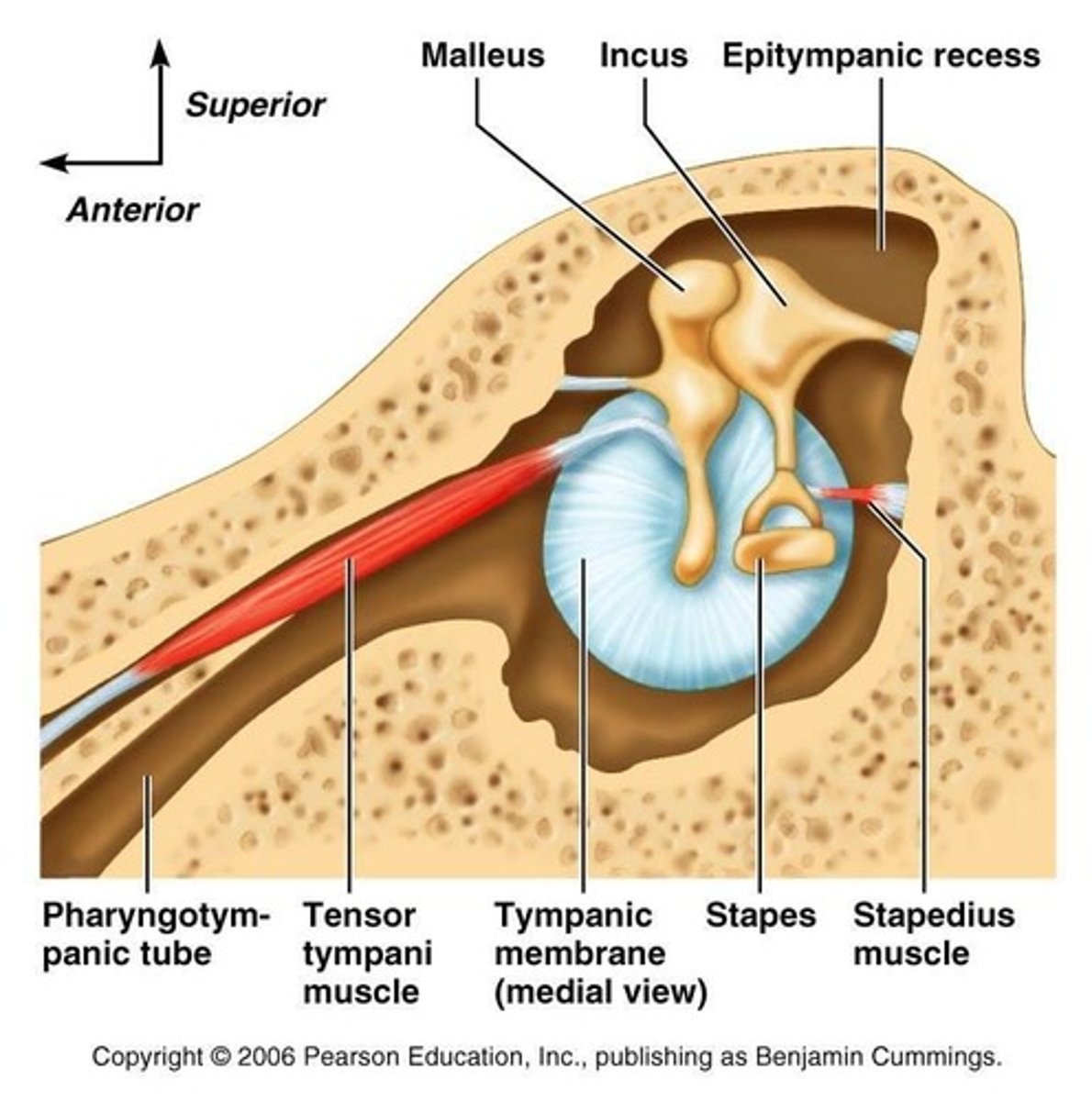
Tensor tympani
O: Wall of auditory tube and wall of its own canal
I: Handle of malleus
N: Mandibular division of trigeminal nerve
A: Dampens down vibrations of tympanic membrane
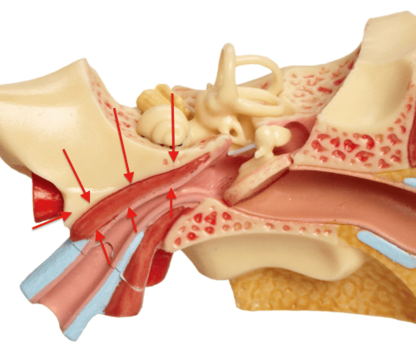
Stapedius
O: Pyramid (bony projection on posterior wall of middle ear)
I: Neck of stapes
N: Facial nerve
A: Dampens down vibrations of stapes
Occipitofrontalis (Occipital belly)
O: Highest nuchal line of occipital bone
I: Epicranial aponeurosis
N: Facial nerve
A: Moves scalp on skull and raises eyebrows
Occipitofrontalis (Frontal belly)
O: Skin and superficial fascia of eyebrows
I: Epicranial aponeurosis
N: Facial nerve
A: Moves scalp on skull and raises eyebrows
Orbicularis oculi (Palpebral part)
O: Medial palpebral ligament
I: Lateral palpebral raphe
N: Facial nerve
A: Closes eyelids and dilates lacrimal sac
Orbicularis oculi (Orbital part)
O: Medial palpebral ligament and adjoining bone
I: Loops return to origin
N: Facial nerve
A: Throws skin around orbit into folds to protect eyeball
Corrugator supercilii
O: Superciliary arch
I: Skin of eyebrow
N: Facial nerve
A: Vertical wrinkles of forehead, as in frowning
Compressor nasi
O: Frontal process of maxilla
I: Aponeurosis of bridge of nose
N: Facial nerve
A: Compresses mobile nose cartilage
Dilator naris
O: Maxilla
I: Ala of nose
N: Facial nerve
A: Widens nasal aperture
Procerus
O: Nasal bone
I: Skin between eyebrows
N: Facial nerve
A: Wrinkles skin of nose
Orbicularis oris
O: Maxilla, mandible, and skin
I: Encircles oral orifice
N: Facial nerve
A: Compresses lips together
Levator labii superioris aleque nasi
O: Arise from bones and fascia around oral aperture and insert into substance of lips
I: Arise from bones and fascia around oral aperture and insert into substance of lips
N: Facial nerve
A: Separate lips
Levator labii superioris
O: Arise from bones and fascia around oral aperture and insert into substance of lips
I: Arise from bones and fascia around oral aperture and insert into substance of lips
N: Facial nerve
A: Separate lips
Zygomaticus major
O: Arise from bones and fascia around oral aperture and insert into substance of lips
I: Arise from bones and fascia around oral aperture and insert into substance of lips
N: Facial nerve
A: Separate lips
Levator anguli oris
O: Arise from bones and fascia around oral aperture and insert into substance of lips
I: Arise from bones and fascia around oral aperture and insert into substance of lips
N: Facial nerve
A: Separate lips
Risorius
O: Arise from bones and fascia around oral aperture and insert into substance of lips
I: Arise from bones and fascia around oral aperture and insert into substance of lips
N: Facial nerve
A: Separate lips
Depressor anguli oris
O: Arise from bones and fascia around oral aperture and insert into substance of lips
I: Arise from bones and fascia around oral aperture and insert into substance of lips
N: Facial nerve
A: Separate lips
Depressor labii inferioris
O: Arise from bones and fascia around oral aperture and insert into substance of lips
I: Arise from bones and fascia around oral aperture and insert into substance of lips
N: Facial nerve
A: Separate lips
Mentalis
O: Arise from bones and fascia around oral aperture and insert into substance of lips
I: Arise from bones and fascia around oral aperture and insert into substance of lips
N: Facial nerve
A: Separate lips
Buccinator
O: Outer surface of alveolar margins of maxilla and mandible and pterygomandibular ligament
I: N/a
N: Facial nerve
A: Compresses cheeks and lips against teeth
Masseter
O: Zygomatic arch
I: Lateral surface ramus of mandible
N: Mandibular division of trigeminal nerve
A: Elevates mandible to occlude teeth
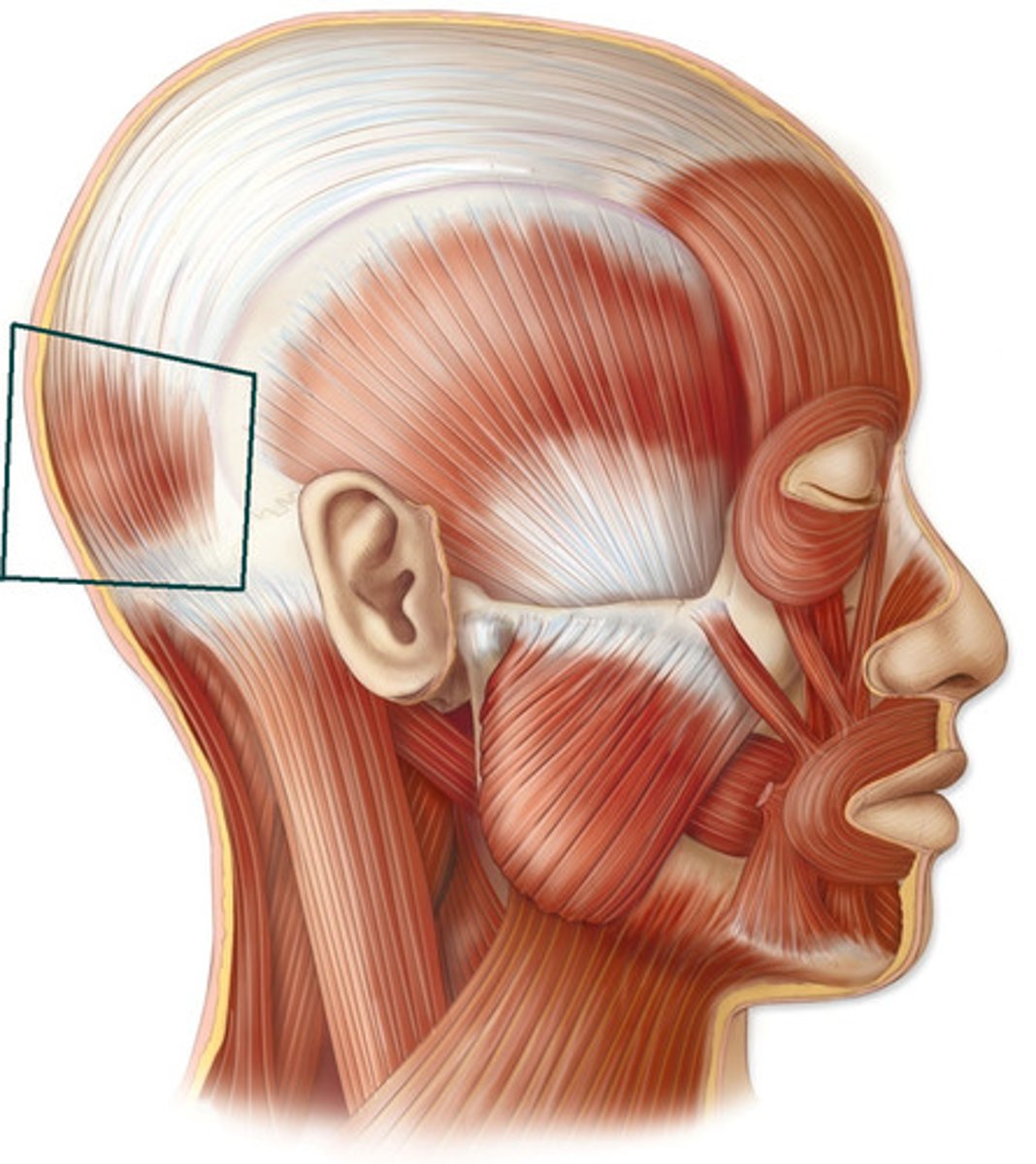
Temporalis
O: Floor of temporal fossa
I: Coronoid process of mandible
N: Mandibular division of trigeminal nerve
A: Anterior and superior fibers elevate mandible; posterior fibers retract mandible
Lateral pterygoid (two heads)
O: Greater wing of sphenoid and lateral pterygoid plate
I: Neck of mandible and articular disc
N: Mandibular division of trigeminal nerve
A: Pulls neck of mandible forward
Medial pterygoid (two heads)
O: Tuberosity of maxilla and lateral pterygoid plate
I: Medial surface angle of mandible
N: Mandibular division of trigeminal nerve
A: Elevates mandible
Platysma
O: Deep fascia over pectoralis major and deltoid
I: Body of mandible and angle of mouth
N: Facial nerve cervical branch
A: Depresses mandible and angle of mouth
Sternocleidomastoid
O: Manubrium sterni and medial third of clavicle
I: Mastoid process of temporal bone and occipital bone
N: Spinal part of accessory nerve and C2 and 3
A: Two muscles acting together extend head and flex neck, one muscle rotates head to opposite side
Digastric (Posterior belly)
O: Mastoid process of temporal bone
I: Intermediate tendon is held to hyoid by fascial sling
N: Facial nerve
A: Depresses mandible or elevates hyoid bone
Digastric (Anterior belly)
O: Body of mandible
I: Intermediate tendon is held to hyoid by fascial sling
N: Nerve to mylohyoid
A: Depresses mandible and elevates hyoid bone
Stylohyoid
O: Styloid process
I: Body of hyoid bone
N: Facial nerve
A: Elevates hyoid bone
Mylohyoid
O: Mylohyoid line of body of mandible
I: Body of hyoid bone and fibrous raphe
N: Inferior alveolar nerve
A: Elevates floor of mouth and hyoid bone or depresses mandible
Geniohyoid
O: Inferior mental spine of mandible
I: Body of hyoid bone
N: Ansa cervicalis: C1, 2, and 3
Sternohyoid
O: Manubrium sterni and clavicle
I: Body of hyoid bone
N: Ansa cervicalis: C1, 2, and 3
A: Depresses hyoid bone
Sternothyroid
O: Manubrium sterni
I: Oblique line on lamina of thyroid cartilage
N: Ansa cervicalis: C1, 2 and 3
A: Depresses larynx
Thyrohyoid
O: Oblique line on lamina of thyroid cartilage
I: Lower border of hyoid bone
N: 1st cervical nerve
A: Depresses hyoid bone or elevates larynx
Omohyoid (Inferior belly)
O: Upper margin of scapula and suprascapular ligament
I: Intermediate tendon is held to clavicle and first rib by fascial sling
N: Ansa cervicalis: C1, 2, and 3
A: Depresses hyoid bone
Scalenus anterior
O: Transverse processes of 3rd, 4th, 5th, and 6th cervical vertebrae
I: 1st rib
N: C4, 5, and 6
A: Elevates 1st rib; laterally flexes and rotates cervical part of vertebral column
Scalenus medius
O: Transverse processes of upper six cervical vertebrae
I: 1st rub
N: Anterior rami of cervical nerve
A: Elevates 1st rib; laterally flexes and rotates cervical part of vertebral column
Scalenus posterior
O: Transverse processes of lower cervical vertebrae
I: 2nd rib
N: Anterior rami of cervical nerve
A: Elevates 2nd rib; laterally flexes and rotates cervical part of vertebral column
Longitudinal (Intrinsic muscle of the tongue)
O: Median septum and submucosa
I: Mucous membrane
N: Hypoglossal nerve
A: Alters shape of tongue
Genioglossus
O: Superior genial spine of mandible
I: Blends with other muscles of tongue
N: Hypoglossal nerve
A: Protrudes apex of tongue through mouth
Hyoglossus
O: Body and greater comu of hyoid bone
I: Blends with other muscles of the tongue
N: Hypoglossal nerve
A: Depresses tongue
Styloglossus
O: Styloid process of temporal bone
I: Blends with other muscles of the tongue
N: Hypoglossal nerve
A: Draws tongue upward and backward, narrows oropharyngeal isthmus
Tensor veli palatini
O: Spine of sphenoid, auditory tube
I: With muscles of other side, forms palatine aponeurosis
N: Nerve to medial pterygoid from mandibular nerve
A: Tenses soft palate
Levator veli paalatini
O: Petrous part of temporal bone, auditory tube
I: Palatine aponeurosis
N: Pharyngeal plexus
A: Raises soft palate
Palatoglossus
O: Palatine aponeurosis
I: Side of tongue
N: Pharyngeal plexus
A: Pulls root of tongue upward and backward narrows palatopharyngeal folds medially
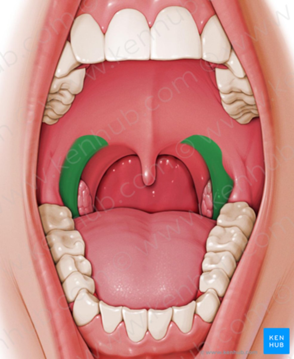
Palatopharyngeus
O: Palatine aponeurosis
I: Posterior border of thyroid cartilage
N: Pharyngeal plexus
A: Elevates wall of pharynx, pulls palatopharyngeal folds medially
Musuclus uvulae
O: Posterior border of hard palate
I: Mucous membrane of uvula
N: Pharyngeal plexus
A: Elevates uvula