STEP 2 Concepts
 Call Kai
 Learn
Learn Practice Test
 Spaced Repetition
 Match
Match Flashcards
 Knowt Play
1/354

There's no tags or description
Looks like no tags are added yet.
Name | Mastery | Learn | Test | Matching | Spaced | Call with Kai | Chat |
|---|
No analytics yet
Send a link to your students to track their progress
355 Terms
An 82-year-old woman is brought to the physician by her granddaughter because of a 6-week history of increasing forgetfulness. She is a retired schoolteacher and lives independently. Her granddaughter is concerned because on several occasions she has left the stove on when she went to bed, During conversations with her granddaughter, she has difficulty remembering past events and seems unconcerned about her memory lapses. The patient describes trouble sleeping through the night and has had a decreased appetite resulting in a 4.5-kg (10-lb) weight loss over the past month. She has a history of similar sympton,s 2 and 5 years ago that were successfully treated with medication. She appears unkempt and has poor personal hygiene. Her temperature is 37°C (98.6°F), blood pressure is 110/70 mm 'Hg, and pulse is 80/niin and regular. Mental status examination sho~s psychomotor retardation, a flat affect, impaired ability to recall past events, and trouble repeating three numbers in sequence. She is unable to recall the names of recent presidents. Her serum urea nitrogen concentration is 25 mg/dl, and serum creatinine concentration is 1. 7 n,g/dl. Whats her diagnosis?
This case describes an elderly woman exhibiting symptoms consistent with major depressive disorder, characterized by cognitive impairment (memory lapses), changes in appetite, sleep disturbances, and psychomotor retardation. The patient's history of similar symptoms treated with medication suggests recurrent episodes of depression.
32-year-old woman with infertility, irregular 35–50-day menstrual cycles, obesity, and acne. TSH, LH, FSH, testosterone, and DHEAS are normal. What is the most likely diagnosis?
Diagnosis: Polycystic ovary syndrome (PCOS)
Key clues
Infertility from chronic anovulation
Irregular, long cycles: every 35–50 days
Obesity
Acne = clinical hyperandrogenism
Normal TSH, LH, FSH, testosterone, and DHEAS do not exclude PCOS
Flashcard takeaway:
Reproductive-age woman with oligomenorrhea + signs of androgen excess + infertility → think PCOS, even when serum androgen levels are normal.
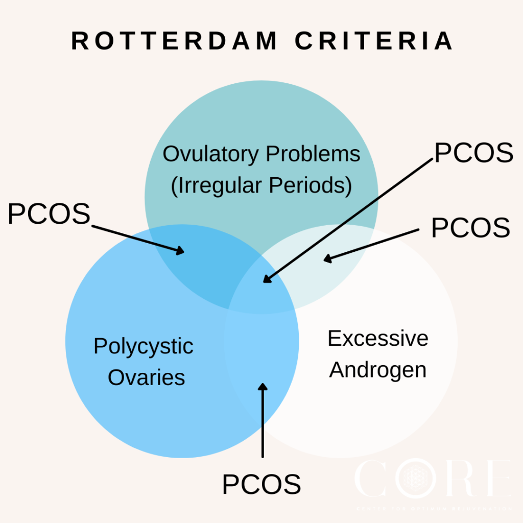
Need 2 out of the 3 rotterdam criteria
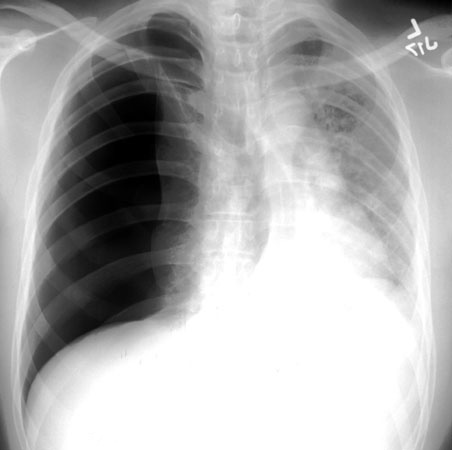
24-year-old man with sudden, worsening right-sided chest pain and dyspnea. Vital signs are normal. Chest x-ray is below. What is the next step in management?
Patient has a right-sided pneumothorax
Answer:
Chest tube placement (tube thoracostomy) because he has a symptomatic, likely large spontaneous pneumothorax.
Key point:
Stable but large/symptomatic pneumothorax → chest tube
Small and minimally symptomatic → observation with oxygen
Tension pneumothorax with hypotension or instability → immediate needle decompression, then chest tube
52-year-old man with obesity, polyuria, nocturia, weight loss, and a random glucose of 280 mg/dL. He has newly diagnosed type 2 diabetes mellitus. BP 160/85. Which serum concentration is most likely increased?
A. Glucagon
B. Insulin
C. Ketones
Answer:
Insulin concentration — early type 2 diabetes causes insulin resistance, so pancreatic β cells compensate by producing more insulin.
Key point:
Early type 2 DM: ↑ insulin and ↑ C-peptide
Later type 2 DM: β-cell failure can cause insulin levels to decrease
Weight loss occurs because glucose cannot be used effectively, leading to breakdown of fat and muscle.
A 66-year-old woman suddenly loses the right half of her visual field in both eyes. Strength, sensation, and speech are normal. Central vision is preserved. Which artery is most likely occluded?
Answer:
Left posterior cerebral artery (PCA)
Key clues:
Contralateral homonymous hemianopia
Macular sparing
No major motor weakness
A 70-year-old right-handed man can write a complete sentence but is unable to read the sentence immediately afterward. He also has a right homonymous hemianopia. Which artery is most likely occluded?
Answer:
Left posterior cerebral artery (PCA)
Key clue:
Alexia without agraphia = can write but cannot read.
67-year-old man with sudden vertigo, nausea, nystagmus, right-sided Horner syndrome, right palate weakness, decreased pain sensation on the right face and left body, and right-sided limb ataxia. Which artery is occluded?
Answer:
Right posterior inferior cerebellar artery (PICA) → lateral medullary (Wallenberg) syndrome
Key clues:
Vertigo/nystagmus → vestibular nuclei
Ipsilateral Horner syndrome
Ipsilateral facial pain-temperature loss
Contralateral body pain-temperature loss
Dysphagia/palate weakness → nucleus ambiguus
Ipsilateral ataxia
Memory tip: PICA = “Don’t PICA horse that can’t eat” → dysphagia and hoarseness from nucleus ambiguus injury.
The fastest distinctions are: ACA = leg, MCA = face/arm, PCA = vision, PICA = swallowing, AICA = face/hearing, and basilar = locked-in syndrome.
A 72-year-old right-handed man develops sudden weakness and numbness of the right face and arm. His right leg is only mildly affected. He speaks fluently, but his words are nonsensical, and he cannot understand commands. He also has loss of the right visual field. Which artery is most likely occluded?
Answer:
Left middle cerebral artery (MCA)
Key clues:
Face/arm > leg weakness
Aphasia from dominant hemisphere involvement
Contralateral homonymous hemianopia
A 68-year-old woman develops sudden weakness of the left face and arm. She ignores people standing on her left side and only eats food from the right side of her plate. She does not recognize that her left arm is weak. Which artery is most likely occluded?
Answer:
Right middle cerebral artery (MCA)
Key clues:
Face/arm > leg weakness
Contralateral neglect
Lack of awareness of deficits
A 74-year-old man develops sudden weakness and loss of sensation affecting his right leg more than his right arm. He is unusually quiet, has little motivation to speak, and develops urinary incontinence. Which artery is most likely occluded?
Answer:
Left anterior cerebral artery (ACA)
Key clues:
Leg > arm weakness
Abulia or personality changes
Urinary incontinence
A 61-year-old man develops sudden vertigo, vomiting, and difficulty walking. Examination shows left facial paralysis, decreased tearing, loss of taste over the anterior tongue, and hearing loss in the left ear. Pain sensation is decreased over the left face and right body. Which artery is occluded?
This is Lateral Pontine Syndrome
Answer:
Left anterior inferior cerebellar artery (AICA)
Key clues:
Ipsilateral facial paralysis
Hearing loss
Decreased tearing and salivation
Loss of taste from anterior two-thirds of tongue
Memory tip: Facial droop or hearing loss → AICA.
A 65-year-old man develops sudden weakness of the right arm and leg with loss of vibration and position sensation on the right side. When he sticks out his tongue, it deviates to the left. Which artery is occluded?
Answer:
Left anterior spinal artery
Key clues:
Contralateral motor weakness
Contralateral loss of vibration and proprioception
Ipsilateral tongue weakness
Tongue deviates toward the lesion
A 59-year-old man suddenly becomes unable to move his arms or legs or speak. He remains fully conscious and communicates by blinking and moving his eyes vertically. Which artery is most likely occluded?
Answer:
Basilar artery
Diagnosis:
Locked-in syndrome
Key clues:
Quadriplegia
Inability to speak
Preserved consciousness
Preserved blinking and vertical eye movements
A 63-year-old man with long-standing hypertension develops sudden weakness of the left face, arm, and leg. Sensation, language, visual fields, and attention are normal. Which brain structure is most likely affected?
Answer:
Right posterior limb of the internal capsule
Cause:
Lacunar infarction from small-vessel disease.
Key clue:
Pure motor weakness without cortical findings.
A 70-year-old woman with diabetes and hypertension develops sudden numbness involving the left face, arm, and leg. Strength, speech, visual fields, and coordination are normal. Which structure is most likely affected?
Answer:
Right thalamus
Key clue:
Pure sensory loss involving the entire contralateral side without cortical findings.
A 69-year-old man with poorly controlled hypertension develops weakness and severe incoordination of the right arm and leg. He has no aphasia, neglect, visual-field loss, or sensory deficits. What is the most likely type of stroke?
Answer:
Lacunar infarction causing ataxic hemiparesis
Common locations:
Internal capsule
Pons
Key clue:
Weakness plus disproportionate ipsilateral limb incoordination without cortical signs.
A 32-year-old woman develops a persistent cough after a viral URI. Chest x-ray is normal, but she has bilateral end-expiratory wheezing and mildly decreased peak expiratory flow. What is the next step in pharmacotherapy?
Answer:
Inhaled short-acting β₂-agonist, such as albuterol.
Diagnosis:
Acute bronchitis with transient bronchospasm
Key clues:
Cough following a viral URI
Wheezing and decreased peak flow → bronchospasm
Normal chest x-ray → pneumonia unlikely
Antibiotics are not indicated
Acute bronchitis with wheezing may improve with a β₂-agonist bronchodilator.
A 37-year-old woman with chronic rheumatic mitral stenosis has progressive exertional dyspnea, an opening snap, a late diastolic murmur at the apex, and a right ventricular lift. Which pressure is most likely increased?
Answer:
Pulmonary artery pressure due to secondary pulmonary hypertension.
Mechanism:
Mitral stenosis → ↑ left atrial pressure → ↑ pulmonary venous and capillary pressure (↑ PCWP) → pulmonary hypertension → right ventricular hypertrophy/lift.
Flashcard takeaway:
Mitral stenosis causes increased LA pressure, PCWP, and pulmonary artery pressure, with decreased left ventricular filling and cardiac output.
An asymptomatic 47-year-old man has a total cholesterol level of 225 mg/dL and no major cardiovascular disease history. What is the most appropriate next step?
Answer:
Obtain a complete lipid profile to measure LDL, HDL, and triglycerides, then calculate his cardiovascular risk.
Why the others are wrong
A) Cardiac diet: Lifestyle counseling is appropriate, but first determine the actual lipid pattern.
C) Exercise stress test: Do not screen an asymptomatic person with a stress test simply because cholesterol is mildly elevated.
D) Cholestyramine + niacin: Premature and no longer a typical first-line combination.
E) Pravastatin: You do not yet know his LDL level or calculated cardiovascular risk.
Key point:
A mildly elevated total cholesterol alone is not enough to start a statin. Treatment decisions require the full lipid panel and assessment of overall cardiovascular risk.
A 25-year-old man with IV drug use has 2 months of progressive headache, low-grade fever, weight loss, and neck stiffness. Examination shows papilledema and CN VI palsy. CT shows ventricular enlargement. CSF shows elevated opening pressure, low glucose, high protein, and lymphocytic pleocytosis. What is the most likely diagnosis?
Answer:
Cryptococcal meningitis, likely due to undiagnosed HIV.
Key clues:
IV drug use → increased risk for HIV
Subacute/chronic meningitis
Markedly increased intracranial pressure → papilledema and CN VI palsy
Hydrocephalus
CSF: lymphocytes ↑, protein ↑, glucose ↓
Important correction: Cryptococcus often has very few CSF white cells in advanced AIDS, but that is not required. It can still produce lymphocytic pleocytosis. Both cryptococcal and tuberculous meningitis can cause hydrocephalus, but the HIV-risk clue makes Cryptococcus more likely here.
A 44-year-old woman, gravida 4 para 4, has progressive leakage of small amounts of urine while running. Examination shows a second-degree cystourethrocele. What is the most likely cause?
Answer:
Stress urinary incontinence
Key clues:
Leakage with running or increased intra-abdominal pressure
History of multiple vaginal deliveries
Cystourethrocele indicates weakened pelvic-floor support and urethral hypermobility
Flashcard takeaway:
Urine leakage with coughing, sneezing, laughing, or exercise → stress incontinence.
A 62-year-old woman hospitalized after a hip fracture develops sudden dyspnea, tachycardia, and blood-tinged sputum. V/Q scan shows multiple segmental ventilation-perfusion mismatches. She is hemodynamically stable. What is the next step?
Answer:
C) Heparin therapy
Diagnosis:
Acute pulmonary embolism from immobilization after hip fracture.
Why:
A high-probability V/Q scan confirms PE. Because her blood pressure is stable, treat with therapeutic anticoagulation, not thrombolysis. Thrombolytics such as urokinase are reserved mainly for PE with hemodynamic compromise.
Step 2 rule:
Stable PE → anticoagulation
Unstable PE with hypotension/shock → thrombolysis
No additional pulmonary angiography is needed after a diagnostic V/Q scan.
A 67-year-old heavy smoker has reproducible right calf pain after walking two blocks, shiny skin, decreased hair growth, absent distal pulses, and an ABI of 0.6. He has no rest pain or ulcers. In addition to smoking cessation, what is the next step?
Answer:
A) Daily structured exercise program
Diagnosis:
Peripheral artery disease causing intermittent claudication
Why:
His exertional pain that resolves with rest and ABI <0.9 indicate PAD. Stable claudication is initially treated with structured walking exercise and cardiovascular risk-factor management. Revascularization is reserved for lifestyle-limiting symptoms that persist despite medical therapy and exercise, or for limb-threatening ischemia.
Flashcard rule:
Exertional claudication only → exercise first
Persistent disabling symptoms despite treatment → angioplasty/revascularization
Rest pain, ulcer, or gangrene → urgent vascular evaluation/revascularization
More: The clues are:
Pain occurs predictably after walking two blocks → intermittent claudication
Symptoms have progressed over 3 months
Shiny skin and hair loss → chronic poor arterial supply
ABI of 0.6 confirms PAD
No rest pain, ulcers, gangrene, cold limb, weakness, or sudden pulse loss
So the best next step is A) a structured daily exercise program, along with smoking cessation and cardiovascular risk reduction. Exercise encourages improved muscle efficiency and collateral blood flow, increasing the distance he can walk before pain begins.
Why not arteriography?
Arteriography maps the exact location of arterial narrowing, but knowing the precise anatomy would not change the initial treatment. Imaging is generally done when:
Symptoms remain functionally limiting despite exercise and medical therapy, and
Angioplasty or bypass is being considered.
When you would urgently “find the clot”
An acute arterial occlusion presents suddenly with the 6 Ps:
Pain, pallor, pulselessness, paresthesia, paralysis, and poikilothermia/coldness.
That requires urgent vascular evaluation and revascularization. This patient instead has a stable, viable limb with exertional symptoms.
Exam rule:
Stable claudication → exercise and risk-factor modification first.
Rest pain, ulcers/gangrene, or failed conservative therapy → vascular imaging and possible revascularization.
A 30-year-old woman has gained 7 lb after 4 months of lithium therapy for bipolar disorder. Which laboratory test should be obtained next?
Answer:
E) Serum thyroid-stimulating hormone concentration
Why:
Lithium can inhibit thyroid hormone synthesis and release, causing hypothyroidism. Her new weight gain is the clue.
Flashcard takeaway:
Lithium + weight gain, fatigue, cold intolerance, constipation → check TSH
Lithium + polyuria/polydipsia → check renal function and consider nephrogenic diabetes insipidus
Both thyroid and renal function require routine monitoring, but the symptom in this vignette points specifically to the thyroid
A 62-year-old woman with BP 250/135 mm Hg develops acute headache, confusion, and bilateral papilledema. Which medication should be given initially
Answer:
E) IV nitroprusside
Diagnosis:
Hypertensive emergency with hypertensive encephalopathy
Key clues:
Severe hypertension
Confusion and headache → acute neurologic end-organ damage
Papilledema → markedly elevated pressure with retinal injury
She needs a rapidly titratable IV antihypertensive. Among these choices, nitroprusside is the best option. Oral clonidine or nifedipine cannot be safely titrated, and nitroglycerin is preferred for acute coronary syndrome or pulmonary edema.
Clonidine: used orally for severe hypertension without acute organ damage.
Hydralazine: less predictable; commonly tested in severe hypertension during pregnancy.
Nifedipine: rapid short-acting nifedipine can decrease BP too abruptly.
Nitroglycerin: more useful when severe hypertension accompanies acute coronary syndrome or pulmonary edema; her lungs are clear and there is no chest pain.
Step 2 rule:
Hypertensive emergency → lower mean arterial pressure by no more than about 25% during the first hour, rather than immediately normalizing the BP, to avoid organ hypoperfusion.
A 30-year-old woman has intermittent episodes of headache, palpitations, sweating, irritability, pallor, and hypertension. Where is the lesion most likely located?
Answer:
A) Adrenal gland — pheochromocytoma of the adrenal medulla
Key clues:
Episodic headache, sweating, and palpitations
Hypertension
Pallor from catecholamine-induced vasoconstriction
Symptoms occur in sudden “spells”
A pheochromocytoma secretes excess catecholamines and usually arises from chromaffin cells in the adrenal medulla.
Flashcard takeaway:
Episodic headache + sweating + palpitations + hypertension → pheochromocytoma → adrenal medulla.
The pulse increase when standing is not the main clue; her blood pressure does not significantly drop, so this is not true orthostatic hypotension.
A healthy 24-year-old woman with regular menses has a mildly tender 5-cm adnexal mass. Her last menstrual period was 3 weeks ago, and a pregnancy test is negative. What is the next step?
Answer:
A) Repeat pelvic examination in 2 weeks
Most likely diagnosis:
A functional ovarian cyst, likely a corpus luteum cyst during the luteal phase.
Why:
She is young, stable, and only mildly symptomatic. Functional cysts commonly regress after the next menstrual period, so reassess after menstruation before pursuing invasive testing.
Flashcard takeaway:
Premenopausal patient + small, mildly tender adnexal mass discovered late in the menstrual cycle → repeat examination after the next menses. Tumor markers, CT, and laparoscopy are unnecessary unless the mass persists, enlarges, or appears suspicious.
A 72-year-old man with long-standing type 2 diabetes has creatinine 3.8 mg/dL and 3+ proteinuria. His prostate is mildly enlarged, but postvoid residual is only 10 mL. Which treatment would most likely have prevented progression of his renal disease?
Normal Creatinine
Adult Men: 0.7 to 1.3 mg/dL (62 to 115 µmol/L)
Adult Women: 0.6 to 1.1 mg/dL (53 to 97 µmol/L)
Answer:
D) Oral enalapril therapy
Diagnosis:
Diabetic nephropathy
Why:
ACE inhibitors dilate the efferent arteriole, decreasing intraglomerular pressure and proteinuria, which slows progression of diabetic kidney disease.
Key clue:
The very low postvoid residual means his kidney disease is not due to BPH obstruction.
Flashcard takeaway:
Diabetes + significant proteinuria → ACE inhibitor/ARB for renal protection.
An asymptomatic 67-year-old woman has confirmed hypercalcemia: total calcium 11.9 mg/dL and elevated ionized calcium, with normal albumin, renal function, and hemoglobin. What is the most appropriate next step?
Answer:
B) Measure serum parathyroid hormone (PTH)
Why:
PTH is the first test after confirming true hypercalcemia because it separates:
Elevated/inappropriately normal PTH → primary hyperparathyroidism
Suppressed PTH → investigate malignancy, vitamin D excess, granulomatous disease, or other causes
Her relatively low phosphorus also supports a possible PTH-mediated process.
Why not a bone scan or skeletal survey?
Those tests look for skeletal disease but do not determine the initial cause of hypercalcemia. First check PTH; further testing depends on whether it is elevated or suppressed.
Flashcard rule:
Confirmed hypercalcemia → measure PTH first.
A 67-year-old man with a mechanical aortic valve develops high fever, headache, a new murmur, and focal neurologic deficits. Which complication is he at greatest risk for?
Answer:
A) Brain abscess
Why:
This is prosthetic-valve infective endocarditis. Infected vegetations can break off and travel to the brain as septic emboli, causing cerebral infection and abscess formation.
Key clues:
Prosthetic heart valve
High fever and murmur
Focal deficits: hemiparesis and Babinski sign
No neck stiffness → meningitis is less likely
Flashcard takeaway:
Infective endocarditis + focal neurologic deficits → septic cerebral emboli → brain abscess or embolic stroke.
An 18-year-old man has hypertension, muscular upper extremities, diminished lower-extremity pulses, a systolic murmur heard over the chest and back, and left ventricular hypertrophy. What is the next step in management?
Answer:
E) Operative treatment
Diagnosis:
Coarctation of the aorta
Key clues:
Upper-extremity hypertension
Weak/delayed femoral and distal pulses
Systolic murmur radiating to the back
LVH from longstanding pressure overload
Because he already has significant hypertension and cardiac effects, the fixed aortic obstruction requires correction with surgical repair or catheter-based intervention—not observation or antihypertensive medication alone.
Flashcard rule:
Young patient + arm hypertension + weak leg pulses → coarctation → repair the obstruction.
A 57-year-old woman with rheumatoid arthritis treated with long-term prednisone develops sudden severe low-back pain while lifting her grandson. She has focal lumbar tenderness but no radiation, weakness, sensory loss, or abnormal reflexes. What is the most likely diagnosis?
Answer:
E) Osteoporotic vertebral compression fracture
Key clues:
Long-term glucocorticoid use → secondary osteoporosis
Sudden pain after minor strain/lifting
Focal vertebral tenderness
No radicular symptoms or neurologic deficits
Negative straight-leg raise
Flashcard takeaway:
Chronic steroids + acute focal back pain after minimal trauma → vertebral compression fracture.
A 38-year-old woman develops increased BUN and creatinine 1 month after a kidney transplant. Biopsy confirms acute rejection despite cyclosporine and prednisone. What is the most effective treatment?
Answer:
B) Increase the corticosteroid dosage—typically high-dose IV glucocorticoids.
Why:
Acute cellular rejection occurs weeks to months after transplantation and is treated initially by intensifying immunosuppression with corticosteroids.
Flashcard takeaway:
Acute transplant rejection → high-dose steroids
Steroid-resistant or recurrent rejection → anti–T-cell therapy
Do not remove the graft or stop cyclosporine as the first step.
A 23-year-old man with sickle cell disease has severe low-back pain rated 8/10, nausea, poor oral intake, and no relief with ibuprofen. Creatinine is elevated at 2.6 mg/dL. In addition to oxygen and IV fluids, what is the next pharmacologic step?
Answer:
E) Intravenous morphine
Diagnosis:
Acute vaso-occlusive pain crisis
Why:
Severe sickle cell pain requires rapid treatment with a parenteral opioid. IV medication is preferred because he has severe pain, nausea, and decreased oral intake.
Why not the others?
NSAIDs—celecoxib or ketorolac: avoid due to acute kidney injury
Oral oxycodone: not ideal for severe pain with poor oral intake
Meperidine: its neurotoxic metabolite accumulates in renal dysfunction and can cause seizures
Flashcard rule:
Severe vaso-occlusive crisis → IV opioid. Avoid NSAIDs and meperidine when renal function is impaired.
A 77-year-old woman taking aspirin for arthritis develops tinnitus and ear pain. Which acid–base pattern is most likely?
Winter’s formula:
Expected PCO₂ = (1.5 × HCO₃⁻) + 8 ± 2
Diagnosis:
Salicylate toxicity
Why:
Salicylate toxicity causes a mixed respiratory alkalosis and anion-gap metabolic acidosis, so the pH may appear nearly normal. Tinnitus is the key clinical clue.
Flashcard rule:
Aspirin + tinnitus + near-normal pH with low bicarbonate and elevated anion gap → mixed respiratory alkalosis and AG metabolic acidosis.
A 67-year-old man develops an acutely painful, warm, swollen knee. X-ray shows chondrocalcinosis. What is the underlying mechanism? and treatment?
Answer:
A) Deposition of calcium pyrophosphate dihydrate crystals
Diagnosis:
Acute CPP crystal arthritis (pseudogout)
Key clues:
Older adult
Acute monoarthritis, commonly involving the knee
Chondrocalcinosis = cartilage calcification, most commonly from CPP crystal deposition.
Synovial fluid would show rhomboid-shaped, weakly positively birefringent crystals
Flashcard rule:
Hot swollen knee + chondrocalcinosis → pseudogout → calcium pyrophosphate crystals.
Treatment of acute pseudogout (CPPD):
One affected joint: arthrocentesis followed by intra-articular glucocorticoid injection
Multiple joints: NSAID or colchicine
Unable to take either → short course of oral glucocorticoids
Flashcard rule:
Acute CPPD → NSAID, colchicine, or steroids. Aspirate an acutely hot, swollen joint to confirm crystals and exclude infection.
Unlike gout, there is no medication that dissolves CPP crystals.
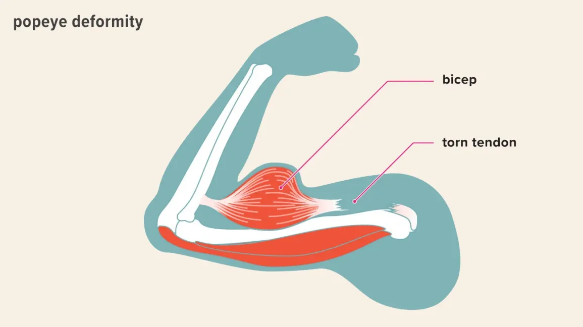
A 57-year-old man develops a painful, mobile mass in the anterior upper arm after lifting heavy boxes. He has chronic shoulder pain, pain with shoulder flexion, and painful elbow flexion, but normal elbow extension and intact distal neurovascular function. What is the diagnosis? and treatment?
Answer:
C) Biceps tendon rupture, most likely rupture of the proximal long head of the biceps tendon.
Key clues:
Sudden onset after heavy lifting
Older patient with chronic shoulder disease
Anterior upper-arm bulge = “Popeye” deformity
Pain or weakness with elbow flexion/supination
Normal elbow extension because the triceps is intact
Flashcard rule:
Heavy lifting + sudden upper-arm bulge in an older adult → proximal biceps tendon rupture.
Treatment:
Usually conservative management:
Rest and avoid painful lifting
Ice and NSAIDs/acetaminophen
Physical therapy to restore shoulder and elbow strength
A proximal long-head biceps rupture usually does not require surgical repair because the short head remains attached and function is largely preserved. Surgery, usually biceps tenodesis, is considered for persistent pain/cramping, substantial weakness, associated shoulder pathology, or high-demand patients.
Flashcard rule:
Proximal biceps rupture → conservative treatment.
Distal biceps rupture → surgical repair, especially in active patients, because it causes major supination weakness.
A 78-year-old woman with atrial fibrillation and a ventricular rate of 160/min has hypotension, severe hypoxemia, and pulmonary edema. What is the next step?
Answer:
F) Immediate synchronized electrical cardioversion
Why:
She has hemodynamically unstable atrial fibrillation:
BP 82/50 mm Hg
Acute pulmonary edema with crackles
Oxygen saturation 88% despite supplemental oxygen
Dizziness and respiratory distress
Unstable tachyarrhythmia requires immediate synchronized cardioversion rather than rate-control medication.
Why not medications?
Diltiazem, esmolol, and amiodarone take longer and may worsen hypotension. Albuterol does not address the cause—the wheezing is from pulmonary edema, or “cardiac asthma.”
Flashcard rule:
AF with hypotension, ischemia, altered mental status, shock, or pulmonary edema → synchronized cardioversion.
A 26-year-old woman develops sudden palpitations. Her pulse is 190/min and regular, blood pressure is 118/72 mm Hg, and she has no chest pain, confusion, or pulmonary edema. ECG shows a regular narrow-complex tachycardia with no clearly visible P waves. Vagal maneuvers do not terminate the rhythm. Diagnosis? What is the next step?
Stable SVT
Answer:
Rapid IV adenosine
Diagnosis:
Paroxysmal supraventricular tachycardia, usually AVNRT
Classic clues:
Sudden onset and termination
Regular rhythm
Narrow QRS
Hemodynamically stable
Vagal maneuvers unsuccessful
Stable, regular narrow-complex tachycardia is treated with vagal maneuvers followed by adenosine. Unstable tachycardia requires synchronized cardioversion.
Rule:
Stable + regular + narrow = adenosine
Do not choose adenosine for an irregularly irregular rhythm such as atrial fibrillation.
A 68-year-old man with a prior myocardial infarction develops palpitations. His pulse is 160/min and regular, and his blood pressure is 125/75 mm Hg. He is alert and has no chest pain or pulmonary edema. ECG shows a regular wide-complex tachycardia with AV dissociation. Diagnosis? What is the next step?
Answer:
IV amiodarone
Diagnosis:
Stable monomorphic ventricular tachycardia with a pulse
Classic clues:
Wide-complex, regular tachycardia
Prior MI or structural heart disease
Still has a pulse
No hypotension, shock, ischemia, or heart failure
Amiodarone is an antiarrhythmic option for stable wide-complex ventricular tachycardia. If the patient becomes unstable, perform synchronized cardioversion instead.
Rule:
Stable VT with a pulse = antiarrhythmic such as amiodarone
Unstable VT with a pulse = synchronized cardioversion
A hospitalized patient becomes unresponsive and pulseless. The monitor shows ventricular fibrillation. CPR is initiated, and the patient receives repeated defibrillation and epinephrine, but ventricular fibrillation persists. What medication should be administered next?
Answer:
IV/IO amiodarone
Diagnosis:
Refractory ventricular fibrillation or pulseless ventricular tachycardia
Amiodarone or lidocaine is given for VF/pulseless VT that persists despite CPR, repeated defibrillation, and epinephrine.
Rule:
Pulseless VF/VT = defibrillate first; amiodarone comes later.
A 70-year-old man develops dizziness and hypotension after an inferior myocardial infarction. His pulse is 30/min, and ECG shows complete heart block. IV atropine does not improve his heart rate. What is the next step?
Answer:
Transcutaneous cardiac pacing
Classic clues:
Severe bradycardia
Hypotension, confusion, shock, chest pain, or heart failure
Atropine unsuccessful
Immediate stabilization is needed
For symptomatic bradycardia with cardiopulmonary compromise, pacing is appropriate when atropine is ineffective; transvenous pacing may follow.
Rule:
Unstable bradycardia → atropine → transcutaneous pacing if unsuccessful
A 72-year-old man has recurrent syncope. ECG shows regular P waves and regular QRS complexes, but the atrial and ventricular rhythms have no relationship to one another. No reversible medication, electrolyte, or ischemic cause is identified. What is the definitive treatment?
Answer:
Permanent pacemaker placement
Diagnosis:
Third-degree AV block
Also choose a permanent pacemaker for Mobitz type II second-degree AV block, high-grade AV block, or complete heart block when there is no reversible cause—even when symptoms are minimal.
Rule:
Mobitz I: often observe unless symptomatic
Mobitz II: permanent pacemaker
Third-degree block: permanent pacemaker
Symptomatic sick sinus syndrome: permanent pacemaker
Fast distinction
Stable regular narrow tachycardia: adenosine
Stable wide-complex VT with pulse: amiodarone
Unstable tachycardia: synchronized cardioversion
Pulseless VF/VT: defibrillation, then amiodarone later
Unstable bradycardia unresponsive to atropine: temporary pacing
Mobitz II or complete heart block: permanent pacemaker
A 72-year-old woman with long-standing poorly controlled hypertension develops progressive exertional dyspnea, bibasilar crackles, and an S₃. Echocardiography shows normal valves and an ejection fraction of 58%. What is the most likely diagnosis?
Answer:
B) Diastolic dysfunction — heart failure with preserved ejection fraction (HFpEF)
Why:
Chronic hypertension causes left ventricular hypertrophy and stiffening, so the ventricle cannot relax and fill normally. This raises left-sided filling pressures, causing pulmonary congestion despite a preserved ejection fraction. HFpEF is defined by heart-failure symptoms with an LVEF of at least 50%.
Key clues:
Older woman with chronic hypertension
Exertional dyspnea and crackles
Preserved EF: 58%
No JVD or peripheral edema → predominantly left-sided failure
Flashcard rule:
Heart-failure symptoms + EF ≥50% + long-standing hypertension → diastolic dysfunction/HFpEF.
- HFpEF describes the ejection fraction: EF is preserved, usually ≥50%.
High-output heart failure describes the total cardiac output: the heart is pumping an abnormally large amount of blood but still cannot meet the body’s demand.
A 37-year-old woman has progressive dyspnea, blood-tinged sputum, JVD, a loud S₁, a high-pitched extra sound just after S₂, and a low-pitched diastolic murmur at the apex radiating to the axillary line. What is the diagnosis?
Answer:
F) Mitral stenosis
Key clues:
Loud S₁
High-pitched sound after S₂ = opening snap
Low-pitched diastolic rumble at the apex
Hemoptysis from increased pulmonary venous pressure
JVD suggests advanced pulmonary hypertension causing right-sided strain.
Step 2 takeaway:
Loud S₁ + opening snap + apical diastolic rumble = mitral stenosis.
The radiation toward the axilla can distract you toward mitral regurgitation, but mitral regurgitation is holosystolic, not diastolic.
A 57-year-old man develops unilateral leg swelling 48 hours after prostatectomy. On low molecular weight heparin and received pneumatic compassion stockings during surgery. Duplex ultrasound shows occlusion of the popliteal and superficial femoral veins. What is the next step?
Answer:
D) Therapeutic heparin
Diagnosis:
Acute proximal deep venous thrombosis
Why:
Popliteal and femoral vein thrombosis = proximal DVT
Prophylactic LMWH is no longer enough once a DVT is confirmed
Begin full-dose anticoagulation to prevent clot extension and pulmonary embolism
Heparin works immediately; warfarin does not
Why not tPA or thrombectomy?
He is stable with normal pulses and no limb-threatening ischemia. Thrombolysis also carries a high bleeding risk shortly after surgery.
Flashcard rule:
Confirmed proximal DVT → therapeutic anticoagulation with heparin.
The “superficial femoral vein” is still part of the deep venous system.
Yes—he still needs therapeutic heparin.
Why despite already receiving LMWH?
The LMWH was a low prophylactic dose intended to reduce the chance of DVT after surgery. It does not guarantee prevention. Now that ultrasound confirms a proximal DVT, he must be escalated to full-dose therapeutic anticoagulation to prevent clot extension and pulmonary embolism. Most proximal DVTs are treated with anticoagulation alone.
Because he recently had surgery, IV unfractionated heparin may be preferred in this older-style question because it has a short duration and can be rapidly stopped or reversed if postoperative bleeding occurs.
Why do normal pulses not change the answer?
Pulses evaluate arterial blood flow:
DVT = blocked vein → swelling, but pulses usually remain normal.
Arterial occlusion = blocked artery → absent pulses, pallor, coolness, severe pain.
So, normal pulses confirm that the limb still has arterial perfusion; they do not rule out a DVT.
Flashcard rule:
DVT while on prophylactic LMWH → switch to therapeutic-dose anticoagulation. Normal pulses are expected because DVT is venous, not arterial.
A 57-year-old man with non–small cell lung cancer diagnosed 1 year ago develops fever, worsening dyspnea, and cough. He previously received chemotherapy and radiation, but his last treatment was 2 months ago. Chest x-ray now shows an increased right perihilar mass and a new right middle-lobe infiltrate. His WBC count is elevated with neutrophilia. What is the most likely cause of his pneumonia?
Answer:
Airway obstruction secondary to carcinoma → postobstructive pneumonia
Why:
Chemotherapy and radiation do not automatically mean the cancer is gone. The phrase “increased perihilar mass” means the tumor is still present and has likely progressed.
Central lung tumor → bronchial obstruction → secretions become trapped distal to the blockage → bacterial growth → pneumonia in that lung segment.
Why not chemotherapy-related immunosuppression?
Treatment ended 2 months ago
He is not neutropenic
He has neutrophilic leukocytosis
The infiltrate is near/distal to the enlarging mass
Flashcard rule:
Lung cancer treatment history does not equal remission. Enlarging central mass + localized infiltrate → postobstructive pneumonia.
An 82-year-old woman has recurrent episodes of cough producing copious green sputum for 5 years. She had repeated episodes of pneumonia during childhood. She does not smoke. Examination shows digital clubbing, and chest x-ray shows bilateral basilar linear scarring. What is the most likely diagnosis? Treatment?
Answer:
C) Bronchiectasis
Treatment:
Bronchiectasis = clear the secretions with chest physiotherapy; treat infectious exacerbations with antibiotics.
In this patient with recurrent copious green sputum, the best Step 2 answer would usually be airway clearance plus antibiotics for an acute bacterial exacerbation.
Why:
Repeated lung infections can permanently damage and dilate the bronchi. The abnormal airways cannot clear mucus effectively, causing a cycle of retained secretions → recurrent infections → additional airway damage. Classic findings are chronic cough, large amounts of purulent sputum, recurrent respiratory infections, and clubbing.
Why not A) α₁-antitrypsin deficiency?
α₁-antitrypsin deficiency primarily causes early-onset emphysema, especially in the lower lungs. Expect progressive dyspnea, obstructive lung disease, hyperinflation, and possibly liver disease—not copious purulent sputum as the major presentation.
Why not D) Cystic fibrosis?
CF can cause recurrent infections and bronchiectasis, but it generally presents much earlier and often includes other clues:
Chronic sinus disease
Pancreatic insufficiency or malabsorption
Poor growth
Male infertility
Lifelong respiratory symptoms
An 82-year-old with no gastrointestinal or lifelong systemic manifestations is far more likely to have acquired bronchiectasis from previous infections.
Why not I) Sarcoidosis?
Sarcoidosis more commonly causes a dry cough, dyspnea, bilateral hilar lymphadenopathy, and systemic manifestations. It does not typically cause recurrent episodes of copious green sputum.
Flashcard rule:
Recurrent infections + copious purulent sputum + clubbing + scarring → bronchiectasis.
The recurrent infections should make you think bronchiectasis first; CF is only one possible underlying cause, usually with an earlier and multisystem presentation.
A 57-year-old woman has 6 months of progressive exertional dyspnea, bibasilar crackles, interstitial markings on chest x-ray, decreased diffusion capacity, decreased FVC, and an increased FEV₁/FVC ratio. What is the next diagnostic step?
Answer:
D) High-resolution CT scan of the chest
Most likely process:
Interstitial lung disease, possibly idiopathic pulmonary fibrosis.
Why:
Her PFTs show a restrictive pattern:
↓ FVC
Normal or ↑ FEV₁/FVC
↓ DLCO due to impaired gas transfer
High-resolution CT best characterizes the interstitial disease and looks for patterns such as basilar, subpleural reticulation and honeycombing, which suggest usual interstitial pneumonia.
Why not rheumatoid factor?
She has no joint or systemic findings suggesting rheumatoid arthritis. Autoimmune testing can help identify a cause of ILD, but it does not define the lung pattern as well as HRCT.
rule:
Progressive dyspnea + dry crackles + restrictive PFTs + interstitial markings → high-resolution chest CT.
Because “interstitial lung disease” is a category, not one diagnosis. HRCT helps distinguish whether the process is mainly inflammatory or fibrotic, and the treatments are very different.
Examples
UIP/IPF pattern: basal, peripheral reticulation, traction bronchiectasis, and honeycombing
→ treat with antifibrotic therapy such as nintedanib or pirfenidone, which slows further loss of lung function.Autoimmune-associated ILD/NSIP pattern: more ground-glass opacity and less honeycombing
→ evaluate for diseases such as rheumatoid arthritis or systemic sclerosis; treatment may include mycophenolate, rituximab, azathioprine, or corticosteroids, depending on the underlying disease.Hypersensitivity pneumonitis pattern: mosaic attenuation, air trapping, or centrilobular nodules
→ identify and remove the exposure, such as birds, mold, or occupational antigens; severe inflammatory disease may also receive corticosteroids.
HRCT can also show whether the pattern is diagnostic enough to avoid a lung biopsy or whether further testing—autoimmune labs, bronchoscopy, or biopsy—is necessary.
Step 2 takeaway:
Restrictive PFTs establish ILD; HRCT identifies the pattern and directs the cause-specific treatment.
A 57-year-old woman with severe obesity (BMI 57) has marked daytime sleepiness, nocturia, leg swelling, hypertension, resting hypoxemia, and P pulmonale on ECG. Chest x-ray is normal. What is the most likely cause of her nocturia?
Answer:
E) Obesity-hypoventilation syndrome, likely with coexisting obstructive sleep apnea.
Why:
Severe obesity causes nocturnal hypoventilation and upper-airway obstruction → hypoxemia and pulmonary hypertension → right-heart strain, explaining the P pulmonale and leg edema. Obesity-hypoventilation syndrome is associated with daytime hypoxemia/hypercapnia, sleepiness, pulmonary hypertension, and cor pulmonale.
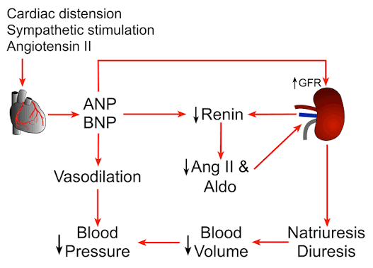
During obstructive episodes, strong inspiratory effort creates very negative intrathoracic pressure, which stretches the atria and increases atrial natriuretic peptide (ANP):
↑ ANP → sodium and water excretion → nocturia.
Flashcard rule:
Severe obesity + daytime sleepiness + hypoxemia + P pulmonale/edema + nocturia → obesity-hypoventilation/OSA. Nocturia results from increased ANP.
A previously healthy 23-year-old woman has 4 months of intermittent loose stools and abdominal cramping that improves after defecation. She has no fever, bleeding, weight loss, abnormal examination findings, or laboratory abnormalities. What mechanism is responsible?dd
Answer:
D) Smooth muscle hypersensitivity
Diagnosis:
Irritable bowel syndrome with diarrhea (IBS-D)
The answer choice is describing visceral hypersensitivity with abnormal intestinal motility. The bowel is structurally normal but reacts excessively to normal stretching and contractions, causing cramping and altered stool frequency. Abdominal pain related to defecation with diarrhea and no alarm findings is characteristic of IBS.
Why not the others?
Bacterial overgrowth: usually occurs with bowel surgery, anatomic abnormalities, or motility disorders and may cause bloating, malabsorption, weight loss, or vitamin deficiencies.
Endomysial antibodies: suggest celiac disease; expect malabsorption clues such as weight loss, iron-deficiency anemia, nutrient deficiencies, or bulky greasy stools.
Medication adverse effect: ginger can cause GI upset, but the classic pain relieved by defecation with normal testing points more strongly to IBS.
Stimulant laxative use: no laxative use is reported, and there are no electrolyte abnormalities or evidence of surreptitious use.
Flashcard rule:
Chronic abdominal pain relieved by defecation + altered bowel habits + no alarm signs → IBS from visceral/smooth-muscle hypersensitivity.
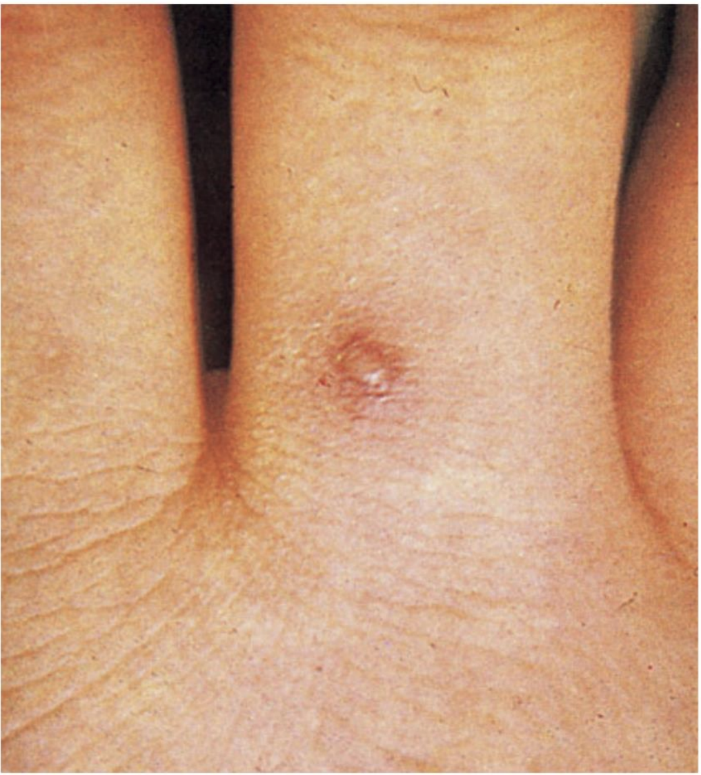
A 24-year-old woman develops migratory pain in multiple joints, a swollen knee, and several tender pustules on her extremities shortly after the onset of menstruation. Which medication is most effective?
Answer:
B) Ceftriaxone
Diagnosis:
Disseminated gonococcal infection (DGI) causing the arthritis-dermatitis syndrome.
Classic clues:
Migratory polyarthralgia
Tenosynovitis or joint swelling
Few pustular lesions on the distal extremities
Young, sexually active patient
Dissemination can occur around menstruation
Genital symptoms may be completely absent
Why ceftriaxone?
DGI results from hematogenous spread of Neisseria gonorrhoeae and requires parenteral ceftriaxone. Test for gonorrhea and chlamydia; add doxycycline if chlamydia has not been excluded.
Why not tetracycline?
Tetracyclines can treat chlamydia but are not adequate treatment for disseminated gonorrhea.
Flashcard rule:
Migratory joint pain + tenosynovitis/swollen joint + scattered pustules → disseminated gonorrhea → ceftriaxone.
A 62-year-old man has had 1 year of progressive difficulty initiating urination and terminal dribbling. He now has painful pelvic pressure and cannot urinate. Laboratory studies show elevated creatinine, hyperkalemia, glucose of 230, and low bicarbonate. What is the most likely cause of his kidney injury?
Answer:
Bladder outlet obstruction from benign prostatic hyperplasia → postrenal AKI
Key clues:
Older man
Hesitancy and terminal dribbling
Acute painful urinary retention
Hyperkalemia and metabolic acidosis
Urine backs up and reduces filtration
Next step:
Immediate bladder decompression with a Foley catheter.
Rule:
Cannot get urine out = postrenal.
With obstruction:
The kidneys excrete less H⁺ and ammonium.
Acid accumulates in the blood.
Bicarbonate binds and buffers that acid.
Therefore, the measured serum bicarbonate decreases.
His hyperkalemia also worsens this because elevated potassium suppresses renal ammonia production, further limiting acid excretion. Obstructive nephropathy can therefore produce a hyperkalemic, often normal-anion-gap metabolic acidosis, similar to type 4 renal tubular acidosis.
A 70-year-old woman has had vomiting and diarrhea for 3 days. She is dizzy when standing and has dry mucous membranes, tachycardia, and decreased urine output. Laboratory studies show a BUN-to-creatinine ratio greater than 20, low urine sodium, and hyaline casts. What is the cause of her kidney injury?
Answer:
Volume depletion causing decreased renal perfusion → prerenal AKI
Key clues:
Vomiting, diarrhea, bleeding, or poor intake
Hypotension or orthostatic symptoms
Kidney tissue initially remains intact
Kidney conserves sodium and water
Concentrated urine and hyaline casts
Treatment:
Restore circulating volume with IV isotonic fluids.
Rule:
Not enough blood arriving at the kidney = prerenal.
Common causes:
Dehydration from vomiting or diarrhea
Hemorrhage
Sepsis or severe hypotension
Heart failure
Cirrhosis
Because the kidneys sense low blood flow, they try to conserve sodium and water, producing concentrated urine.
A 45-year-old woman develops fever, a diffuse rash, and increased creatinine 2 weeks after beginning an antibiotic. Urinalysis shows sterile pyuria and WBC casts. What is the most likely diagnosis?
Answer:
Drug-induced acute interstitial nephritis → intrinsic renal AKI
Mechanism:
A medication-triggered immune reaction causes inflammation of the renal interstitium and tubules.
Key clues:
New medication, especially an antibiotic, NSAID, or proton-pump inhibitor
Fever, rash, or eosinophilia
Sterile pyuria
WBC casts
Increased creatinine
The full fever-rash-eosinophilia triad is often absent, so the medication timeline and urine findings are especially important.
Treatment:
Stop the causative medication.
most commonly triggered by medications such as:
Antibiotics
NSAIDs
Proton-pump inhibitors
Possible findings:
Fever
Rash
Eosinophilia
Sterile pyuria
WBC casts
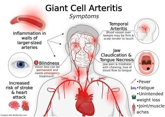
A 67-year-old woman has new severe headaches, temporal tenderness, fever, weight loss, and aching of the shoulders and neck. ESR is markedly elevated. She develops sudden, painless monocular vision loss with pallor and edema of the optic disc. What is the underlying mechanism?
Answer:
C) Arterial inflammation
Diagnosis:
Giant cell arteritis associated with polymyalgia rheumatica
Mechanism
Granulomatous inflammation affects medium and large arteries, including branches of the carotid artery. Inflammation narrows or occludes the posterior ciliary arteries, reducing blood flow to the optic nerve and causing arteritic anterior ischemic optic neuropathy with sudden permanent vision loss.
Key clues
Age >50
New headache and tender temporal area
Fever, fatigue, weight loss
Shoulder and neck aching → polymyalgia rheumatica
Very high ESR
Sudden monocular vision loss
Why not arterial embolization?
An embolus can cause sudden monocular blindness, but it would not explain the preceding weeks of temporal headaches, constitutional symptoms, shoulder aching, temporal tenderness, and markedly elevated ESR.
Immediate management
Give high-dose glucocorticoids immediately—with visual loss, typically IV methylprednisolone—before waiting for temporal artery biopsy. Treatment is urgent to prevent blindness in the other eye.
Flashcard rule:
Older patient + headache + temporal tenderness + PMR symptoms + high ESR + vision loss → giant cell arteritis from arterial inflammation.
A 78-year-old woman with chronic leukemia and significant weight loss develops acute confusion, lateral nystagmus, an abnormal Romberg test, and an ataxic gait. Finger-to-nose testing is normal. Supplementation with which vitamin would most likely have prevented her condition?
Answer:
B) Vitamin B₁ (thiamine)
Diagnosis:
Wernicke encephalopathy due to malnutrition
Classic triad
Confusion
Eye abnormalities: nystagmus or ophthalmoplegia
Gait ataxia
She does not drink alcohol, but malignancy and substantial weight loss can cause severe malnutrition and thiamine deficiency.
Why is finger-to-nose normal?
Her main problem is gait and balance, not limb coordination. Wernicke encephalopathy commonly causes truncal/gait ataxia, so a patient may walk unsteadily while still completing finger-to-nose testing normally.
Why not vitamin B₁₂?
Vitamin B₁₂ deficiency more commonly causes:
Paresthesias
Loss of vibration and proprioception
Sensory ataxia
Weakness or hyperreflexia
Macrocytic anemia
It does not classically cause the combination of acute confusion + nystagmus + gait ataxia.
Step 2 rule:
Malnutrition or alcoholism + confusion + nystagmus + ataxia → Wernicke encephalopathy → give thiamine, ideally before glucose.
A 38-year-old man with nephrotic syndrome due to minimal change disease becomes weak and oliguric while receiving sodium restriction and furosemide. His blood pressure drops from 110/70 supine to 90/60 sitting, and he no longer has edema. Urinalysis shows heavy proteinuria but no hematuria. What is the most likely cause?
Answer:
D) Intravascular volume depletion causing prerenal AKI
Why?
Nephrotic syndrome causes albumin loss → reduced plasma oncotic pressure → fluid leaves the blood vessels and enters the tissues. Furosemide and sodium restriction then remove additional fluid.
He has been overdiuresed:
Orthostatic fall in blood pressure
Weakness
Oliguria
No remaining edema
Less circulating blood reaches the kidneys → decreased GFR → oliguria. Diuretics and nephrotic syndrome are both recognized causes of reduced effective circulating volume and prerenal kidney injury.
Why not the others?
Acute interstitial nephritis: Furosemide can cause it, and WBCs are a distractor, but you would expect stronger inflammatory clues such as fever, rash, eosinophilia, or WBC casts. His orthostatic hypotension strongly indicates hypovolemia.
Acute ischemic renal failure/ATN: This can develop if hypoperfusion persists, but the current presentation is early prerenal underperfusion.
Renal vein thrombosis: Nephrotic syndrome increases clot risk, but renal vein thrombosis more often causes flank pain, hematuria, and acute kidney dysfunction.
Worsening nephrotic syndrome: Would generally cause worsening edema, not absent edema with orthostatic hypotension.
Flashcard rule:
Nephrotic syndrome + aggressive diuresis + orthostatic hypotension + oliguria → intravascular volume depletion → prerenal AKI.
A 72-year-old woman has 8 hours of crushing chest pain radiating to her neck and arms. She is tachycardic and hypotensive with elevated JVP, bilateral crackles, an S₃ gallop, hypoxemia, and peripheral edema. What is the most likely diagnosis?
Answer:
D) Cardiogenic shock due to acute myocardial infarction
Why?
Her acute MI has caused severe left ventricular pump failure:
↓ cardiac output → hypotension, tachycardia, dizziness
Blood backs up → elevated JVP, pulmonary edema/crackles, S₃, and leg edema
Cardiogenic shock after MI is characterized by hypotension and inadequate cardiac output with pulmonary and systemic venous congestion.
Why not papillary muscle rupture?
Papillary muscle rupture causes acute severe mitral regurgitation, usually with a new systolic murmur and rapid pulmonary edema. This vignette gives no murmur and instead emphasizes generalized pump failure with an S₃.
Why not the others?
Cardiac tamponade: hypotension and elevated JVP, but usually clear lungs
Hypovolemia: hypotension with low JVP and clear lungs
Acute aortic insufficiency: diastolic murmur, often associated with acute aortic pathology
Papillary muscle rupture: acute MR with a new systolic murmur
Flashcard rule:
Acute MI + hypotension + elevated JVP + crackles/S₃ = cardiogenic shock.
A 67-year-old man has fever, dysuria, lethargy, tachycardia, tachypnea, and hypotension. Urinalysis shows numerous leukocytes and gram-negative bacilli. He also has bandemia and thrombocytopenia. Blood cultures are ordered, and IV saline is started. What should be given next?
Diagnosis:
Septic shock from a gram-negative urinary tract infection/urosepsis
Key clues
Infection source: dysuria + pyuria + gram-negative bacilli
Systemic infection: fever and marked bandemia
Organ dysfunction: confusion/somnolence
Shock: BP 88/60 mm Hg
Low platelets may reflect sepsis-associated consumption/DIC
Give empiric broad-spectrum IV antibiotics immediately after cultures are collected, as long as obtaining cultures does not delay treatment. Septic shock requires antimicrobial therapy ideally within 1 hour.
Why not platelets?
A platelet count of 70,000/mm³ without active bleeding is not, by itself, an indication for platelet transfusion. The thrombocytopenia is a complication of the sepsis; treating the infection is the priority.
Why not the others?
Methylprednisolone: steroids are not first-line; corticosteroids may be considered only if shock remains refractory despite adequate fluids and vasopressors.
Endotoxin antibody: not standard treatment.
Whole blood: no major hemorrhage or severe anemia.
Flashcard rule:
Suspected infection + hypotension + altered mental status → septic shock → IV fluids and immediate broad-spectrum IV antibiotics.
What routine vaccines should be reviewed before military deployment
MMR, varicella, Tdap, influenza, polio, hepatitis A, and typhoid. Additional vaccines depend on the destination and exposure risk.
Which vaccines may be added for specific military deployments?
Yellow fever, anthrax, smallpox
Which deployed personnel especially need hepatitis B vaccination?
Healthcare personnel or medics with potential blood exposure. Also ensure vaccination in anyone with sexual exposure risk.
What should be done after close exposure to invasive meningococcal disease?
Give prompt chemoprophylaxis with ceftriaxone, rifampin, or ciprofloxacin
Trap: Ceftriaxone treats suspected meningococcal meningitis; ciprofloxacin and rifampin are primarily remembered as prophylaxis options.
A service member returns from Egypt with painless hematuria. Diagnosis?
Schistosoma haematobium infection. Eggs lodge in the urinary tract and cause dysuria or hematuria
A returning traveler has portal hypertension after freshwater exposure in an endemic region. Diagnosis?
Schistosoma mansoni infection causing periportal fibrosis and portal hypertension.
How is schistosomiasis exposure usually acquired?
Skin contact with contaminated freshwater containing cercariae released by snails.
Prevention: Avoid swimming or wading in freshwater in endemic regions (Africa/middle East, South America and the Caribbean, or Asia).
A sailor or service member exposed to contaminated water develops fever, conjunctival suffusion, and jaundice. Diagnosis?
Leptospirosis.
What is Weil disease?
Severe leptospirosis causing jaundice, acute kidney injury, and hemorrhagic manifestations.
What is the treatment for leptospirosis?
Mild disease: Usually doxycycline.
Severe disease: IV penicillin, ceftriaxone, or cefotaxime.
Doxycycline prophylaxis may be considered for short-term, very high-risk exposure but is not routine for every exposure.
nonpasteurized milk or livestock exposure followed by undulating fever and drenching sweats suggests what? and what is the treatment?
Brucellosis
Doxycycline plus rifampin, generally for a prolonged course to reduce relapse.

Exposure to cattle or sheep giving birth followed by fever, pneumonia, hepatitis, or culture-negative endocarditis suggests what?
Q fever caused by Coxiella burnetii.
Key distinction: Pneumonia and culture-negative endocarditis strongly favor Q fever over brucellosis.
Treatment: Doxycycline for symptomatic acute infection.
Rodent exposure followed by fever, myalgias, and rapidly developing noncardiogenic pulmonary edema suggests what?
Hantavirus pulmonary syndrome, classically associated with deer-mouse exposure.
Cleaning a barn, cabin, or enclosed area contaminated by aerosolized rodent urine or feces.
Hemoptysis and a widened mediastinum after possible spore exposure suggest what? Treatment?
Inhalational anthrax causing hemorrhagic mediastinitis.
important virulence factors of Bacillus anthracis?
Poly-D-glutamate capsule
Protective antigen
Edema factor
Lethal factor
How is severe systemic or inhalational anthrax treated?
Hospitalization with combination IV antimicrobial therapy, often including a fluoroquinolone, plus consideration of anthrax antitoxin.
Trap: Oral doxycycline alone is not adequate treatment for severe systemic anthrax.
Which medications may be used for malaria prophylaxis?
Atovaquone-proguanil, doxycycline, mefloquine, chloroquine in sensitive regions, primaquine, or tafenoquine.
Which malaria prophylaxis medication should be avoided in patients with major psychiatric disorders?
Mefloquine because of potentially serious neuropsychiatric effects.
What must be checked before primaquine or tafenoquine?
A quantitative G6PD level because these medications can cause severe hemolysis in G6PD deficiency (will mainly be present in males).
Which malaria species form dormant hepatic hypnozoites?
Plasmodium vivax and P ovale.
Which malaria species causes quartan fever approximately every 72 hours (3 days)?
Plasmodium malariae.
Which malaria species causes the most severe disease?
Plasmodium falciparum, which may cause cerebral malaria, severe anemia, pulmonary edema, kidney failure, shock, or death.
What other postinfectious complication is associated with Campylobacter?
Reactive arthritis, especially in genetically susceptible patients
Bloody inflammatory diarrhea after poultry, egg, reptile, or turtle exposure suggests what? treatment?
Nontyphoidal Salmonella.
Supportive
Fever, weight loss, pancytopenia, massive hepatosplenomegaly, and darkened skin after tropical travel suggest what?
Visceral leishmaniasis, commonly caused by Leishmania donovani and transmitted by sandflies.
What might be seen on tissue biopsy in visceral leishmaniasis? Treatment?
Back: Intracellular amastigotes within macrophages.
Treatment: Liposomal amphotericin B is commonly used.
What medications treat Chagas disease?
Benznidazole or nifurtimox
A returning traveler develops fever, headache, myalgias, meningitis, flaccid paralysis, or encephalitis after mosquito exposure. Diagnosis?
What is the CSF findings
West Nile virus
Lymphocytic pleocytosis, elevated protein, and usually normal glucose.
Very high fever, severe myalgias or “break-bone pain,” thrombocytopenia, bleeding, and capillary leak suggest what?
What test is associated with it and what do you see?
Treatment?
Severe dengue.
Test: positive tourniquet test
Petechiae appear below an inflated blood-pressure cuff due to capillary fragility.
Treatment: Careful supportive fluid management and acetaminophen. Avoid aspirin and NSAIDs until dengue is excluded because of bleeding risk.
A peacekeeper develops fever, petechiae, bleeding, vomiting, or diarrhea after contact with sick people, dead bodies, or wild animals in Africa. What should be suspected?
How is it transmitted?
Treatment?
Ebola or Marburg virus disease. Immediately isolate the patient and contact public-health authorities.
Direct contact with infected blood, body fluids, contaminated objects, or infected animals—not routine airborne transmission.
Intensive supportive care is essential. Inmazeb and Ebanga are approved monoclonal-antibody treatments for Ebola disease caused by Zaire ebolavirus. There is currently no approved specific therapy for Marburg virus disease.
Fever and severe joint pain resembling dengue but without prominent hemorrhage suggest what?
Chikungunya. Joint pain may be severe and prolonged.
Which congenital findings are classically associated with Zika infection during pregnancy?
Microcephaly and intracranial calcifications, along with other neurologic and ocular abnormalities.
Fever followed by jaundice, hepatic injury, and possible hemorrhage after travel to an endemic region suggests what?
Yellow fever. Treatment is supportive; prevention is through vaccination and mosquito precautions.
A 7-year-old boy has worsening daytime urinary accidents and bed-wetting four nights per week. He also passes occasional hard, painful stools. He has no dysuria, abnormal examination findings, or urinalysis abnormalities. What is the next step in management?
Answer:
A) Administer an osmotic laxative, such as polyethylene glycol.
Diagnosis:
Constipation-associated bladder dysfunction
Why constipation causes urinary incontinence
A stool-filled rectum can:
Press against the bladder and reduce its functional capacity
Trigger involuntary bladder contractions
Interfere with coordinated bladder emptying
This can cause both daytime accidents and nighttime bed-wetting. Constipation and daytime urinary symptoms should be treated before starting treatment directed specifically at nocturnal enuresis.
When the other answers would be right
B) Desmopressin
Choose this for monosymptomatic nocturnal enuresis:
Bed-wetting only
No daytime urinary symptoms
No untreated constipation
Normal urinalysis
It is especially useful when rapid, short-term dryness is desired, such as for sleepovers or camp, or when an alarm is impractical. Relapse is common after the medication is stopped.
Remember:
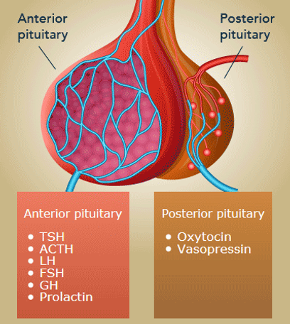
Desmopressin: synthetic ADH acting on renal V₂ receptors → increases water reabsorption and decreases nighttime urine production.
Desmopressin challenge:
Urine concentrates → central DI (not enough ADH in the posterior pituitary)
Urine stays dilute → nephrogenic DI
Urine concentrates with water deprivation alone → primary polydipsia
C) Bed-wetting alarm
Choose this for a motivated child and family with persistent monosymptomatic nocturnal enuresis, especially when long-term improvement is preferred.
First treat:
Constipation
Daytime urinary accidents
Urinary tract infection
Other underlying conditions
This child is not ready for an alarm yet because he has daytime wetting and constipation.
D) Renal ultrasonography
Choose imaging when there are clues to structural or neurologic disease, such as:
Recurrent febrile UTIs
Abnormal urinalysis
Continuous urine leakage
Weak or abnormal urinary stream
Abnormal genital or neurologic examination
Persistent symptoms despite appropriate initial treatment
This child has a normal examination and urinalysis with a clear reversible cause.
E) Reassurance that this is normal behavior
Reassurance may be sufficient for a younger child with occasional isolated nighttime wetting, no daytime accidents, and no concerning symptoms.
This patient is 7, has frequent nighttime wetting, worsening daytime incontinence, and painful hard stools, so the constipation needs treatment rather than reassurance alone.
Child with daytime and nighttime wetting + hard, painful stools → treat constipation first with an osmotic laxative.
A 35-year-old man with untreated HIV and a CD4 count of 170/mm³ develops progressive dyspnea, fever, and a nonproductive cough. Oxygen saturation is low, LDH is elevated, and chest imaging shows diffuse bilateral ground-glass opacities. What is the most likely organism? Treatment? When do we prophylaxis?
Answer:
Pneumocystis jirovecii
Treatment:
TMP-SMX; add glucocorticoids for significant hypoxemia.
Prophylaxis:
TMP-SMX when CD4 is below the indicated threshold.
Memory:
PCP = pulmonary disease below 200.
Current nuance: prophylaxis is indicated at CD4 <100 regardless of viral load, or at CD4 100–200 with detectable HIV RNA; people beginning ART who have CD4 <200 are also generally covered.
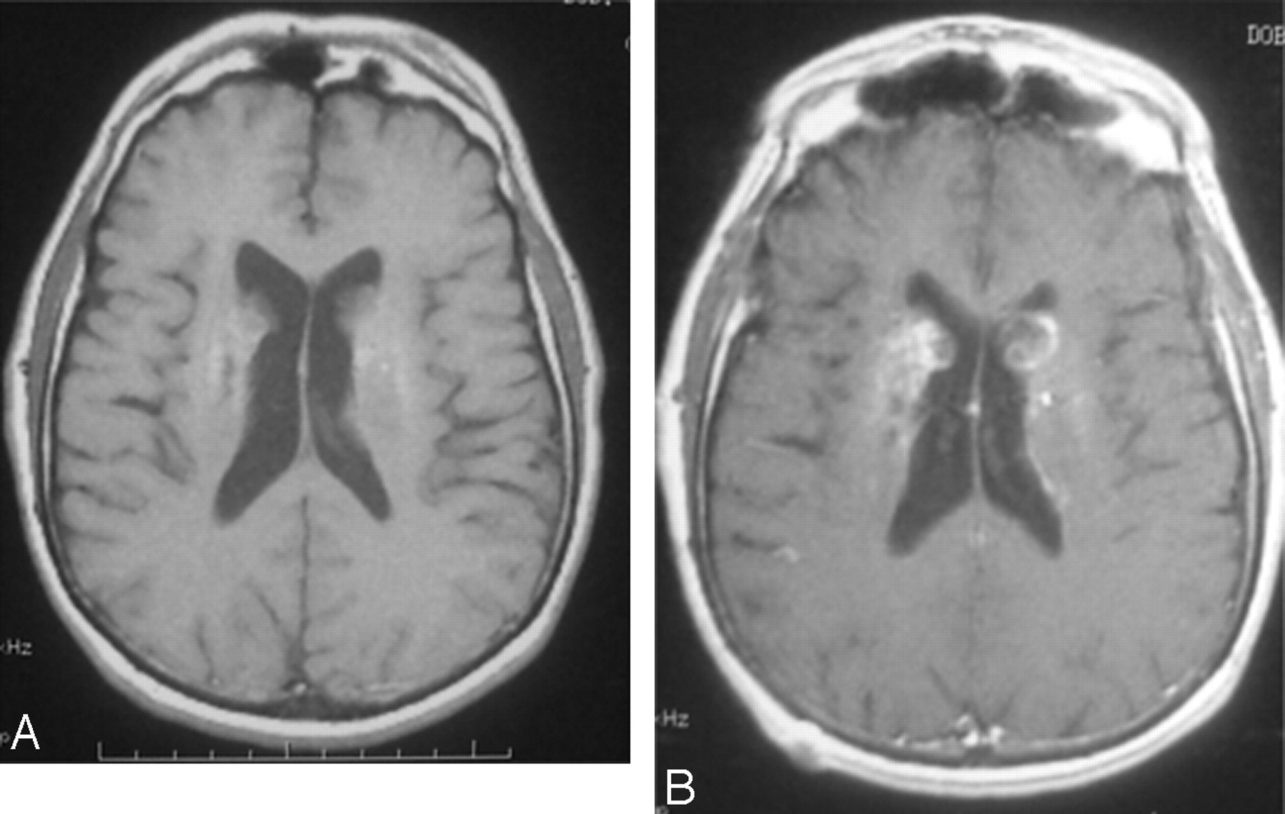
A woman with HIV, a CD4 count of 70/mm³, headache, confusion, and a new seizure has multiple ring-enhancing brain lesions with surrounding edema. Toxoplasma IgG is positive. What is the diagnosis? Treatment and Prophylaxis?
What will the CT show?
Answer:
Toxoplasma encephalitis
Treatment:
Pyrimethamine + sulfadiazine + leucovorin.
Prophylaxis:
TMP-SMX when CD4 is <100 and Toxoplasma IgG is positive.
Memory:
Multiple rings = Toxo.
A single dominant ring-enhancing lesion should raise concern for primary CNS lymphoma.
A patient with AIDS and a CD4 count of 60/mm³ has several weeks of headache, confusion, and blurred vision. Lumbar puncture shows markedly increased opening pressure, low glucose, elevated protein, and lymphocytes. What is the likely organism? Treatment?
Answer:
Cryptococcus neoformans
Key clue:
Subacute meningitis with markedly elevated intracranial pressure.
Treatment:
Amphotericin B + flucytosine initially, followed by fluconazole.
Memory:
Crypto creates pressure.
Routine fluconazole prophylaxis is not given to every asymptomatic person with HIV in the United States.
A patient with advanced HIV develops weeks of progressive weakness, visual-field loss, and cognitive changes. MRI shows multiple nonenhancing white-matter lesions without mass effect. There is no fever or headache. What is the cause? Treatment?
Answer:
JC virus causing progressive multifocal leukoencephalopathy
Treatment:
Start or optimize ART to restore immune function.
Memory:
PML = progressive deficits, multiple white-matter lesions, little inflammation or mass effect.
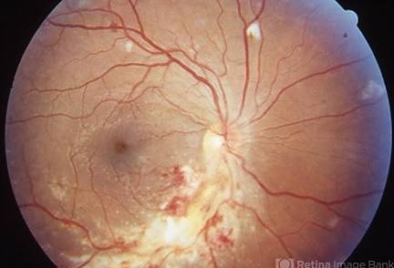
A patient with a CD4 count of 25/mm³ develops floaters, blurry vision, and painless visual-field loss. Fundoscopy shows retinal hemorrhages with fluffy white exudates. What is the diagnosis? Treatment?
Answer:
CMV retinitis
Classic appearance:
“Pizza-pie” or “cottage cheese and ketchup” retina.
Treatment:
Valganciclovir or IV ganciclovir, depending on severity.
Memory:
CMV = can’t see below 50.
A patient with untreated AIDS and a CD4 count of 30/mm³ has persistent fever, night sweats, weight loss, diarrhea, anemia, and markedly elevated alkaline phosphatase. Blood cultures grow acid-fast organisms. What is the diagnosis? Treatment?
Answer:
Disseminated Mycobacterium avium complex
Treatment:
A macrolide plus ethambutol.
Memory:
MAC attacks the whole body below 50.
Modern prophylaxis nuance: azithromycin prophylaxis is generally reserved for CD4 <50 when the patient is not receiving effective ART, remains viremic, or lacks a fully suppressive regimen. It is not routinely needed when effective ART is started immediately.
A patient with HIV and a CD4 count of 140/mm³ develops several weeks of profuse watery diarrhea, abdominal cramps, and weight loss. Stool testing shows acid-fast oocysts. What organisms should be considered?
Answer:
Cryptosporidium or Cystoisospora belli
Distinction:
Cryptosporidium: small acid-fast oocysts; treatment centers on ART and supportive care
Cystoisospora: larger oval acid-fast oocysts; responds to TMP-SMX
Memory:
Low CD4 + chronic watery diarrhea = protozoa.
Primary Cystoisospora prophylaxis with TMP-SMX is considered for CD4 <200 when living in or traveling to endemic areas.