The Muscular System
1/40
There's no tags or description
Looks like no tags are added yet.
Name | Mastery | Learn | Test | Matching | Spaced | Call with Kai |
|---|
No analytics yet
Send a link to your students to track their progress
41 Terms
Muscle
A tissue type that is able to contract/shorten for movement
Function of Muscle
- Producing movement
- Maintaining posture
- Stabilizing joints
- Generating heat
Properties of Muscles
- Able to contract (shorten)
- Extensible (able to be stretched)
- Elastic (able to return to original length after being stretched)
Different Types of Muscle
1. Smooth
2. Cardiac
3. Skeletal
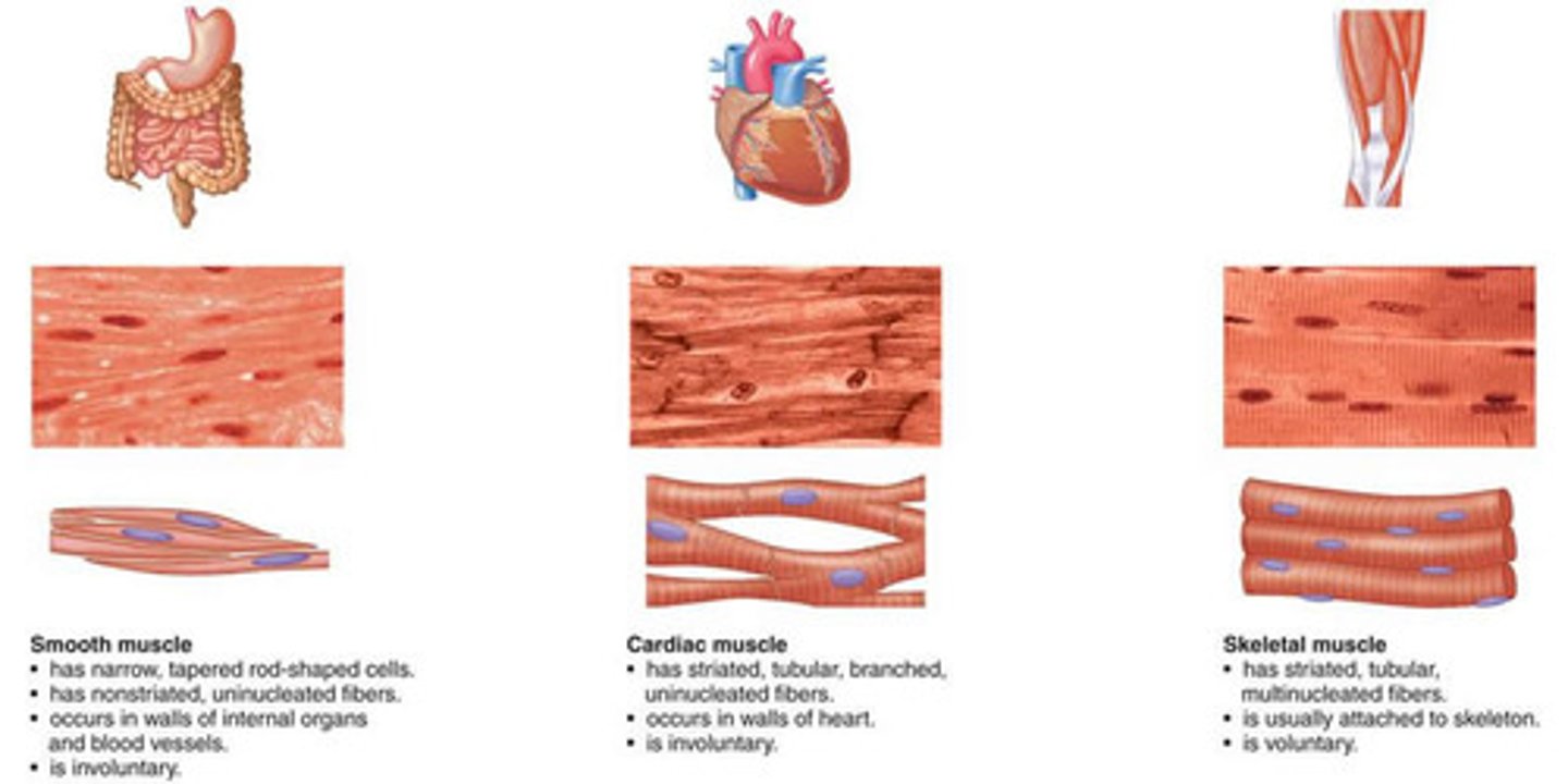
Skeletal Muscle
are muscles which are attached to the skeleton through tendons
- make up 40% of human body mass
- are voluntary and are mainly responsible for locomotion
- Contractions of skeletal muscle brings movement at joints
- Gives the body form & contours
- Maintain posture
- contains layers separated by dense connective tissue
• Allows for support and protection against the force of contraction for movement
• Supplies a pathway for blood vessels and nerves
• Amount increases with age - leads to decreased muscular strength when older
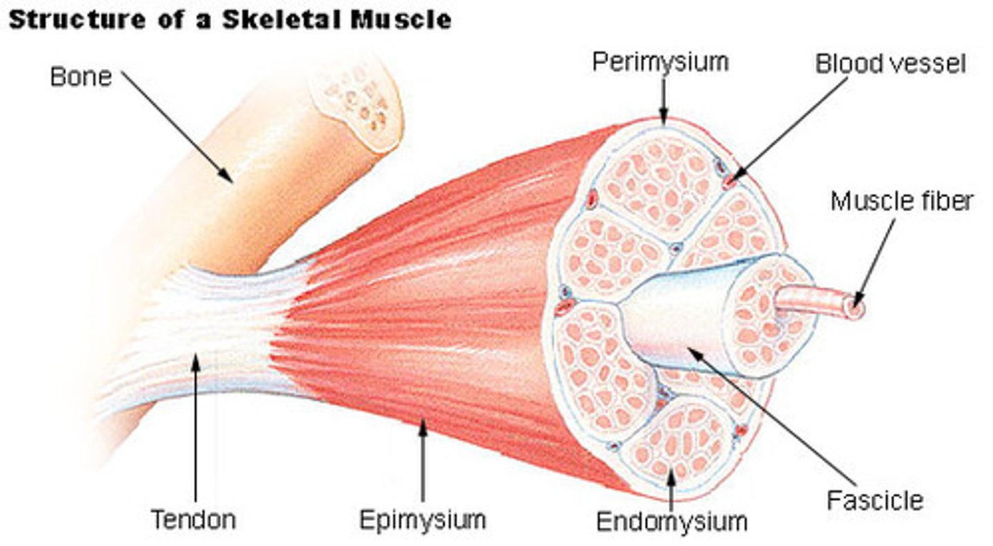
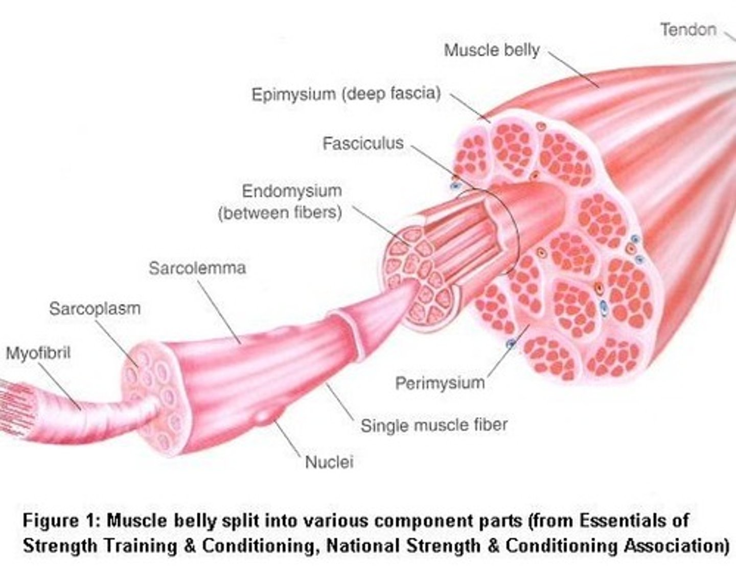
Structure of Skeletal Muscle
- Muscle Fibers (cells)
- Endomysium - Dense Connective tissue that covers individual fibers
- Fasciculi - muscle fibre bundles
• Contains between 10-100 muscle fibres
- Perimysium
• surrounds each fasciculi so it can act as an individual unit
- Epimysium holds the bundles together
• Towards the end of the bundle the epimysium tapers to form a tendon which attaches the muscle to a bone
Organelles in a Muscle Cell
1. Nucleus
2. Sarcosome
3. Sarcolemma
4. Sarcoplasm
Organelles in a Muscle Cell - Nucleus
- Multi-nucleated
• skeletal muscle is subjected to intense demands during exercise & other energy-dependent activates
• Multiple nuclei in each muscle cell allows for enhanced oxygen uptake and tissue repair
Organelles in a Muscle Cell - Sarcosome
a specialised mitochondria
- Generates energy during muscle contractions
Organelles in a Muscle Cell - Sarcolemma
- Is a thin plasma membrane around the cell
- Does normal cell membrane functioning
- is directly involved in synaptic transmission of action potential propagation and exciting-contraction coupling
Organelles in a Muscle Cell - Sarcoplasm
- Cytoplasm within the sarcolemma
- Stores and releases calcium ions
Muscle Movement
Muscles can only contract
- They pull bones together
- Muscles that move the skeleton come in pairs
Agonist
the muscle that causes the desired effect
- works with antagonist
Antagonist
the muscle that has the opposite effect
- works with agonist
Synergists
muscles that help the agonist indirectly by steadying a joint
- prevent unwanted movement
- assists the agonist muscle for a specific action at a joint
- works with fixator
Fixator
A muscle that contracts to immobilise a joint
- works with synergist
Terminology for Movement
- Origin
- Insertion
- Belly
Origin
the end of the muscle that is fixed to the stationary bone
- opposite insertion
Insertion
the attachment of the other end of the muscle to the movable bone
- opposite origin
Belly
the fleshy part of the muscle between the origin and insertion
Terminology for Movement - Elbow
The scapula and humerus are the stationary bones in this movement
- The radius and ulna are the bones which need to 'move'
- Biceps:
• Origin - Scapula/humerus
• Insertion - radius
- Triceps:
• Origin - Scapula/Humerus
• Insertion - Ulna
- Flexion:
• bicep is contracted (agonist)
• tricep is relaxed (antagonist)
- Extension:
• bicep is relaxed (antagonist)
• tricep is contracted (agonist)
Muscle Tone
Muscle tone is maintaining partial contraction of skeletal muscle to tighten a muscle but not produce movement
- Achieved by many different fibres taking turns to contract
- Muscle tone holds many of our body parts in position
• E.g. the head droops when we fall asleep due to loss of muscle tone
- Posture depends on muscle tone holding the body in a certain position
- Floppy baby syndrome, or hypotonia, is a way of describing a baby with low muscle tone
Arrangement of a Muscle
1. Muscle
2. Muscle Fibre Bundles
3. Myofibrils
4. Myofilaments
Arrangement of a Muscle - Muscle Fibre Bundles
- Muscle cells/fibres are held together in bundles known as fasciculi
- Each bundle or fasciculi contains between 10 - 100 muscle cells/fibres
- in each fibre are myofibrils
Arrangement of a Muscle - Myofibrils
Cylindrical organelles
- They lie parallel to each other and run the length of the fibre
- Is found in sarcoplasm in each fibre
- Surrounding the myofibril there is a network of tubules and channels called the sarcoplasmic reticulum where calcium is stored
• Calcium is release during contractions
- Each myofibril is made up many smaller units called myofilaments
Arrangement of a Muscle - Myofilaments
the actual units involved in contraction
- have symmetrical, alternating patterns of thick and thin elements
- The arrangement of thick and thin filaments gives a banded or striated appearance to the muscle
• striations allow myofibrils to be divided into functional repeating segments called sarcomeres
- When supplied with ATP and is activated by a nerve impulse the filaments slide past each other
• Shortens the myofibril
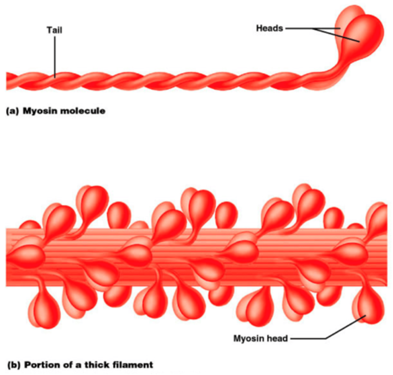
Thick Myofilaments
composed mainly of the protein myosin
- have a rod-like tail ending with two globular heads
Thin Myofilaments
composed mainly of the protein actin
- contain active sites where myosin binds to during contraction
- Also contains two protein filaments:
• tropomyosin - prevents the muscles from contracting at the wrong time
• Troponin - promotes muscle contraction

Sliding Filament Theory
The theory that suggests that actin and myosin slide over each other to shorten the myofibril
- causes muscle contractions
Sarcomere Structure During Contractions
- Movement of thick and thin filaments relative to one another causes the lengthening (relaxing) and shortening (contracting) of the sarcomere
- When the muscle is not contracting, thin strands of a protein molecule (tropomyosin) wrap around the actin filaments
• blocks the actin binding sites
• troponin are attached to the tropomyosin
- Nerve signals send signals to the muscle cell when the muscle needs to move
- Surrounding the myofibril there is a network of tubules and channels called the sarcoplasmic reticulum in which calcium is stored
Structure of the Sarcomere
1. Z-Disc/Line
2. A-Band
3. I-Band
4. M-Line
Structure of the Sarcomere - Z-Disc/Line
- Defines the boundaries of a sarcomere
• one sarcomere extends from one Z-disc to the next
- is composed of large protein dics and serve as an anchoring site for contractile proteins
- Z-lines are pulled closer together during the muscle contraction
Structure of the Sarcomere - M-Line
is the centre of a sarcomere
- contains proteins that function as anchoring sites for the myosin molecules
- provides elasticity to muscles due to the presence of elastic fibres
Structure of the Sarcomere - A-Band
Covers the length of the thick filament (Myosin) in a sarcomere
- The amount of actin found in the A-band depends on how much the sarcomere is contracted
- The size of the A-band remain constant during a muscle contraction
- At the ends of A-bands the filaments overlap
• H-zone is area of A-band that is not overlapped
• Is lighter because not overlapping
Structure of the Sarcomere - I-Band
The zone of a sarcomere that is not covered by Myosin molecules
- Is the distance between successive thick filaments
- Contains only thin filaments
- The amount of actin found in the I-band depends on how much the sarcomere is contracted
Steps in Sliding Filament Theory
1. Nerve Activation
2. Hydrolysis
3. Power Stroke
4. Detachment
5. Reset
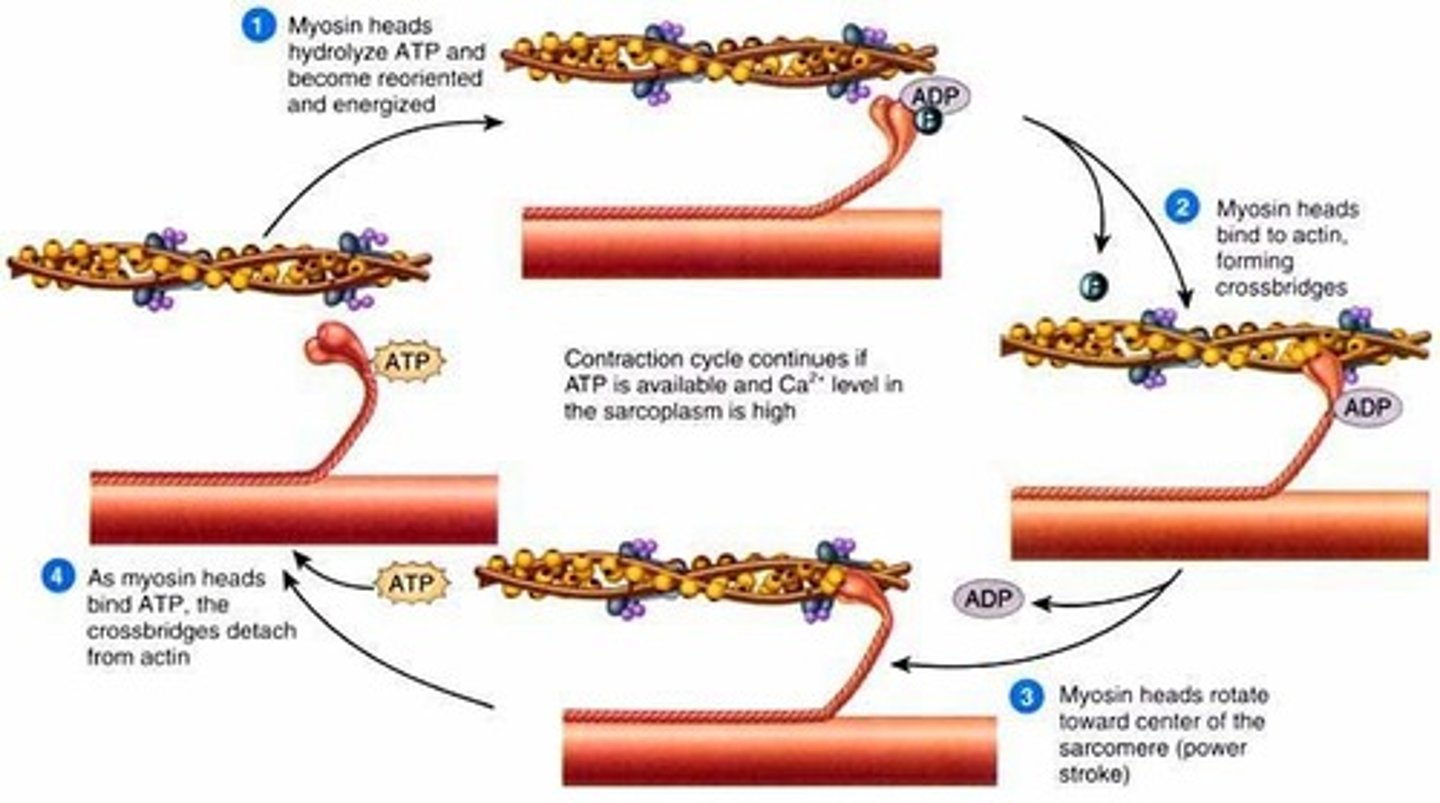
Steps in Sliding Filament Theory - Nerve Activation
When a nerve signal reaches the muscle cell calcium is released from sarcoplasmic reticulum
- Calcium causes a conformational change in the tropomyosin molecule
• shifts position to expose binding sites of the actin proteins
Steps in Sliding Filament Theory - Hydrolysis
ATP is hydrolyzed in the heads of molecules of myosin
- causes a change in the shape of the head and binds to actin filaments
- The myosin heads bind to the binding sites of the actin proteins
- forms a cross-bridge as the inorganic phosphate is released
Steps in Sliding Filament Theory - Power Stroke
ADP is released from ATP (releases energy)
- causes initiation of a power stroke
- Is where the thin filament gets pulled closer toward the midline of a sarcomere
Steps in Sliding Filament Theory - Detachment
A new ATP molecule binds to the myosin head causing the separation of the actin-myosin cross-bridge
- The ATP is hydrolysed to ADP and inorganic phosphate (hydrolysis)
- the energy transferred from the ATP to the myosin head causes it to "cock" back
Steps in Sliding Filament Theory - Reset
The myosin head leaves the binding site and returns to its original position
- another molecule of ATP is attached and the process repeats