EEG for non cranial/ARTERIAL procedures (CEA, AAA, CABG)
 Call Kai
 Learn
Learn Practice Test
 Spaced Repetition
 Match
Match Flashcards
 Knowt Play
1/67

There's no tags or description
Looks like no tags are added yet.
Name | Mastery | Learn | Test | Matching | Spaced | Call with Kai | Chat |
|---|
No analytics yet
Send a link to your students to track their progress
68 Terms
What is a carotid endarerectomy or CEA
a surgery performed to restore normal blood flow to the brain - specifically by removing plaque buildup in the carotid artery in a patient’s neck and removing constriction to blood flow in this vessel. A CEA is performed to prevent future strokes.
The right common carotid artery originates from the _____ artery. The left common carotid artery originates from the ____ arch.
brachiocephalic, aortic
What are the vessels involved in a CEA?
common carotid
internal carotid (which feeds into the Circle of Willis), which then bifurcates into the MCA and ACA (although the MCA is what is involved for the CEA)
external carotid
_____ areas of the brain are the border zone regions where blood supply is (increased/decreased). These areas do NOT generate their own blood supply due to distance from feeder vessels, and can be at greater risk of infarct or stroke during CEA procedures.
Watershed, decreased
What are the cranial nerves at risk due to proximity to the surgical incision site?
IX - glossophyaryngeal (which innervates the stylopharyngeus muscle or the soft palate)
X - vagus (which is the recurrent laryngeal nerve or RLN that innervates the vocalis muscle)
XII - hypoglossal which innervates the intrinsic tongue muscles
What is carotid stenosis or carotid artery disease?
Narrowing of the vessel (that’s what stenosis is defined as!) as the result of plaque in the carotid arteries
What is a transient ischemic attack (TIA) or mini-stroke?
When a cerebral artery is partially or temporarily blocked
What is a stroke?
When a cerebral artery is COMPLETELY blocked, cutting off blood flow to the brain.
the blood clots that result from carotid stenosis can travel up from the carotid and to the cerebral artery in the brain, and when it lodges there, this can result in stroke.
What is a shunt?
A small plastic tube that diverts blood flow around the section of the carotid artery where the surgeon is removing the plaque
What is our primary modality for CEA - SSEPs or EEG?
EEG
For CEA EEG, a minimum of ___ channels is needed with a vascular montage
8
Signal changes are more likely to be noted in the (contralateral/ipsilateral) to the side of the surgery, but can also be noted bilaterally.
ipsilateral
What is the criteria for alert, significant change in our EEG?
greater than 50% decrease in amplitude of 8-15 Hz activity
all EEG progressively diminished in amplitude, approaching isoelectric pattern
What is our secondary modality for CEA - EEG or SSEPs?
SSEPs
monitor median and posterior nerve
For SSEPs in CEA, how many cortical channels do we have?
2-3
For CEA SSEPs, signal changes are more likely to be noted in the side that is (ipsilateral/contralateral) to the side of surgery.
contralateral
What is the SSEP alert criteria (refresher)?
50% decrease in amplitude, 10% increase in latency
Signal changes for most patients with CEA SSEPs occur in the first (20/30) seconds, 80% in (1/2) minutes or less
20, 1
For most surgeries, TIVA is the (preferred/standard) anesthetic regimen, and this is (consistent/not consistent) with what we need for monitorable CEA EEG.
not consistent - in higher dosages, propofol and additional sedatives will have a profound effect on the EEG. If possible for the anesthesia team to avoid using propofol altogether, this will allow for reliable EEG to be maintained and avoid burst suppression.
What are the surgical steps for a CEA? Which is most likely to cause a stroke - putting on the clamp or taking it off? What is the sequence of clamping for the common, external, and internal carotid?
neck incision
exposing carotid
heparin (4-5 min, an anticoagulant or blood thinner used to prevent blood clots), look at EEG and SSEPs
placing vessel loops in preparation to clamp
CLAMP ON: sequence of clamping goes from internal carotid, common carotid, and then external carotid
open carotid
possibly place a shunt
remove plaque
close carotid
CLAMP OFF (stop timer): this is when a stroke becomes most likely
doppler (ultrasound to measure blood flow in the blood vessels, works by bouncing high frequency waves off red blood cells)
protamine administered
If you see (ipsilateral/contralateral) EEG and (ipsilateral/contralateral) SSEP changes, the surgeon will place a (clamp/shunt) so the brain receives proper perfusion.
ipsilateral, contralateral, shunt (helps keep blood flowing through alternative pathway - outside of the vessel - during procedure)
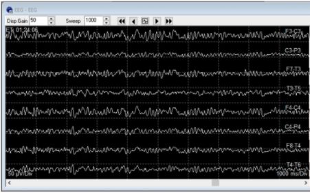
Baseline EEG should look like this image here. Once the clamp goes onto the carotid artery and the timer has started, if the brain has adequate ____ without the need of a shunt, then the EEG should continue appearing stable and (symmetric/asymmetric)
perfusion, symmetric
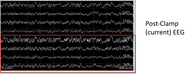
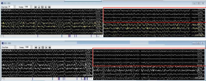
When the anticoagulant heparin is administered and we take our baseline, the EEG looks stable and symmetric (first picture). Is this true for when the clamp was added (second picture)? The surgery is taking place on the left CEA.
No - we can see a flatlining of response in the odd-numbered electrodes specifically (indicating the change is happening on the ipsilateral side, and constitutes a true EEG change). After the shunt is placed, we can see this reperfusion reflected on the EEG.

During a CEA, EEG is unreliable when it is in a ____ ____ pattern. If this pattern is noted during this procedure, it is important to relay this info to the surgical team and then request that anesthetic levels be adjusted for optimal EEG monitoring - especially if the surgeon has made the decision to add a (shunt/clamp) based on IONM signals.
burst suppression, shunt
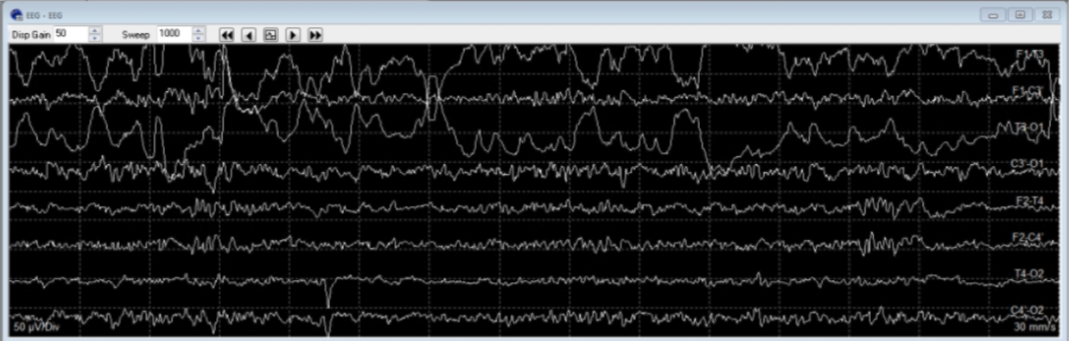
The common electrode showing artifact here is:
T3
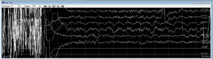
Amplifer saturation can be caused by:
Bovie (electrocautery)
For CEA SSEPs, we take our signals from which two nerves?
median nerve (left and right)
posterior tibial nerve (left and right)
Within the first minute of clamping for a right-sided CEA procedure, there was a change noted in EEG - namely, slower frequencies noted in the (left/right) sided EEG channels, followed by attenuation in (left/right) SSEP signals. Surgeon was informed and began placing (another clamp/shunt). Signals returned to normal limits of baselines after 4 mins of initial signal changes.
right (since changes are ipsilateral for EEG), contralateral or left side SSEP signals (median and posterior tibial nerve). shunt
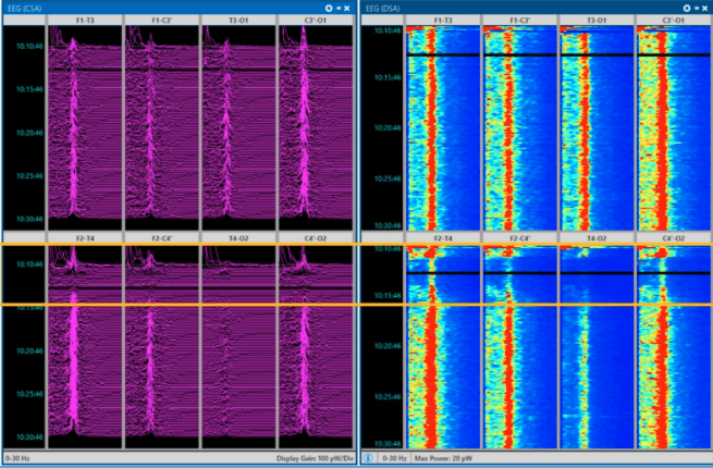
In CSA and DSA EEG, we can see (slowing/speeding up) during clamping that returns when the shunt is in place.
slowing
To detect changes, we use (raw/processed) EEG signals as primary source rather than rely on spectral edge frequency since you may miss an immediate critical change. The utility of looking at the (raw/processed) EEG is to reflect back on how long the EEG was in alert status.
raw, processed
prior to the CEA surgery, it’s a good idea to ask the surgeon if they are selectively shunting the patient - why? Immediate and timely updates on IONM signals are also critical following vessel clamping and (unclamping/shunting). IP should be made aware of surgical stages so they can focus on interpretations during critical periods that require prompt communication. Be ready for immediate changes during critical stages.
to avoid risk of continuous brain ischemia while also preventing mechanical complications like dislodging debris which can lead to stroke (if a clot travels to the brain), scraping artery wall, or getting into the surgical field). unclamping.
Though we focus on ipsilateral EEG and contralateral SSEP signals during CEAs, it is possible to see ____ changes with extreme blockage in collateral blood flow.
global
What is a transcarotid artery revascularization or TCAR - name steps?
minimally invasive procedure involving a soft, flexible sheath placed directly into the carotid artery and connected to a system that will reverse the flow of blood away from the brain
blood flow in the carotid is temporarily reversed to prevent emboli or debris/clot from traveling to the brain and causing a stroke during procedure. The patient’s blood then flows through a filter outside the body, and the filtered blood is then returned through a tube in the patient’s upper leg.
A stent is placed in the artery to reduce the risk of stroke due to narrowing of the artery (stenosis). After the stent is successfully placed, the blood flow reversal is turned off and blood flows in its normal direction. The incision is sewn up with minimal to no visible scarring.
What is a transcarotid artery revascularization or TCAR - what does it treat, and what two things does it combine? What does it reduce the risk of?
carotid artery disease, combines endarterectomy and stenting. reduces stroke risk.
What are the vessels involved in a TCAR?
common carotid
internal carotid (which feeds into circle of Willis and then bifurcates to become MCA and ACA, though ACA is what is involved in this procedure)
external carotid
We do also have watershed zones.
What is the primary modality for TCAR - SSEPs, or EEG?
EEG is the primary modality with a minimum of 8 channels, we use a vascular EEG montage.
Signal changes are more likely to be noted in the side that is ipsilateral to the side of the surgery, but can also be indicated bilaterally.
Do we monitor UN for SSEPs?
No - median nerve. Same idea as for CEA (two or three cortical channels, changes in contralateral side to surgery)
What are the steps of a TCAR?
small incision at base of neck or clavicle
carotid artery exposure
sheath placed in carotid
flow reversal initiated
stent deployed
flow restored, sheath removed, incision closed
We don’t want to use too much of a (sedative/narcotic) with EEG since that could impair our signals, but so could too much _____ agent.
sedative (propofol), inhalational
When monitoring EEG to look for any possible surgical change, we should make particular note of EEG ____ and (symmetry/asymmetry)
suppression, asymmetry
On a right-sided TCAR, we would expect to see any attenuations in SSEPs that we could attribute to surgical cause be on the (left/right) side.
left (contralateral) - let surgeon know so that unclamping can be performed if needed
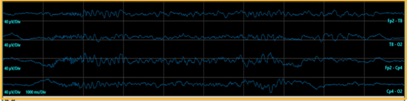
What is this pattern referred to as?
asymmetry on EEG between periods of suppression
What are the 3 types of aortic aneurysms?
ascending thoracic
descending thoracic
abdominal
What is an abdominal aortic aneurysm repair?
surgery performed to repair an abnormal widening of the abdominal aorta (also known as an aneurysm)
if the size of the aneurysm exceeds 55 mm, the risk of rupture becomes greater than the risk of surgery, and an AAA is performed to prevent rupture
if the aneurysm WERE to rupture, blood would fill the abdominal cavity and cause death within minutes
What is Marfan syndrome?
genetic disorder that affects the body’s connective tissue, or blood vessels. Patients with this syndrome are at greater risk of developing aneurysms.
With the temporary interruption of aortic perfusion during a repair, there is the risk of spinal cord ischemia and paraplegia. Which artery provides a major portion of blood supply to the spinal cord
Artery of Adamkiewiez (perfuses lower 2/3 of the spinal cord)
Besides the AoA, additional feeder vessels, including the intercostal arteries that branch off the aorta, play a crucial role in perfusing the ____ regions of the thoracic cord (hint: thoracic!)
watershed
Post-op risk of paraplegia has been reported to be as high as 50% when the ischemic time is greater than 45 mins. true or false?
true
What are the surgical steps for an AAA or abdominal aneurysm repair?
general anesthesia is used
incision down the center of abdomen (below breastbone to below the navel) and across thw abdomen as the second incision - underneath the left arm across to the center of the abdomen and down to below the navel
administer heparin
sigmental feeder vessels clamped
body cooling 32 degrees C or 17 degrees C (neuroprotective hypothermia that slows down metabolism and reduces oxygen demand)
clamps are placed on the aorta above and below the site of the aneurysm to stop blood flow
aortic incision
placing graft inside aorta
close aorta around graft
possible viable intercostal arteries or ICAs re-implanted
remove clamps
patient warming
close abdomen
For an AAA, which is the primary modality - SSEPs or MEPs?
MEPs (when clamping sigmental feeder vessels especially)
What are the steps of an EVAR or endovascular aortic repair?
use general or regional anesthesia
two incisions are made in the groin to access bilateral femoral arteries
fluoroscopy is used to insert needle into femoral arteries
guide wire is threaded to aneurysm site
sheath is placed over the guide wire
contrast dye is injected to see the position of the aneurysm + surrounding blood vessels
stent-graft is then inserted through the femoral artery and up to the side of the aneurysm.
stent graft is expanded and attached to the wall of the aorta
dye is injected again to check for any blood leaking out into the aneurysm area
if NO blood is leaking, instruments are removed, bilateral incision sites are closed
What are the different modalities for an AAA, and what are their alert criteria (talk about MEPs only)
MEP utilized during clamping of sigmental or intercostal arteries to predict potential ischemia of spinal cord
MEP signal changes can help provide info to the surgeon on whether patient would benefit from reimplantation of the intercostal arteries after aortic repair to provide collateral blood flow to thoracic watershed region of spinal cord (abdominal aortic repair)
Talk about SSEPs with regards to AAA
attenuation is expected during patient cooling and can assist with identifying the optimal temperature for circulatory arrest (corticals > subcorticals > erbs)
time of signal reappearance can provide more info on the post-operative outcome
Talk about EEG with regards to AAA
EEG should be monitored during profound body cooling in preparation for circulatory arrest, since some patients may still have a small amount of residual EEG activity even at 14 C
complete electrical silence identified with an isoelectric EEG pattern helps the surgical team decide when the patient is sufficiently cooled (neuroprotective state)
return of EEG is noted after patient warming - time of reappearance of signals provides more info on post-op outcome
assist with determining optimal amount of time patient can tolerate circulatory arrest
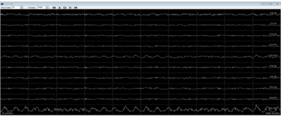
What is this EEG known as?
isoelectric
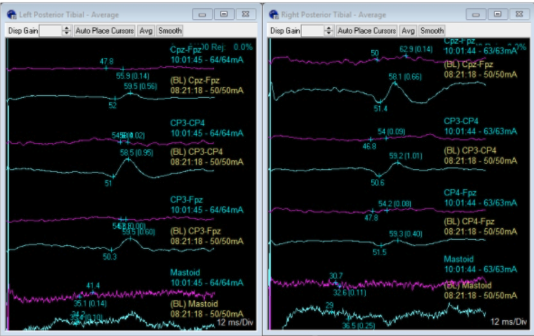
Since blue is baseline trace and purple is the current trace, we can say that we are seeing (bilateral/unilateral) SSEP decrease in PTN.
bilateral - purple trace for cortical, subcortical, and peripheral is much flatter
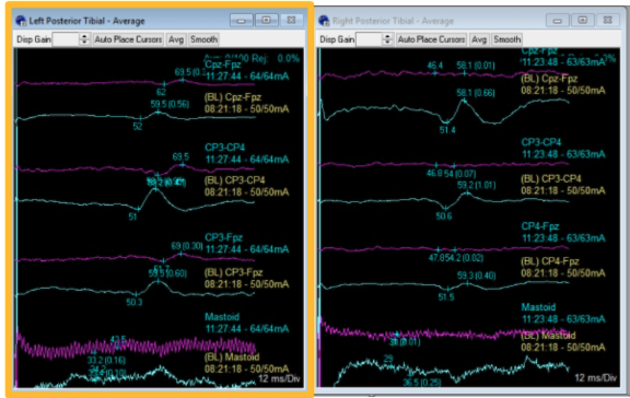
This picture not only reflects a decrease in amplitude of the signals (purple is current trace, and blue is baseline), but also an increased ____.
latency
What is the primary objective of monitoring AAAs?
provide timely warning of new physiologic imbalance and assess patient response to corrective action
monitor arteries that branch from the aorta and provide vital blood flow to thoracic (watershed) region of the spinal cord
postop neurological function is presumed to be intact if signals return to baseline at the end of the procedure
surgeries with IOM decreased overall cost of care for patients
What is a coronary artery bypass graft?
Surgery to restore normal blood flow to obstructed coronary artery by way of bypass graft placement
To treat the blocked or narrowed artery, a piece of healthy blood vessel from elsewhere in the patient’s body is used to bypass the blockage. The bypass graft connects to an area above and below blockage, so blood flow can be restored to heart muscle.
What is the difference between vein and artery graft?
vein graft: vein is harvested from the leg or wrist, and then stitched from the aorta to the right coronary artery
artery graft: artery is harvested from the chest and stitched from the subclavian artery to the left coronary artery
normal heart has less fat and plaque build-up within the vessels
What are the steps of surgery for a CABG?
make incision on either leg or wrist to harvest blood vessels for grafts
close leg/wrist incision
place electrodes after the harvest
chest incision from just below Adam’s apple to just above the navel
sternum will be cut in half and separated with a retractor
we can either use a bypass machine, or not
close sternum with small wires
drains placed to prevent fluid buildup around heart
skin closure
What are the steps of CABG with bypass machine (from this section only?)
surgeon will temporarily stop the heart to sew grafts into place
heart is hooked up to bypass machine
once bypass machine is running the heart will be stopped by an injection of cold solution
graft is placed
bypass machine is turned off
bypass machine will be removed, and heart should restart on its own
if heart does not restart, small shock will be administered
temporary pacemaker may be placed, if needed, for recovery
What are the steps of CABG without bypass machine?
heart continues to function and pump blood through procedure
bypass machine kept on standby just in case
graft placed
examine grafts to make sure they function properly
Shifts in latency for MN and PTN can occur during ____ and constitute a ____/alert criteria.
cooling, change.
Isoelectric pattern from EEG is considered (normal/alert) criteria.
alert
For CABG, our signals are utilized to assess effective body warming and the amount of time it takes for signals to reemerge when body temperature is normalized. true or false?
false - body COOLING
Since we use a limited 2 channel EEG montage to assess the depth of anesthesia for spinal procedures, it is difficult to identify symmetry of the hemispheres - so it is best to describe them as _____ and (continuous/discontinuous)
limited and continuous
Increased sedation can cause ____ suppression or isoelectric patterns on an EEG.
burst