Exam 3 Psych
1/103
Earn XP
Description and Tags
Name | Mastery | Learn | Test | Matching | Spaced | Call with Kai |
|---|
No analytics yet
Send a link to your students to track their progress
104 Terms
why is sex not a physiological drive?
sex is not essential for the survival of the individual
there are similarities however:
arousal, satiation
role of hormones
involvement in specific brain areas
phases of the sexual response cycle
excitement phase
plateau phase
orgasm phase
resolution period
refractory phase
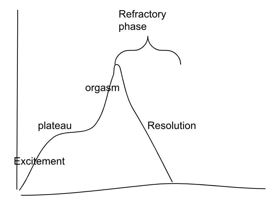
refractory phase
amount of time that a male requires before being able to reach orgasm again
coolidge effect
quicker return to sexual arousal for a male when a new female is introduced, shortening of refractory phase for men when new gal becomes available
observed in many species
helps explain cheap sperm, expensive eggs theory
analogous to sensory specific satiety - the more of a specific food a person eats, the less appealing the food becomes, encourages varied diet
cheap sperm expensive eggs theory
a male can potentially have a large number of children quickly by mating with different females→ more of his genes passed on to the next generation
the major sex hormones
androgens- male
estrogen
progesterone
androgens
responsible for male characteristics and fxn
testosterone
necessary for male sexual behaviors but requires only small amt
females initiate more at midcycle, may be more impt than estrogen
result of sexual activity in both males and females (inc caused by behavior)
estrogen
responsible for female characteristics and fxns
progesterone
controls reproductive fxns including sexual receptivity and desire in females
oxytocin
promotes sexual arousal and orgasm (males and females)
brain structures involved in sexual activity
medial preoptic area of hypothalamus
ventral medial nucleus of hypothalamus
medial amygdala
paraventricular nucleus of hypothalamus
sexual dimorphic nucleus of preoptic area of hypothalamus
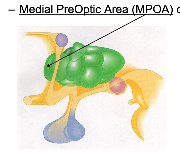
medial preoptic area of hypothalamus and sex
MPOA
stimulation in rates inc copulation in both males and females
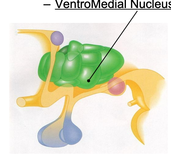
ventral medial nucleus of hypothalamus and sex
VMN
active during copulation
destruction reduces responsiveness of females to males and vigor of males mating
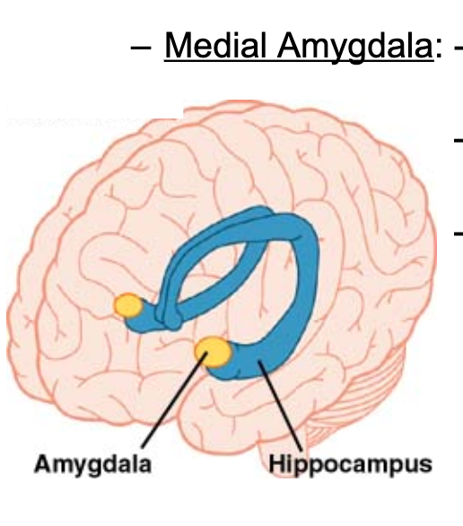
medial amygdala and sex
active during copulation in both males and females
stimulation→ dopamine release in MPOA
involved in recognizing and responding to opposite sex
paraventricular nucleus of hypothalamus and sex
PVN
involved in sexual performance in males
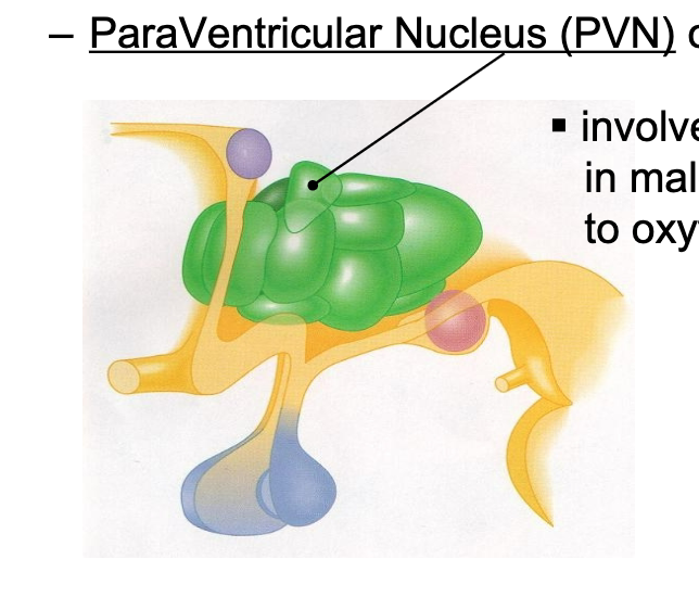
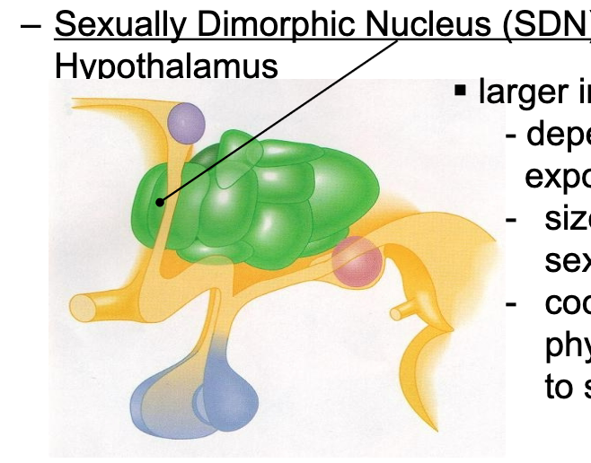
sexual dimorphic nucleus of preoptic area of hypothalamus and sex
SDN
larger in male rats
size depends on prenatal exposure to testosterone
size related to level of sexual activity
coordinates behavioral and physiological responses to sensory cues
dopamine role in sex
inc in medial preoptic area of hypothalamus
Also involved in mesolimbic pathway( NAcc, MFB and VTA)
inc in ventral tegmental area in males during ejaculation
inc in nucleus accumbens (with coolidge effect)
serotonin role in sex
inhibitory effect on sexual drive
inc following orgasm- contributes to refractory period
sensory stimuli and sex
tactual, auditory, visual, olfactory, pheromones
visual sensory stimuli and sex
body symmetry is another important determinant of sexual attractiveness
may be indicative of genetic fitness
olfactory sensory stimuli and sex
major histocompatibility complex (MHC) is a group of genes involved in immune system function
many vertebrates prefer mates with dissimilar MHC genes→ involved in better disease resistance in offpsring
unclear role in humans → relationship btwn MHC type of pleasantness of odor from sweat
pheromones and sex
airborne chemicals released by an animal that has a physiological or behavioral effect on another animal of the same species
female gypsy moth attract mate from 2 miles away
pheromones detected by vomeronasal organ (VNO) → sends signals to MPOA and VMN
microscopic in size, unclear if it is functional in humans
possible human pheromones- AND(male) and EST (female)
recently been cast in doubt
pheromonal influence of sexual behavior of humans is unclear and controversial
emotion and psych
relationship btwn emotional experiences and body’s resonses is unclear and controversial
james lange theory of emotion
late 1800s
perception of specific patterns of physiological arousal affecting specific emotional experience
i feel sad bc I cry (rather than reverse)
faced criticism afterwards by cannon
cannons criticism
criticism of james lange thoery (1920)
autonomic nervous system responds the same way for different emotions
perception of physiological responses cannot account for the variety of emotional experiences
schachter and singers congitive theory of emotion
1962
identification of emotions relies on a cognitive assessment of the external stimulus situation → physiological arousal contributes only to the intensity of emotion
emotion impact on autonomic system
the autonomic nervous system is activated differently depending on the emotion
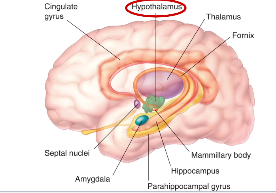
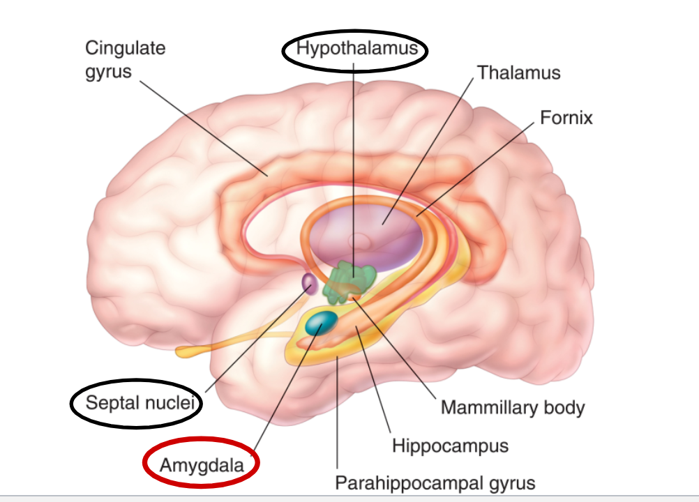
limbic system
complex network of cortical and subcortical structures
hypothalamus
septal nuclei
amygdala
anterior cingulate cortex and anterior insular cortex
what 2 things play a role in emotion
cognitive assessment of external stimuli and physiological feedback
hypothalamus and emotion
electrical stimulation in animals produces threatening or defensive behaviors
in humans it evokes feelings of rage, fear or pleasure
septal nuclei and emotion
evokes feelings of pleasure (sexual)
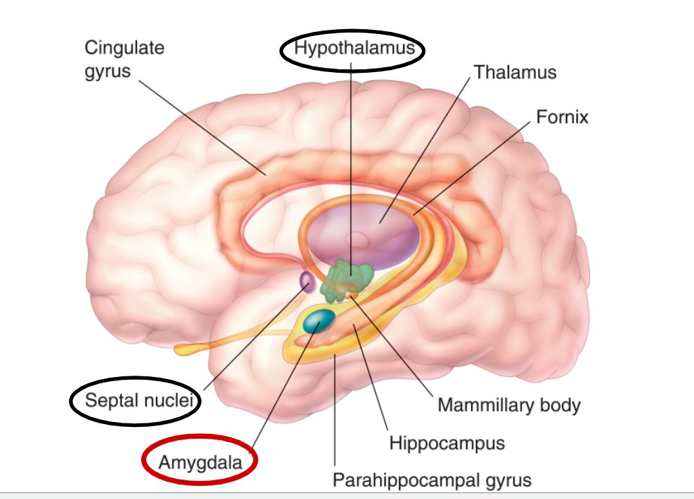
amygdala and emotion
involved in perception of facial expressions of emotion (esp fear)
damage- removes fear and aggression, peeps do not respond emotionally to rewards and punishments
stimulation- produces fear and aggression
major role in fear and anxiety
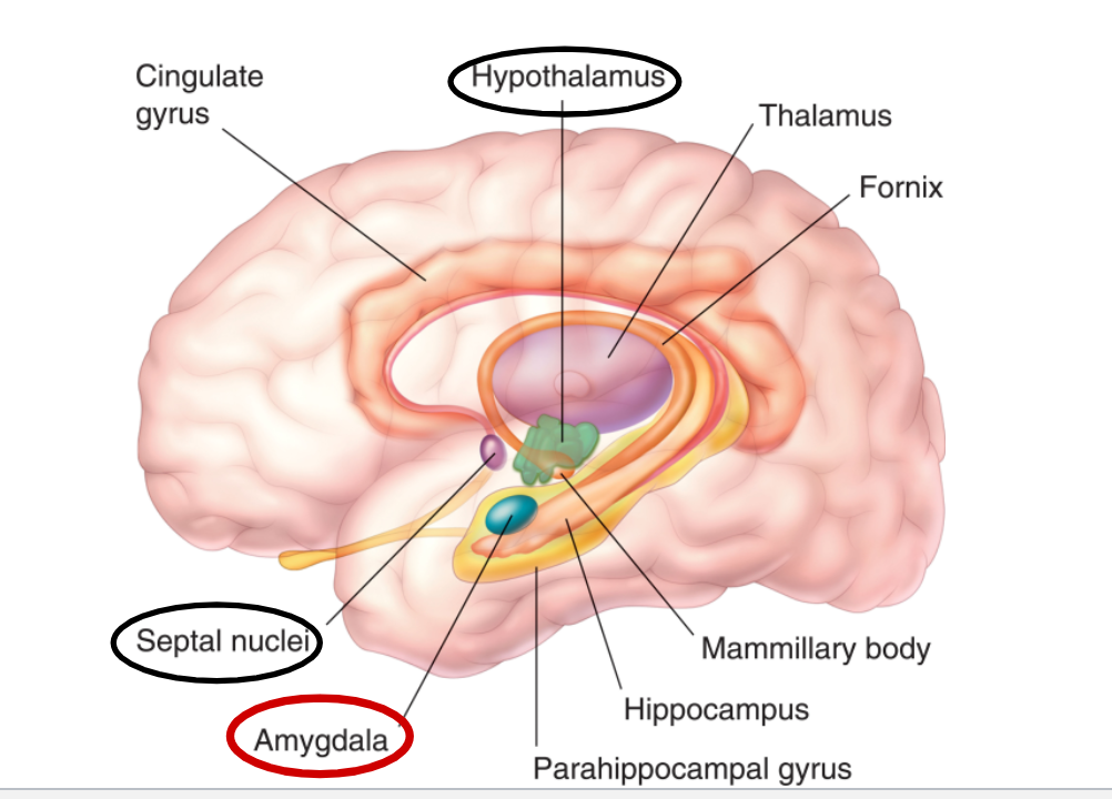
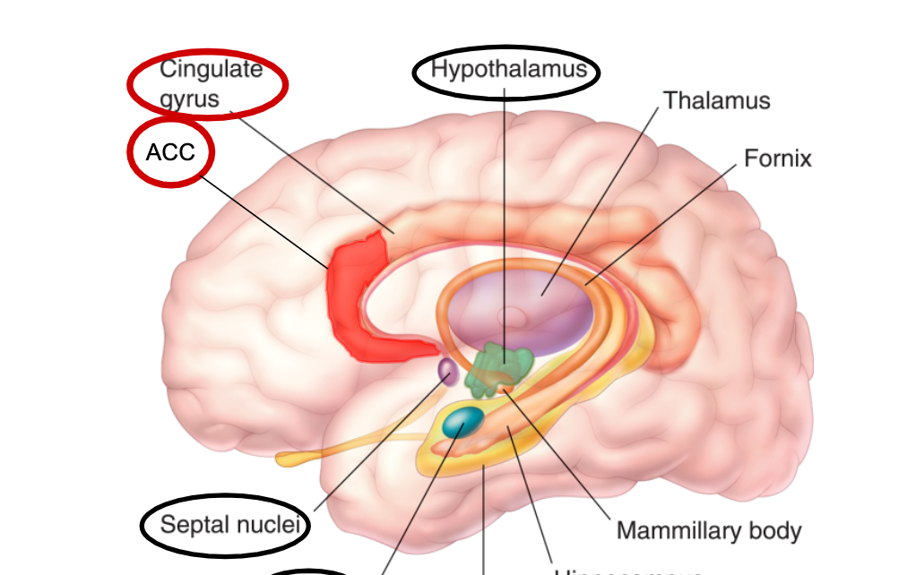
Anterior cingulate cortex + anterior insular cortex and emotion
Anterior cingulate cortex
wraps around corpus callosum
uses input from AIC to generate action via prefrontal cortex
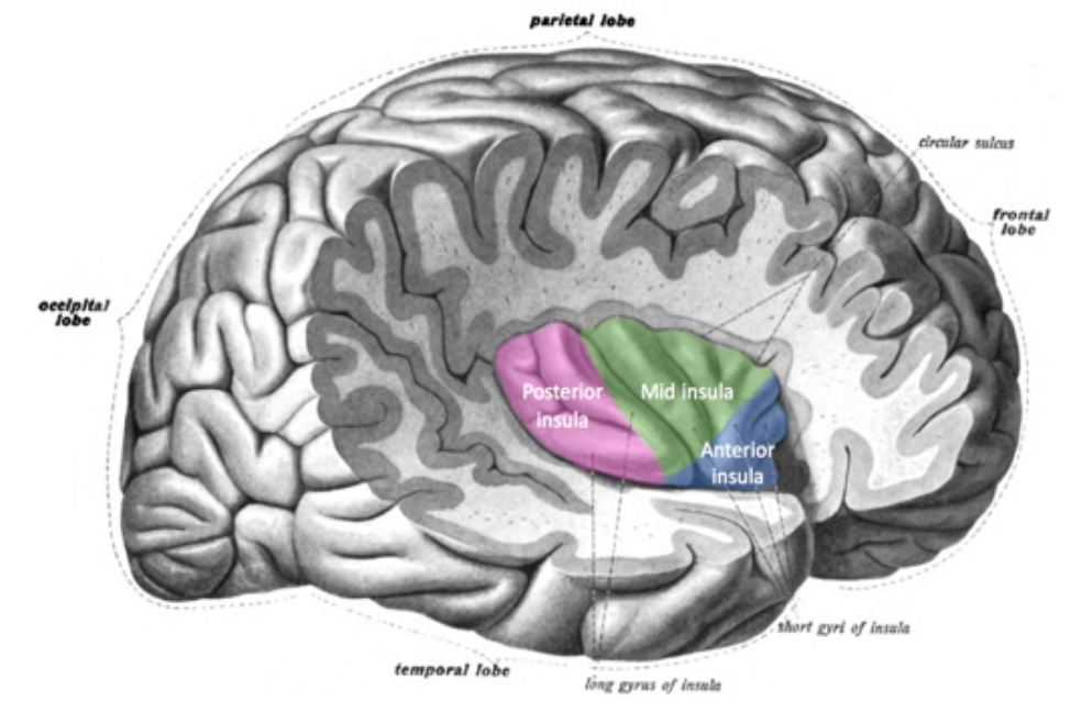
Anterior insular cortex
deep in brain, insula
awareness of emotion
both form network with amygdala and hypothalamus
both involved in all basic emotions and with empathy
conscious experience of emotion
prefrontal cortex and emotion
connections with limbic system
uses emotional info from limbic system for decision making
damage- blunts emotional response, impaired ability to process and anticipate consequences of behavior and use that information to modify behavioral choices
hemispheric asymmetry
hemispheres differ in
structure (brocas area larger on L)
function performed (L more involved in linguistics)
right more involved with facial expression and emotion→ difficulty recognizing emotions in others following brain damage
Sensory system
set of components of the PNS and CNS involved in acquiring and processing of specific sensory info
sensation- acquistion of sensory info
perception- interpretation of sensory info
receptor
a cell that is suited by its structure and fxn to respond to a specific form of energy
often a specialized neuron
transducer- device that converts energy from one form to another
stimulus
specific energy form for which the receptor is specialized for
hearing
stimulus- vibration in conducting medium
types of sound
pure tones
complex sound
pure tones
single frequency
number of alternating compression and decompression(cycles) that occur per unit time
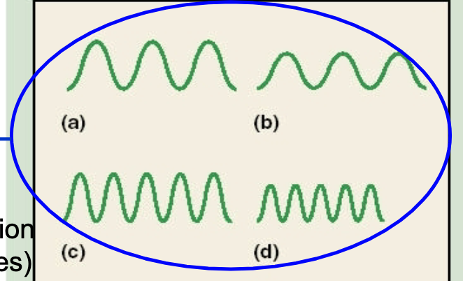
complex sounds
combination of 2+ frequencies
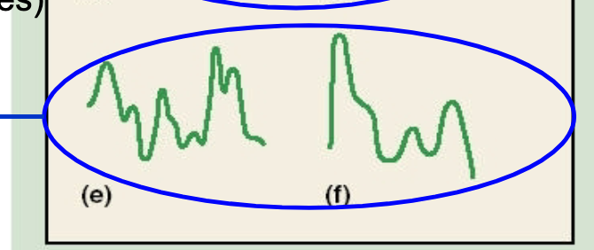
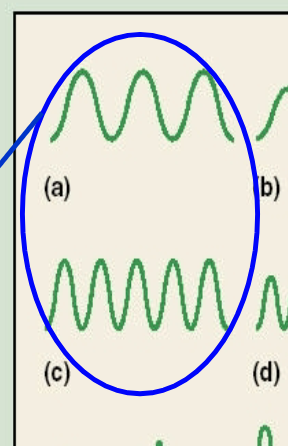
pitch
experience of the frequency of sound
same frequency= same pitch
unequal amount of waves (higher or lower frequency) = diff pitch
image= low frequency vs high frequency results in different pitch
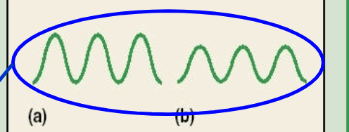
loudness
experience of the intensity (physical energy of a sound)
unequal intensity= unequal loudness
same amplitude height= same loudness
different amplitude height = diff loudness
image= different amplitude height= difference in loudness
auditory apparatus
outer ear
middle ear
inner ear
outer ear
pinna- impt for sound location in space
external auditory canal- resonation chamber, amplify intensity of sound
middle ear
tympanic membrane
auditory ossicles- transfer/amplify vibrations from ear drum to oval window on cochlea
inner ear
semicircular canals
basilar membrane- vibrate when oval window vibrates
tectorial membrane on top of basilar
hair cells stick into tectorial membrane→ organized in rows
cochlea
organ of corti
auditory nerve
auditory pathways
auditory nerve→ coclear nuclei (filter raw data)→ superior olive→ inferior colliculus (startle reflex)→ medial geniculate nucleus of thalamus→ primary auditory cortex
primary auditory cortex
1st cortical region recieving info from sound, involved in analyzing simplest aspects of sound
topographically organized- aadjacent neurons in cortex receive info from adjacent receptor locations in basilar membrane
map of basilar mem
secondary auditory cortex
anterior- apex of basilar membrane corresponds to here
posterior- base (closest to oval window) of basilar membrane corresponds to here
frequency theory
basilar mem vibrates in synchrony with a sound → auditory nerve axons fire at same frequency
problem- individual neurons can fire at no more than 1000 Hz but we can hear up to 20,000 Hz
volley theory- groups of neurons can follow the freq of a sound whereas a single nueron cannot (summation)
place theory
sounds with diff freqinduce peaks of max vibration in diff places on basilar membrane
high freq- basilar mem vibrate more at base
low freq- basilar mem vibrates more at apex
problem- with sounds below 200Hz the whole membrane vibrates equally
frequency place theory
frequency part- synchrony of firing rate of auditory nerve axons with sound frequency→ pitch perception of sounds up to 200Hz
place part- place of max vibration on basilar mem→ pitch perception of sounds greater than 200 Hz
perception of complex sound
we mostly hear in complex sounds rather than just pure tones
use fourier analysis to analyze sound
fourier analysis- Any complex sound can be broken down into pure tones (component frequencies)
basilar membrane of cochlea acts as fourier analysis
cocktail party effect- isolate meaningful auditory stimulus from loud environment
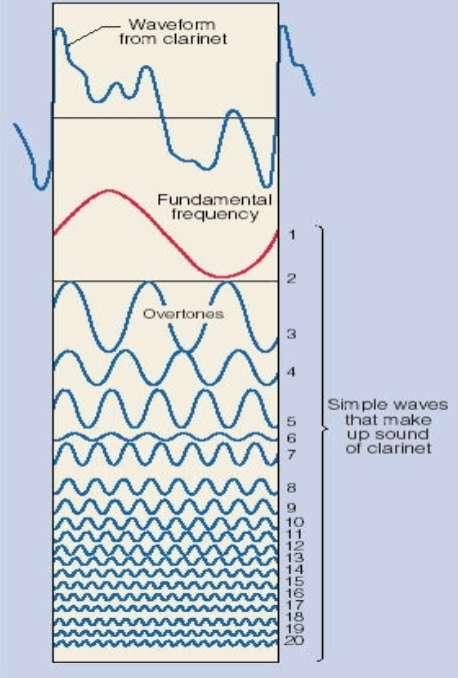
fourier analysis
any complex sound can be broken down into pure tones
basilar mem of cochlea acts as fourier analysis
cocktail party effect
isolate meaningful auditory stimulus from loud environment
outer hair cells of ear involved; mute certain sounds so you can filter sound
3 types of binaural cues for sound localization
Superior olive is the hub!
Phase difference- a sound arriving from one side of the body is at a different phase of the sound wave at each ear (low freq sounds)
Intensity differences- the head creates a sound shadow-> near ear receives a slightly more intense sound than far ear
Difference in time of arrival- sound reaches near ear slightly before the far ear
the one monaural cue
Head-Related Transfer Function (HRTF): frequency alterations to a sound as it passes through the pinna and auditory canal, as well as through the head and other parts of the body.
vision
stimulus- visible light from the electromagnetic spectrum
electromagnetic spectrum order
radio, microwave, infrared, visible light, ultraviolet, x ray, gamma ray
cornea
curved and thick transparent membrane in front of the eye that accounds for 80% of the ability to form images
lens
series of transparent onion-like layers
shape can be changed by contractions of ciliary muscles
retina
the neural tissue and photoreceptors located on the inner surface of the posterior portion of the eye
3 layers
photoreceptor layer, bipolar layer and ganglion layer
photoreceptors
rods and cones
contain 1 protein and 1 vitamen
rods
contain rhodopsin (photopigment), very sensitive to light
detect diff levels of light and dark, not colors
most concentrated away from fovea
cones
contain lodopsin (photopigment)
require high level of light
3 kinds : red blue green
most concentrated in fovea
horizontal cells
helps further synapsing with other photoreceptors and bipolar cells
bipolar cells
connect photoreceptor layer to ganglion cells
amacrine cells
help further synapsing
ganglion cells
axons cluster together to form optic nerve
retina made up of…
photoreceptors, horizontal cells, bipolar cells, amacrine cells, ganglion cells
fovea
the center of the retina, area of focus, most cones here
light flows in what way in the eye
flows through ganglion, amacrine, bipolar cells, horizontal cells, then photoreceptor cells, neural processing occurs from photoreceptors to the ganglion cells
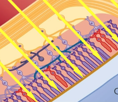
Retina in dark
Na channels OPEN
depolarization.
inhibitory NT released
bipolar cells inhibited as well as ganglion cells
retina in light
light protein and lipid separate
Na channels CLOSE
release of inhibitory NT is reduced
firing rate of bipolar cell to ganglion cell inc
receptive field of retina
area of retina from which a ganglion cells receives input
small- one PR to 1 ganglion cell (fovea- more detail)
large- many PR to 1 ganglion cell (away from fovea- less detail)
round in retina
visual field
the part of the enviornment that is registered on the retina
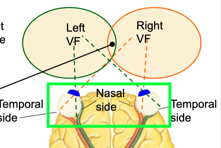
L and R fields overlap = binocular field
if object is on non-overlapping side of L VF= left eye image will form on nasal side and R eye image will form on temporal side
if object is on non-overlapping side of R VF= left eye image will form on temporal side and R eye image will form on nasal side
optic tracts carry info to thalamus→ lateral geniculate nuclei→ primary visual cortex
retinal disparity
Discrepancy in the location of an obj image on the 2 retinas as a function of the obj distance -> detected by the visual cortex-> depth perception
trichromatic theory
von helmholtz and young
All colors are the result of the processing of 3 “pure” colors: red green and blue, each one detected by a specific receptor
Light can mix to create different colors
Problem: yellow also appears to observers as pure color
Summary- 3 types of cones in retina correspond to 3 primary colors; all true but doesn’t explain yellow
opponent process theory
hering
Explains color vision in terms of opposing neural processes in 2 specific receptors
Receptor for red and green -> photochemical
Receptor for blue and yellow-> photochemical
Explains complementary colors and negative color after effect -> flipped color image and different color
2 types of cones correspond to red/green and yellow/blue; can explain rebound effect of negative color after effect
combined theory of color perception
3 types of cones for red green and blue
retina responds in terms of paired colors
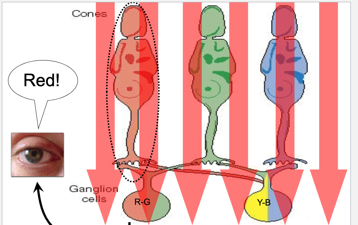
Red light- activates red cone->excites red/green ganglion cell-> interprets as red
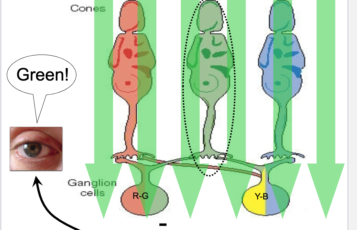
Green light- activates green cone->inhibits red/green ganglion cell -> interprets as green
Blue light- activates blue cone-> inhibits yellow/blue ganglion cell -> interprets as blue
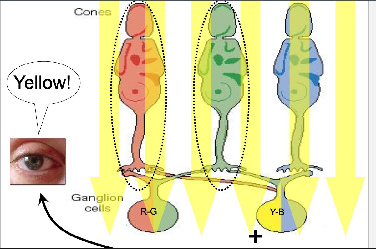
Yellow light- activates red and green cones -> red/green ganglion cell signals cancel each other out, yellow/blue ganglion cell excited -> interprets as yellow
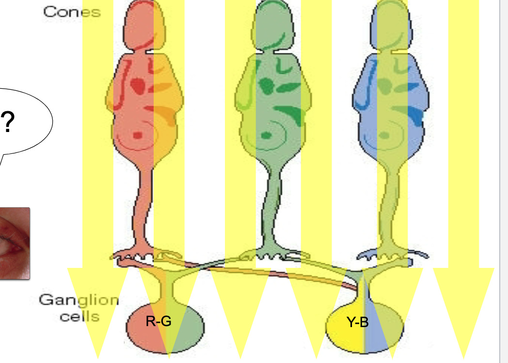
red light color perception
Red light- activates red cone->excites red/green ganglion cell-> interprets as red
green light color perception
Green light- activates green cone->inhibits red/green ganglion cell -> interprets as green
blue light color perception
Blue light- activates blue cone-> inhibits yellow/blue ganglion cell -> interprets as blue
yellow light color perception
Yellow light- activates red and green cones -> red/green ganglion cell signals cancel each other out, yellow/blue ganglion cell excited -> interprets as yellow
R+/G- ganglion cells
red light-> excitatory, green light-> inhibitory
G+/R- ganglion cells
ganglion cells- red light-> inhibitory, green light-> excitory
Y+/B- ganglion cells
yellow light-> excitatory, blue light-> inhibitory
Y-/B+ ganglion cells
yellow light-> inhibitory, blue light-> excitatory
perception of edges
lateral inhibition
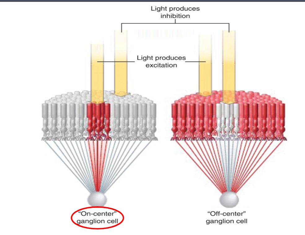
on center and off center ganglion cells
simple cells in visual cortex
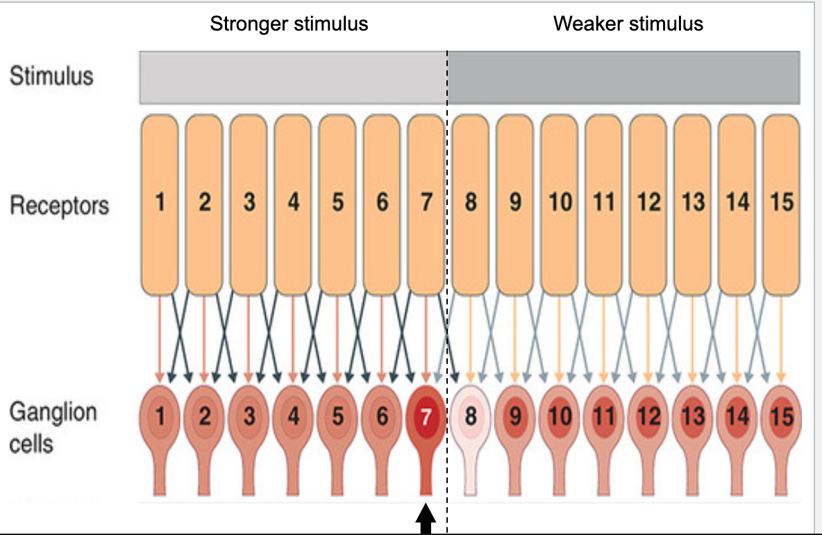
lateral inhibition
ganglion cells inhibit and are inhibited by neighboring cells
explains the mach illusion
Blue arrows are the lateral inhibition; receptors 1-7 are much more impacted than 8-15. This impacts lateral inhibition. Ganglion cell 7 has weaker inhibition due to dec lateral inhibition from receptor 8. Ganglion cell 8 has stronger inhibition due to the strong inhibitory signal from receptor 7.
Because of this, since ganglion cell 7 is less inhibited it is lighter, and ganglion cell 8 is more strongly inhibited and so is darker. This explains the illusion
At edges, ganglion cells get differential amounts of inhibition from darker area and brighter area-> makes edges stand out perceptually
off center and on center ganglion cells
Antagonistic arrangement of receptive fields-> detection of light/dark contrast
microedge detectors
On center- when light shines on center= excitation; light shines on periphery=inhibition
Off center- when light shines on center= inhibition; light shines on periphery=excitation
visual cortex receptive field
rectangular
simple cells of visual cortex
respond to edges at a specific orientation and place on the retina
complex cells of visual cortex
continue to respond (unlike simple cells) when an edge moves to a different location
spatial frequency theory
Visual cortical neurons perform a fourier analysis of the luminosity variations of a scene
Neurons in visual cortex do not detect only edges, but have a variety of different sensitivities
Visual world is combination of high and low spatial frequencies
Need neurons that are sensitive to both:
High freq-> edge detection and fine details
Low freq-> detection of textural properties
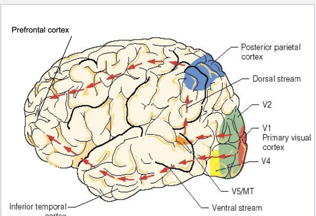
parvocellular system
V1, V2, V4→ ventral stream (what stream- crucial for identifying objects by sight)→ inferior temporal lobe→ prefrontal cortex
Color vision (v4) and detailed obj recognition (inferior temporal lobe)
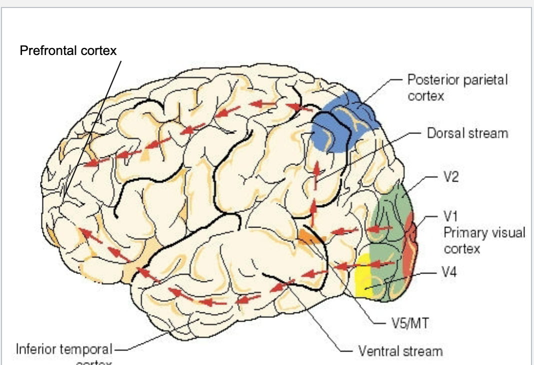
magnocellular system
V1, V2 → median temporal gyrus (V5)→ dorsal stream (where system- for figuring out where objects are in space in relation to others) → posterior parietal cortex→ prefrontal
Brightness, contrast, orientation, movement, depth and location of objects
disorders of visual perception
object agnosia
color agnosia
movement agnosia
neglect